Comparative Genome Analyses of
Streptococcus suis
Isolates from Endocarditis
Demonstrate Persistence of Dual Phenotypic
Clones
Mari Tohya
1, Takayasu Watanabe
1, Fumito Maruyama
2¤b*
, Sakura Arai
1, Atsushi Ota
2,
Taryn B. T. Athey
3, Nahuel Fittipaldi
3,4, Ichiro Nakagawa
2, Tsutomu Sekizaki
1¤a*
1Research Center for Food Safety, Graduate School of Agricultural and Life Sciences, The University of Tokyo, Bunkyo-ku, Tokyo, Japan,2Department of Microbiology, Kyoto University Graduate School of Medicine, Kyoto, Kyoto, Japan,3Public Health Ontario, Toronto, Ontario, Canada,4Department of Laboratory Medicine and Pathobiology, Faculty of Medicine, University of Toronto, Toronto, Ontario, Canada
¤a Current address: Research Center for Food Safety, Graduate School of Agricultural and Life Sciences,
The University of Tokyo, Bunkyo-ku, Tokyo, Japan
¤b Current address: Department of Microbiology, Kyoto University Graduate School of Medicine, Kyoto,
Kyoto, Japan
*[email protected](TS);[email protected](FM)
Abstract
Many bacterial species coexist in the same niche as heterogeneous clones with different
phenotypes; however, understanding of infectious diseases by polyphenotypic bacteria is
still limited. In the present study, encapsulation in isolates of the porcine pathogen
Strepto-coccus suis
from persistent endocarditis lesions was examined. Coexistence of both
encapsulated and unencapsulated
S
.
suis
isolates was found in 26 out of 59 endocarditis
samples. The isolates were serotype 2, and belonged to two different sequence types
(STs), ST1 and ST28. The genomes of each of the 26 pairs of encapsulated and
unencap-sulated isolates from the 26 samples were sequenced. The data showed that each pair of
isolates had one or more unique nonsynonymous mutations in the
cps
gene, and the
encapsulated and unencapsulated isolates from the same samples were closest to each
other. Pairwise comparisons of the sequences of
cps
genes in 7 pairs of encapsulated and
unencapsulated isolates identified insertion/deletions (indels) ranging from one to 10
4bp in
different
cps
genes of unencapsulated isolates. Capsule expression was restored in a
sub-set of unencapsulated isolates by complementation
in trans
with
cps
expression vectors.
Examination of gene content common to isolates indicated that mutation frequency was
higher in ST28 pairs than in ST1 pairs. Genes within mobile genetic elements were mutation
hot spots among ST28 isolates. Taken all together, our results demonstrate the coexistence
of dual phenotype (encapsulated and unencapsulated) bacterial clones and suggest that
the dual phenotypes arose independently in each farm by means of spontaneous mutations
in
cps
genes.
a11111
OPEN ACCESS
Citation:Tohya M, Watanabe T, Maruyama F, Arai S, Ota A, Athey TBT, et al. (2016) Comparative Genome Analyses ofStreptococcus suisIsolates from Endocarditis Demonstrate Persistence of Dual Phenotypic Clones. PLoS ONE 11(7): e0159558. doi:10.1371/journal.pone.0159558
Editor:Patrick M Schlievert, University of Iowa Carver College of Medicine, UNITED STATES
Received:March 15, 2016
Accepted:July 4, 2016
Published:July 19, 2016
Copyright:© 2016 Tohya et al. This is an open access article distributed under the terms of the
Creative Commons Attribution License, which permits unrestricted use, distribution, and reproduction in any medium, provided the original author and source are credited.
Data Availability Statement:All relevant data are within the paper and its Supporting Information files. All genome sequence data files are available from the DNA Data Bank of Japan Sequence Read Archive under accession number DRA004206.
Introduction
Bacteria can adapt to environmental changes through various adaptive strategies. One example
is coexistence of different phenotypes of the same organism at specific niches [
1
].
Polyphenoty-pic infections by a single species have been reported in many bacterial species, including
Pseu-domonas aeruginosa
,
Helicobacter pylori
,
Escherichia coli
, and
Staphylococcus epidermidis
[
1
–
4
]. The ecologies of these infections have shown that the composition of polyphenotypic clones
permit adaptation to various environmental conditions. Coexistence by polyphenotypic clones
is thus thought to be a barrier to treatment and prophylaxis [
2
,
4
–
7
]. On the other hand, the
compositions of coexistable bacteria are affected by antimicrobials and host immunity [
8
,
9
].
Therefore, bacterial population dynamics in response to environmental changes play an
impor-tant role in evasion from host immune system [
10
–
12
].
Variation of surface structures in bacteria is important for adaptation and survival in
vari-able environments. Although capsules are major bacterial extracellular components and show
antiphagocytic effect, the loss of capsules can also be beneficial [
13
–
16
]. Hanage
et al
. reported
that one or more capsule-related genes were not detected by PCR in isolates of
Streptococcus
pneumoniae
from nasopharyngeal swabs and middle ear fluid [
10
]. This finding suggested that
unencapsulation afforded advantages to capsule-less clones in some habitats. In
Streptococcus
pyogenes
, unencapsulated clones isolated from pharyngitis and invasive diseases are known to
be invasive and cause diseases [
16
,
17
]. Similarly, unencapsulated clones of the zoonotic
patho-gen
Streptococcus suis
[
18
–
20
] were isolated from porcine endocarditis lesions [
21
]. The role of
these phenotypic changes has, however, not yet been established. In addition, although
S
.
suis
isolates from different worldwide locations are known to be genetically highly diverse, there is
no information about the genetic diversity of isolates recovered from a single lesion [
22
].
The
S
.
suis
capsule is composed of capsular polysaccharides (CPs), whose biosynthesis is
dependent on the concerted action of enzymes encoded by genes located in the so-called
capsu-lar polysaccharide synthesis (
cps
) locus [
23
–
27
]. Different antigenicities of the
S
.
suis
CPs are
the basis of
S
.
suis
serotyping [
27
,
28
].
S
.
suis
CPs have been shown to play a major role in
pro-tection against host phagocytes [
29
–
31
], and unencapsulated
S
.
suis
clones have been described
as low virulent or completely avirulent, leading to the notion that loss of capsule is unfavorable
for
S
.
suis
virulence. However, the ability to form biofilms and to adhere to epithelial cells and
platelets was previously reported to be stronger in unencapsulated clones than in encapsulated
ones [
32
–
34
], as unencapsulated clones exclusively invaded into epithelial cells [
32
,
34
,
35
].
Therefore, from an ecological perspective, it is plausible that unencapsulated and encapsulated
S
.
suis
clones coexist in the same lesions, and that the presence of both encapsulated and
unen-capsulated cells originated from genetically identical clones facilitate adherence and invasion to
host cells, thereby permitting efficient evasion from the immune system and persistence of the
clonal population in a particular habitat or niche.
In the present study, to determine whether encapsulated and unencapsulated, or dual
pheno-typic
S
.
suis
clones coexist in porcine endocarditis lesions, we collected multiple
S
.
suis
isolates
from each endocarditis lesion and confirm encapsulation status of the isolates. Finally,
compara-tive genome analyses allowed us to precisely define the close relationships between encapsulated
and unencapsulated isolates from the same endocarditis lesion. Our findings suggest that
muta-tions from encapsulated cells to unencapsulated ones occurred independently in each pig or farm.
Materials and Methods
Bacteria, plasmids and growth condition
Fifty-nine heart valve vegetations of porcine endocarditis from 24 farms (
Fig 1A
) were collected
in meat inspection centers in Japan between 2013 and 2014. These samples were stamped onto
Todd-Hewitt (TH, Becton Dickinson, MD, U.S.) agar plates containing 5% horse blood,
fol-lowed by incubation at 37°C for 16 hours under a 5% CO
2atmosphere. Twenty-four colonies
were picked from each sample and purified twice by single colony isolation. Amplification of
the
S
.
suis recN
gene, which serves to confirm species [
36
] was performed by PCR, and the
posi-tive isolates were further examined by PCR permitting serotype estimation [
37
,
38
]. All isolates
were stored in Luria-Bertani (LB, Becton Dickinson) broth containing 30% glycerol at -80°C.
For
S
.
suis
isolates, encapsulation was determined using a co-agglutination test [
21
]. One pair of
encapsulated and unencapsulated
S
.
suis
isolates was selected from each of 26 endocarditis
sam-ples that yielded both encapsulation and unencapsulated phenotypes (a total of 52 isolates,
Table 1
).
Escherichia coli
strains and plasmids used in this study are listed in
S1 Table
[
39
,
40
].
E
.
coli
strains were grown on LB broth at 37°C for 16 hours. When necessary, media were
sup-plemented with spectinomycin (50
μ
g/ml or 100
μ
g/ml for
E
.
coli
and
S
.
suis
, respectively).
Genomic DNA extraction
Genomic DNA was extracted from the 52
S
.
suis
isolates as described previously [
41
]. DNA
concentrations were determined using the Quant-iT PicoGreen dsDNA Assay Kit (Life
Tech-nologies Corporation, CA, U.S.), and quality was checked using a NanoDrop 1000 instrument
(Thermo Fisher Scientific, DE, U.S.).
Multilocus sequence typing (MLST)
The 52
S
.
suis
isolates were examined by MLST, as described previously [
42
]. The sequence
type (ST) was determined by comparing the nucleotide sequences of 7 loci with the data
depos-ited in the
S
.
suis
MLST database (
http://ssuis.mlst.net
).
Whole-genome sequencing
Genomic libraries were prepared using Nextera XT DNA kits (Illumina Inc., CA, U.S.).
Paired-end sequencing was performed using MiSeq Reagent Kit v3 (600-cycles) in the Illumina MiSeq
platform. Quality trimming and filtering of the obtained sequence reads were performed using
CLC Genomics Workbench v8.0.1 (CLC bio, Aarhus, Denmark) with the following
parame-ters: Quality Limit = 0.01, Adapters Trimming = Yes, Remove 5
0Terminal Nucleotides = Yes,
Number of 5
0Terminal Nucleotides to Remove = 20, Remove 3
0Terminal Nucleotides = Yes,
Number of 3
0Terminal Nucleotides to Remove = 5, and Discard Reads below Length = 50.
Construction of phylogenetic trees
The preprocessed reads were
de novo
assembled using A5-miseq, as of May 22th, 2015, with
default parameters [
43
]. A maximum parsimony phylogenetic tree for the 52
S
.
suis
isolates
was constructed using kSNP3 v3.0 with a
k
-mer length of 19 [
44
]. kSNP3 was also used to
con-struct maximum parsimony phylogenetic trees for 12 ST1 isolates with
k
-mer 19 and for 40
ST28 isolates with
k
-mer 21. The Kchooser program in kSNP3 was used to estimate these
opti-mum
k
-mer values. Trees were visualized using FigTree v1.4.2 (
http://tree.bio.ed.ac.uk/
software/figtree
).
Sequence determination of
cps
genes by Sanger sequencing
The nucleotide sequences of the
cps
genes of 7 pairs of encapsulated and unencapsulated
iso-lates (pair no. 1, 2, 3, 9, 10, 11 and 15) were determined as described previously [
21
].
Complementation analysis
In order to construct
cps2E
and
cps2H
expression vectors, we individually amplified the
cps2E
and
cps2H
genes by PCR using genomic DNA of encapsulated
S
.
suis
isolates as template,
prim-ers listed in
S2 Table
, and PrimeSTAR Max DNA Polymerase (TaKaRa Bio, Shiga, Japan).
Expression plasmid pMX1 [
40
] was digested with EcoR
Ι
(TaKaRa Bio), and then fused with
either
cps2E
or
cps2H
fragments using the In-Fusion HD Cloning Kit (TaKaRa Bio). Insert
ori-entation and nucleotide sequences of cloned genes were verified by PCR and Sanger
sequenc-ing. The resultant expression vectors, pCps2E and pCps2H, were introduced into
unencapsulated
S
.
suis
isolates by electroporation. Restoration of capsule expression was
exam-ined by the co-agglutination test as described previously [
21
,
46
].
Detection of nucleotide mutations
Contigs assembled by A5-miseq for the 52 isolates were annotated using Prokka v1.11, run
with default parameters [
47
]. The pan-genomes analysis pipeline (PGAP) v1.02 [
48
], run with
Fig 1. Geographic location of porcine farms examined and results of encapsulated and unencapsulatedS.suisisolates.(A) The map shows the northern east area of the main island of Japan from whichS.suiswas isolated in the present study. The farms are indicated by the following symbols: blank circles and blank triangles: encapsulated and unencapsulatedS.suis, respectively, were exclusively isolated, and filled circles: encapsulated and unencapsulatedS.suiswere both isolated. The farm identifiers (alphabetical characters) and sample Nos. of endocarditis samples are indicated along the above symbols. The map was publicly available from the Geospatial Information Authority in Japan. (B) The heat map shows the encapsulation traits of 24S.suisisolates in each of 59 endocarditis samples, with the following colors: blue for encapsulated, and red for unencapsulated isolates. Untypeable isolates in serotype-specific PCR are indicated by blanks. Encapsulated and unencapsulated isolates were exclusively from 31 (Nos. 27–57) and 2 (Nos. 58 and 59) endocarditis samples, respectively. Sample Nos. 1 to 26 were hereafter used as the No. of the pair of encapsulated and unencapsulated isolates.
default parameters, was used for the identification of single-copy common genes that were
present in the genomes of all isolates. Single-copy common genes were identified
indepen-dently for ST1 and ST28. For identification of mobile genetic elements (MGEs), the positions
of MGEs in the genome of reference strain NSUI002 [
49
] were used. PHAST [
50
] and
Island-Viewer 3 [
51
], run with default parameters, were also used to assess MGE contents. The
pre-processed (as described above) short-read genome data of 12 ST1 and 40 ST28 isolates were
mapped against the P1/7 (ST1) and NSUI002 (ST28) complete genome sequences, respectively,
using CLC Genomics Workbench. Mapping was performed with the following parameters:
Mismatch Cost = 2, Affine Gap Cost = Yes, Insertion Open Cost = 6, Insertion Extend
Cost = 1, Deletion Open Cost = 6, Deletion Extend Cost = 1, Length Fraction = 0.5, Similarity
Fraction = 0.8, Auto-Detect Paired Distance = Yes, and Non-Specific Match Handling = Map
Randomly. Mutation calling was performed using the Basic Variant Detection tool in the CLC
Genomics Workbench with the following parameters: Ignore Broken Pairs = Yes, Minimum
Coverage = 10, Minimum Count = 10, Minimum Frequency (%) = 80, Base Quality
Filter = Yes, Neighborhood Radius = 5, Minimum Central Quality = 30, Minimum
Neighbor-hood Quality = 25, Relative Read Direction Filter = Yes, and Significance = 1.0% [
52
,
53
]. In
each pair of encapsulated and unencapsulated isolates, mutations with nonsynonymous amino
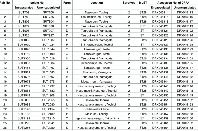
Table 1. S.suisisolates from Japan used in this study.
Pair No. Isolate No. Farm Location Serotype MLST Accession No. of DRA*
Encapsulated Unencapsulated Encapsulated Unencapsulated
1 SUT709 SUT708 A Nasu-gun, Tochigi 2 ST28 DRX045114 DRX045113
2 SUT780 SUT785 B Utsunomiya-shi, Tochigi 2 ST28 DRX045115 DRX045116
3 SUT806 SUT804 A Nasu-gun, Tochigi 2 ST28 DRX045118 DRX045117
4 SUT877 SUT876 C Tsuruoka-shi, Yamagata 2 ST1 DRX045120 DRX045119
5 SUT906 SUT907 D Tsuruoka-shi, Yamagata 2 ST1 DRX045121 DRX045122
6 SUT926 SUT927 D Tsuruoka-shi, Yamagata 2 ST1 DRX045123 DRX045124
7 SUT1006 SUT1007 E Tsuruoka-shi, Yamagata 2 ST28 DRX045125 DRX045126
8 SUT1020 SUT1024 F Shimotsuga-gun, Tochigi 2 ST1 DRX045127 DRX045128
9 SUT1046 SUT1044 G Tanzawa-gun, Iwate 2 ST28 DRX045130 DRX045129
10 SUT1189 SUT1190 G Tanzawa-gun, Iwate 2 ST28 DRX045131 DRX045132
11 SUT1330 SUT1329 H Tsuruoka-shi, Yamagata 2 ST28 DRX045134 DRX045133
12 SUT1337 SUT1334 I Hitachiomiya-shi, Ibaraki 2 ST28 DRX045136 DRX045135
13 SUT1468 SUT1467 G Tanzawa-gun, Iwate 2 ST28 DRX045138 DRX045137
14 SUT1582 SUT1583 J Shonai-shi, Yamagata 2 ST28 DRX045139 DRX045140
15 SUT1596 SUT1607 H Tsuruoka-shi, Yamagata 2 ST28 DRX045141 DRX045142
16 SUT1678 SUT1675 K Mogami-gun, Yamagata 2 ST28 DRX045144 DRX045143
17 SUT1789 SUT1797 L Nasukarasuyama-shi, Tochigi 2 ST28 DRX045145 DRX045146
18 SUT1865 SUT1882 M Nasu-machi, Nasu-gun, Tochigi 2 ST28 DRX045147 DRX045148
19 SUT1914 SUT1908 L Nasukarasuyama-shi, Tochigi 2 ST1 DRX045150 DRX045149
20 SUT2052 SUT2055 N Ishioka-shi, Ibaraki 2 ST28 DRX045151 DRX045152
21 SUT2083 SUT2080 L Nasukarasuyama-shi, Tochigi 2 ST28 DRX045154 DRX045153
22 SUT2148 SUT2154 O Ichihara-shi, Chiba 2 ST28 DRX045155 DRX045156
23 SUT2188 SUT2189 P Moka-shi, Tochigi 2 ST28 DRX045157 DRX045158
24 SUT2199 SUT2210 Q Higashishirakawa-gun, Fukushima 2 ST1 DRX045159 DRX045160
25 SUT2222 SUT2241 N Ishioka-shi, Ibaraki 2 ST28 DRX045161 DRX045162
26 SUT2256 SUT2250 L Nasukarasuyama-shi, Tochigi 2 ST28 DRX045164 DRX045163
*DRA, the DNA Data Bank of Japan (DDBJ) Sequence Read Archive
acid substitutions in the
cps
genes and other single-copy common genes were identified and
were visualized by bar graphs with genome structure of the ST28 reference strain using R
v3.2.2. The
cps
gene cluster was included in this comparison even though
cp
s genes had been
excluded from pool of common genes used in precious analysis due to presence of the indels
spanning over vast nucleotide stretches. The mutation frequency for each single-copy common
gene in each ST was calculated as the rate of the number of nucleotides with a mutation divided
by the total number.
Data access
The genome sequence data obtained in this study were deposited in the DNA Data Bank of
Japan (DDBJ) Sequence Read Archive under accession number DRA004206.
Results
Coexistence of encapsulated and unencapsulated
S
.
suis
in 26
endocarditis samples
A total of 1,416 isolates from 59 endocarditis samples were identified to be
S
.
suis
. In 58 of the
endocarditis samples, PCR genotyping showed that 1,388 isolates were serotype 2, and 4
iso-lates were untypeable, whereas the 24 isoiso-lates from the remaining endocarditis sample were
serotype 16. On the basis of the co-agglutination test, 31 (53%) and 2 (3%) of the 59 samples,
contained encapsulated only, or unencapsulated only isolates, respectively. On the other hand,
both encapsulated and unencapsulated isolates were found in 26 samples (44%). Encapsulated
and unencapsulated phenotypes were observed in 17 out of the 24 farms examined (70.8%)
(
Fig 1A
). The ratio of encapsulated to unencapsulated isolates varied (
Fig 1B
).
Phylogenetic relationships of encapsulated and unencapsulated
S
.
suis
isolates
We selected a pair of encapsulated and unencapsulated
S
.
suis
isolates from each of the 26
endocarditis samples in order to examine their phylogenetic relationships. The STs of
encapsu-lated and unencapsuencapsu-lated isolates were identical in each of 20 pairs of ST28, and 6 pairs of ST1.
Whole-genome short-read data was assembled using A5-miseq (
S3 Table
presents statistics of
the assemblies). Maximum parsimony trees were constructed using genome-wide
single-nucle-otide polymorphisms (SNPs) compared with the reference stains. In the tree of all 52 isolates,
ST1 and ST28 isolates were separately clustered (
Fig 2
). In each pair of ST1 isolates, the
encap-sulated and unencapencap-sulated isolates belonged to the same lineage and were phylogenetically
distinct from other pairs, except for pairs 5 and 6, which were isolated from the same farm D
(
Fig 2
). In each pair of ST28 isolates, the two were phylogenetically closely related to each
other, except for pairs 11 and 15 (
Fig 2
).
Mutations in a
cps
gene cluster and complementation with
cps
expression vectors
Fig 2. A phylogenetic tree for 26 encapsulated and 26 unencapsulatedS.suisisolates.The tree was constructed using the maximum parsimony method for genome-wide SNPs in 52S.suisisolates. The 52 isolates analyzed were composed of 26 encapsulated and unencapsulated isolate pairs from 17 porcine farms, indicated by the following symbols: blank circles for encapsulated, and filled circles for unencapsulated isolates, with pair Nos. and farm identifiers (colored rectangles) along with the symbols. The isolates were separately clustered into two STs, ST1 and ST28; a detailed tree structure in each ST is shown as an enlarged tree. Several ends of the enlarged tree in ST28 are further enlarged. Seventeen colors are used to show distribution of farms in the tree. Only the bootstrap values>50% are indicated on the branches.
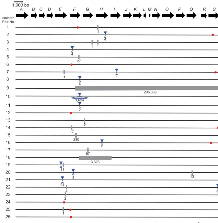
Fig 3. Nonsynonymous mutations and indels in thecpsgene cluster between encapsulated and unencapsulatedS.suisisolates in 26 pairs.The genetic organization of thecpsgene cluster (fromcps2Atocps2S) is shown as a sequence of the arrows located by transcriptional directions with the identifiers such as“A”for“cps2A”. The mutations between encapsulated and unencapsulated isolates in each pair are shown along with the genetic map, with the following symbols: red circles for nonsynonymous mutations, blue triangles for insertions, and gray rectangles for deletions along with the nucleotide length of the mutations.
11, which had mutations in the
cps2E
gene. All transformants showed a positive reaction in the
co-agglutination test using anti-serotype 2 serum, indicating restoration of capsule expression.
Occurrence of mutations in single-copy common genes, in the
cps
gene
cluster, and in MGEs
Mapping results of 52 isolates using CLC Genomics Workbench are listed in
S3 Table
. The
nucle-otide sequences of single-copy common genes and the
cps
gene cluster were compared within
each pair. The analysis was verified by the consistency of the mutation profiles in the Sanger and
MiSeq-based methods. No mutation was found in the
ccpA
gene that regulated the production of
CPs, and nonsynonymous mutations found between the pairs were depicted in
Fig 3
[
54
].
There were differences between ST1 and ST28 pairs in the number of genome-wide
muta-tions identified. Only a few nonsynonymous mutamuta-tions were identified in the ST1 pairs,
whereas the ST28 pairs had a large number of unique nonsynonymous mutations (
Fig 4
,
S4
and
S5
Tables). Indeed, the mean mutation frequencies in all single-copy common genes
Fig 4. The genome structure of the ST28S.suisstrain NSUI002.The following information on the NSUI002 genome is shown from the innermost circle: protein-coding genes transcribed clockwise (1sttrack, blue) and counterclockwise (2nd, blue), common genes that were shared among NSUI002 and all ST28S.
suisisolates, transcribed clockwise (3rd, green) and counterclockwise (4th, green), MGEs (5th, black), and genes in thecpsgene cluster (6th, yellow). The common genes in which synonymous and nonsynonymous mutations were detected are shown in the 7th(purple) and 8th(gray) tracks, respectively. The outermost bar graph for the common genes in the 9thtrack (red) shows the number of pairs in which nonsynonymous mutations were detected. No common gene exhibited a number of pairs more than 15, the maximum value of the graph.
between ST1 and ST28 were calculated to be 2.70 × 10
−7and 3.25 × 10
−5, respectively. At the
nucleotide level, most of the mutations were unique for each pair. All the pairs showed SNPs
within the
cps
gene cluster. In addition, SNPs within a particular MGE (position 1,054,531
–
1,178,032 bp of the reference genome NSUI002) were found in many pairs. This MGE contains
genes related to conjugative transposons (
Fig 4
,
S6 Table
).
The maximum numbers of nonsynonymous mutations per gene were 3 and 15 in the ST1
and ST28 pairs, respectively. Among ST28 pairs, most genes with mutations were part of
MGEs (
Fig 4
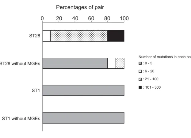
). Therefore, we excluded the single-copy common genes in MGEs and examined
the profiles of mutations. The number of mutations in each pair ranged between 0 to 5, except
for the genes within MGEs, which ranged between 21 to 302 (
Fig 5
). Thus, the mutation
fre-quencies were amended to be 2.95 × 10
−7in ST1, and 3.25 × 10
−6in ST28.
Discussion
In the present study, we demonstrated the coexistence of encapsulated and unencapsulated
S
.
suis
in the same endocarditis lesion. The capsule protects
S
.
suis
cells from the host immune
response [
24
,
30
,
55
,
56
].
S
.
suis
is known, however, to escape the vaccine-immunity by means
of capsule loss [
57
–
59
], such as reported previously for the capsule loss of
Haemophilus
influ-enzae
[
11
,
12
]. Unencapsulation can also afford
S
.
suis
cells some advantages, such as high
adhesion and invasion of host cells [
32
–
35
]. This led us to hypothesize that encapsulated and
unencapsulated
S
.
suis
“
cooperate
”
by expressing both advantageous characteristics at lesions,
Fig 5. The proportion of each pair classified by number of mutations.The number of mutations was calculated in each of 26 pairs, and its distribution is shown in ST1 and ST28 isolates as a percentage of pairs, as follows: gray bars for pairs with 0–5 mutations, blank bars for 6–20, dotted bars for 21–100, and filled bars for 101–300. Percentages are shown in ST1 and ST28 clones when genes on MGEs are excluded from the calculation.
and that each of these phenotypically distinct, but genetically very similar clonal
subpopula-tions provides an advantage that results in persistence of the global population of the clone.
Taken all together, our results seem to support the abovementioned hypothesis. However, we
cannot disregard the alternative hypothesis that the unencapsulated cells in the endocarditis
lesions may not have the capacity to participate in any significant way in inducing heart lesions,
but they simply arise by their faster growth in the particular niche.
We obtained only unencapsulated isolates from 2 samples. Moreover, the ratio of both
phe-notypes varied among 26 samples. It is conceivable that the encapsulated and unencapsulated
cells did not form a completely mixed jumble within the lesions but instead formed different
layers of either encapsulated or unencapsulated cells. Assuming that the layer of
unencapsu-lated cells was cut out and stamped on an agar medium, we can isolate only unencapsuunencapsu-lated
cells from the sample. Indeed, we always succeeded in isolating both phenotypes from the same
lesion by cutting the samples in many pieces for stamp (our unpublished observation). We
col-lected endocarditis samples from many geographically separated farms, and many of these
unrelated samples harbored both encapsulated and unencapsulated phenotypes of the same
clone of
S
.
suis
(
Fig 1A
). The high prevalence may suggest that occurrence of unencapsulated
isolates does not require specific conditions and occur frequently in every farm.
We only detected ST1 and ST28
S
.
suis
isolates in our sample (
Fig 1B
). Both ST1 and ST28
are prevalent in Japan [
60
]. ST1 is also prevalent worldwide and the isolates belonging to ST1
generally express a hemolysin known as suilysin [
61
]. ST28 has become prevalent in several
countries, including the United States of America, Canada, and China [
49
,
62
] and exhibits a
broad range of virulence in porcine groups [
49
]. The very close genetic relationship of the
iso-lates within each ST (
Fig 2
) suggests transmission of each lineage across distant farms by
por-cine transportations, similar to the transmission of
Salmonella
spp. from slaughterhouses to
farms [
63
]. However, we were unable to find any relationships between the geographical
loca-tion of the farms and STs of the isolates (
Fig 1
).
Some bacterial species have been known to cause polyphenotypic infections. Previous
stud-ies reported the coexistence of multiple
P
.
aeruginosa
clones that exhibit different antimicrobial
resistance and colony formation. Because such phenotypic differences were observed after the
establishment of infection and lesion formation, it was suggested that they differentiate
in situ
at lung lesions [
6
,
64
,
65
]. Since encapsulated and unencapsulated
S
.
suis
isolates in most pairs
were phylogenetically closest to each other (
Fig 2
), the encapsulated and unencapsulated
line-ages may have differentiated in the farm, i.e., environment or host body. Bacteria can change
their phenotype to adapt to the surrounding environment, such as genetic change in
P
.
aerugi-nosa
, or transcriptional regulation in
Streptococcus equi
[
66
]. In the present analyses of the
cps
gene cluster, all pairs of encapsulated and unencapsulated isolates had mutations that included
single-nucleotide polymorphisms and indels (
Fig 3
). As reported in a previous study, in which
capsule production by unencapsulated
S
.
suis
isolates was complemented with the intact
cps
genes of reference strains P1/7 and 89
–
1591 [
21
,
46
], we also demonstrated here the
comple-mentation of capsule production in unencapsulated isolates with
cps
expression vectors. This
result suggests that capsule expression by
S
.
suis
in endocarditis lesions was affected by genetic
changes in the
cps
gene cluster.
[
67
]. In the mutation profiles of all single-copy common genes, the spontaneous mutations
unique for a single pair were predominant throughout the genomes (
Fig 4
); however, there
were mutations in the
cps
gene cluster that occurred in multiple pairs. This may have been due
to the limitation of the examined isolates and a bias for the selection of the phenotypes.
Muta-tion frequencies differed between the ST1 and ST28 pairs; however, the difference may be
explained by the large number of mutations concentrated within a MGE in ST28 pairs
(approx-imately 90% of mutations were within a MGE (
Fig 4
,
S6 Table
)). After the exclusion of the
sin-gle-copy common genes within MGEs, the mutation frequency was almost the same to those in
other
Streptococcus
spp. such as
S
.
equi
(5.22 × 10
−7) [
68
],
S
.
pneuomoniae
(1.57 × 10
−6) [
69
]
and,
S
.
pyogenes
(1.1 × 10
−6) [
70
]. Collectively, these findings suggest that mutations may
spon-taneously arise in the
cps
gene cluster; therefore, these mutations may alter the phenotypic
change through capsule expression. On the other hand, the MGE found in ST28 contained two
genomic regions that exhibited high nucleotide sequence similarity with
attL
and
attR
, parts of
a conjugative element, as well as genes related to its transfer [
71
]. Although precise cause why
the MGE carried more mutations than other genome regions remains unknown, we assume
the region may be related to the cause of endocarditis, which will be revealed by genome-wide
association study in the future [
72
].
In summary, we herein demonstrate that different phenotypes of genetically very closely
related
S
.
suis
isolates were from the same endocarditis lesion.
S
.
suis
may adapt to its surrounding
environment through the loss of capsule expression, and endocarditis lesion may be developed by
dual phenotypes of a single clones. Although unencapsulated
S
.
suis
isolates are, in general, easily
phagocytized by immune cells, it is possible that they persist and proliferate in the host by the
assistance of encapsulated cells. In addition, a comparison of ST1 and ST28 suggested that the
iso-lates of different STs employed various mechanisms by which they adapt to the surrounding
envi-ronment. Future studies about coexistence of different phenotypes, which may enable them to
persist in endocarditis lesions, and experimental reproduction of endocarditis with both
pheno-types, will provide insights into the pathogenicity, ecology, diversity and evolution of
S
.
suis
.
Supporting Information
S1 Table.
E
.
coli
strains and plasmids used in this study.
(DOCX)
S2 Table. Primers used to construct
cps
gene expression vectors.
(DOCX)
S3 Table. Mapping and assembly results of isolates.
(XLSX)
S4 Table. Mutations in single-copy common genes of ST28 isolates.
(XLSX)
S5 Table. Mutations in single-copy common genes in ST1 isolates.
(XLSX)
S6 Table. Gene contents of the MGE where the SNPs were most abundant.
(XLSX)
Acknowledgments
and advice. We appreciate Hiroyuki Nakayama, Laboratory of Veterinary Pathology, The
Uni-versity of Tokyo for valuable comments. We thank meat inspection centers of
Utsunomiya-City, Yamagata-Shonai, and Ibaraki-Kensei in Japan for help with collecting porcine
endocar-ditis samples.
Author Contributions
Conceived and designed the experiments: MT TW FM IN TS. Performed the experiments: MT
TW FM SA AO. Analyzed the data: MT TW FM. Contributed reagents/materials/analysis
tools: TBTA NF. Wrote the paper: MT TW FM TBTA NF IN TS.
References
1. Levert M, Zamfir O, Clermont O, Bouvet O, Lespinats S, Hipeaux MC, et al. Molecular and evolutionary bases of within-patient genotypic and phenotypic diversity inEscherichia coliextraintestinal infections. PLoS Pathog. 2010; 6(9):e1001125. Epub 2010/10/14. doi:10.1371/journal.ppat.1001125PMID:
20941353.
2. Van Eldere J, Peetermans WE, Struelens M, Deplano A, Bobbaers H. Polyclonal Staphylococcal endo-carditis caused by genetic variability. Clin Infect Dis. 2000; 31(1):24–30. Epub 2000/07/29. doi:10. 1086/313915PMID:10913391.
3. Romling U, Fiedler B, Bosshammer J, Grothues D, Greipel J, von der Hardt H, et al. Epidemiology of chronicPseudomonas aeruginosainfections in cystic fibrosis. J Infect Dis. 1994; 170(6):1616–1621. Epub 1994/12/01. PMID:7996008.
4. Israel DA, Salama N, Krishna U, Rieger UM, Atherton JC, Falkow S, et al.Helicobacter pylorigenetic diversity within the gastric niche of a single human host. Proc Natl Acad Sci U S A. 2001; 98 (25):14625–14630. Epub 2001/11/29. doi:10.1073/pnas.251551698PMID:11724955.
5. Blaser MJ, Berg DE.Helicobacter pylorigenetic diversity and risk of human disease. J Clin Invest. 2001; 107(7):767–773. Epub 2001/04/04. doi:10.1172/jci12672PMID:11285290.
6. Ashish A, Paterson S, Mowat E, Fothergill JL, Walshaw MJ, Winstanley C. Extensive diversification is a common feature ofPseudomonas aeruginosapopulations during respiratory infections in cystic fibrosis. J Cyst Fibros. 2013; 12(6):790–793. Epub 2013/05/07. doi:10.1016/j.jcf.2013.04.003PMID:
23642644.
7. Sanchez-Romero MA, Casadesus J. Contribution of phenotypic heterogeneity to adaptive antibiotic resistance. Proc Natl Acad Sci U S A. 2014; 111(1):355–360. Epub 2013/12/20. doi:10.1073/pnas. 1316084111PMID:24351930.
8. Temime L, Boelle PY, Opatowski L, Guillemot D. Impact of capsular switch on invasive pneumococcal disease incidence in a vaccinated population. PLoS One. 2008; 3(9):e3244. Epub 2008/09/20. doi:10. 1371/journal.pone.0003244PMID:18802466.
9. Suzuki S, Horinouchi T, Furusawa C. Prediction of antibiotic resistance by gene expression profiles. Nat Commun. 2014; 5:5792. Epub 2014/12/18. doi:10.1038/ncomms6792PMID:25517437.
10. Hanage WP, Kaijalainen T, Saukkoriipi A, Rickcord JL, Spratt BG. A successful, diverse disease-asso-ciated lineage of nontypeable pneumococci that has lost the capsular biosynthesis locus. J Clin Micro-biol. 2006; 44(3):743–749. Epub 2006/03/07. doi:10.1128/jcm.44.3.743-749.2006PMID:16517849.
11. Dworkin MS, Park L, Borchardt SM. The changing epidemiology of invasiveHaemophilus influenzae disease, especially in persons>or = 65 years old. Clin Infect Dis. 2007; 44(6):810–816. Epub 2007/02/
17. doi:10.1086/511861PMID:17304452.
12. Farley MM, Stephens DS, Brachman PS Jr, Harvey RC, Smith JD, Wenger JD. InvasiveHaemophilus influenzaedisease in adults. A prospective, population-based surveillance. CDC Meningitis Surveil-lance Group. Ann Intern Med. 1992; 116(10):806–812. Epub 1992/05/25. PMID:1314530.
13. Hilty M, Wuthrich D, Salter SJ, Engel H, Campbell S, Sa-Leao R, et al. Global phylogenomic analysis of nonencapsulatedStreptococcus pneumoniaereveals a deep-branching classic lineage that is distinct from multiple sporadic lineages. Genome Biol Evol. 2014; 6(12):3281–3294. Epub 2014/12/07. doi:10. 1093/gbe/evu263PMID:25480686.
14. Bellais S, Six A, Fouet A, Longo M, Dmytruk N, Glaser P, et al. Capsular switching in group B Strepto-coccusCC17 hypervirulent clone: a future challenge for polysaccharide vaccine development. J Infect Dis. 2012; 206(11):1745–1752. Epub 2012/09/25. doi:10.1093/infdis/jis605PMID:23002446.
16. Turner CE, Abbott J, Lamagni T, Holden MT, David S, Jones MD, et al. Emergence of a new highly suc-cessful acapsular group AStreptococcusclade of genotypeemm89 in the United Kingdom. MBio. 2015; 6(4):e00622. Epub 2015/07/16. doi:10.1128/mBio.00622-15PMID:26173696.
17. Flores AR, Jewell BE, Fittipaldi N, Beres SB, Musser JM. Human disease isolates of serotype m4 and m22 group A streptococcus lack genes required for hyaluronic acid capsule biosynthesis. MBio. 2012; 3(6):e00413–12. Epub 2012/11/08. doi:10.1128/mBio.00413-12PMID:23131832.
18. Gottschalk M. Streptococcosis. In: Zimmerman J, Karriker LA, Ramirez A, Schwartz KJ, Stevenson GW, editors. Diseases of swine: Wiley Publishers; 2012. pp. 841–855
19. Staats JJ, Feder I, Okwumabua O, Chengappa MM.Streptococcus suis: past and present. Vet Res Commun. 1997; 21(6):381–407. Epub 1997/08/01. PMID:9266659.
20. Lee GT, Chiu CY, Haller BL, Denn PM, Hall CS, Gerberding JL.Streptococcus suismeningitis, United States. Emerg Infect Dis. 2008; 14(1):183–185. Epub 2008/02/09. doi:10.3201/eid1401.070930PMID:
18258107.
21. Lakkitjaroen N, Takamatsu D, Okura M, Sato M, Osaki M, Sekizaki T. Loss of capsule among Strepto-coccus suisisolates from porcine endocarditis and its biological significance. J Med Microbiol. 2011; 60 (Pt 11):1669–1676. Epub 2011/07/23. doi:10.1099/jmm.0.034686-0PMID:21778266.
22. Weinert LA, Chaudhuri RR, Wang J, Peters SE, Corander J, Jombart T, et al. Genomic signatures of human and animal disease in the zoonotic pathogenStreptococcus suis. Nat Commun. 2015; 6:6740. Epub 2015/04/01. doi:10.1038/ncomms7740PMID:25824154.
23. Elliott SD, Tai JY. The type-specific polysaccharides ofStreptococcus suis. J Exp Med. 1978; 148 (6):1699–1704. Epub 1978/12/01. PMID:363973.
24. Smith HE, Damman M, van der Velde J, Wagenaar F, Wisselink HJ, Stockhofe-Zurwieden N, et al. Identification and characterization of thecpslocus ofStreptococcus suisserotype 2: the capsule pro-tects against phagocytosis and is an important virulence factor. Infect Immun. 1999; 67(4):1750–1756. Epub 1999/03/20. PMID:10085014.
25. Smith HE, de Vries R, van't Slot R, Smits MA. Thecpslocus ofStreptococcus suisserotype 2: genetic determinant for the synthesis of sialic acid. Microb Pathog. 2000; 29(2):127–134. Epub 2000/07/25. doi:10.1006/mpat.2000.0372PMID:10906268.
26. Van Calsteren MR, Gagnon F, Lacouture S, Fittipaldi N, Gottschalk M. Structure determination of Streptococcus suisserotype 2 capsular polysaccharide. Biochem Cell Biol. 2010; 88(3):513–525. Epub 2010/06/18. doi:10.1139/o09-170PMID:20555393.
27. Okura M, Takamatsu D, Maruyama F, Nozawa T, Nakagawa I, Osaki M, et al. Genetic analysis of cap-sular polysaccharide synthesis gene clusters from all serotypes ofStreptococcus suis: potential mech-anisms for generation of capsular variation. Appl Environ Microbiol. 2013; 79(8):2796–2806. Epub 2013/02/19. doi:10.1128/aem.03742-12PMID:23416996.
28. Fittipaldi N, Segura M, Grenier D, Gottschalk M. Virulence factors involved in the pathogenesis of the infection caused by the swine pathogen and zoonotic agentStreptococcus suis. Future Microbiol. 2012; 7(2):259–279. Epub 2012/02/14. doi:10.2217/fmb.11.149PMID:22324994.
29. Segura M, Gottschalk M, Olivier M. EncapsulatedStreptococcus suisinhibits activation of signaling pathways involved in phagocytosis. Infect Immun. 2004; 72(9):5322–5330. Epub 2004/08/24. doi:10. 1128/iai.72.9.5322-5330.2004PMID:15322029.
30. Benga L, Fulde M, Neis C, Goethe R, Valentin-Weigand P. Polysaccharide capsule and suilysin con-tribute to extracellular survival ofStreptococcus suisco-cultivated with primary porcine phagocytes. Vet Microbiol. 2008; 132(1–2):211–9. Epub 2008/06/21. doi:10.1016/j.vetmic.2008.05.005PMID:
18565698.
31. Houde M, Gottschalk M, Gagnon F, Van Calsteren MR, Segura M.Streptococcus suiscapsular poly-saccharide inhibits phagocytosis through destabilization of lipid microdomains and prevents lactosyl-ceramide-dependent recognition. Infect Immun. 2012; 80(2):506–517. Epub 2011/11/30. doi:10.1128/ iai.05734-11PMID:22124659.
32. Benga L, Goethe R, Rohde M, Valentin-Weigand P. Non-encapsulated strains reveal novel insights in invasion and survival ofStreptococcus suisin epithelial cells. Cell Microbiol. 2004; 6(9):867–881. Epub 2004/07/27. doi:10.1111/j.1462-5822.2004.00409.xPMID:15272867.
33. Tanabe S, Bonifait L, Fittipaldi N, Grignon L, Gottschalk M, Grenier D. Pleiotropic effects of polysaccha-ride capsule loss on selected biological properties ofStreptococcus suis. Can J Vet Res. 2010; 74 (1):65–70. Epub 2010/04/02. PMID:20357962.
34. Ferrando ML, de Greeff A, van Rooijen WJ, Stockhofe-Zurwieden N, Nielsen J, Wichgers Schreur PJ, et al. Host-pathogen interaction at the intestinal mucosa correlates with zoonotic potential of Strepto-coccus suis. J Infect Dis. 2015; 212(1):95–105. Epub 2014/12/20. doi:10.1093/infdis/jiu813PMID:
35. Tenenbaum T, Papandreou T, Gellrich D, Friedrichs U, Seibt A, Adam R, et al. Polar bacterial invasion and translocation ofStreptococcus suisacross the blood-cerebrospinal fluid barrier in vitro. Cell Micro-biol. 2009; 11(2):323–336. Epub 2008/12/03. doi:10.1111/j.1462-5822.2008.01255.xPMID:
19046337.
36. Ishida S, Tien le HT, Osawa R, Tohya M, Nomoto R, Kawamura Y, et al. Development of an appropriate PCR system for the reclassification ofStreptococcus suis. J Microbiol Methods. 2014; 107:66–70. Epub 2014/09/18. doi:10.1016/j.mimet.2014.09.003PMID:25229648.
37. Okura M, Lachance C, Osaki M, Sekizaki T, Maruyama F, Nozawa T, et al. Development of a two-step multiplex PCR assay for typing of capsular polysaccharide synthesis gene clusters ofStreptococcus suis. J Clin Microbiol. 2014; 52(5):1714–1719. Epub 2014/02/28. doi:10.1128/jcm.03411-13PMID:
24574288.
38. Marois C, Bougeard S, Gottschalk M, Kobisch M. Multiplex PCR assay for detection ofStreptococcus suisspecies and serotypes 2 and 1/2 in tonsils of live and dead pigs. J Clin Microbiol. 2004; 42 (7):3169–3175. Epub 2004/07/10. doi:10.1128/jcm.42.7.3169-3175.2004PMID:15243078.
39. Casadaban MJ, Cohen SN. Analysis of gene control signals by DNA fusion and cloning inEscherichia coli. J Mol Biol. 1980; 138(2):179–207. Epub 1980/04/01. PMID:6997493.
40. Okura M, Osaki M, Fittipaldi N, Gottschalk M, Sekizaki T, Takamatsu D. The minor pilin subunit Sgp2 is necessary for assembly of the pilus encoded by thesrtGcluster ofStreptococcus suis. J Bacteriol. 2011; 193(4):822–831. Epub 2010/12/15. doi:10.1128/jb.01555-09PMID:21148736.
41. Mogollon JD, Pijoan C, Murtaugh MP, Kaplan EL, Collins JE, Cleary PP. Characterization of prototype and clinically defined strains ofStreptococcus suisby genomic fingerprinting. J Clin Microbiol. 1990; 28 (11):2462–2466. Epub 1990/11/01. PMID:1979331.
42. King SJ, Leigh JA, Heath PJ, Luque I, Tarradas C, Dowson CG, et al. Development of a multilocus sequence typing scheme for the pig pathogenStreptococcus suis: identification of virulent clones and potential capsular serotype exchange. J Clin Microbiol. 2002; 40(10):3671–3680. Epub 2002/10/02. PMID:12354864.
43. Tritt A, Eisen JA, Facciotti MT, Darling AE. An integrated pipeline for de novo assembly of microbial genomes. PLoS One. 2012; 7(9):e42304. Epub 2012/10/03. doi:10.1371/journal.pone.0042304PMID:
23028432.
44. Gardner SN, Slezak T, Hall BG. kSNP3.0: SNP detection and phylogenetic analysis of genomes with-out genome alignment or reference genome. Bioinformatics. 2015; 31(17):2877–2878. Epub 2015/04/ 29. doi:10.1093/bioinformatics/btv271PMID:25913206.
45. Tamura K, Peterson D, Peterson N, Stecher G, Nei M, Kumar S. MEGA5: molecular evolutionary genet-ics analysis using maximum likelihood, evolutionary distance, and maximum parsimony methods. Mol Biol Evol. 2011; 28(10):2731–2739. Epub 2011/05/07. doi:10.1093/molbev/msr121PMID:21546353.
46. Lakkitjaroen N, Takamatsu D, Okura M, Sato M, Osaki M, Sekizaki T. Capsule loss or death: the posi-tion of mutaposi-tions among capsule genes sways the destiny ofStreptococcus suis. FEMS Microbiol Lett. 2014; 354(1):46–54. Epub 2014/03/25. doi:10.1111/1574-6968.12428PMID:24654559.
47. Seemann T. Prokka: rapid prokaryotic genome annotation. Bioinformatics. 2014; 30(14):2068–2069. Epub 2014/03/20. doi:10.1093/bioinformatics/btu153PMID:24642063.
48. Zhao Y, Wu J, Yang J, Sun S, Xiao J, Yu J. PGAP: pan-genomes analysis pipeline. Bioinformatics. 2012; 28(3):416–418. Epub 2011/12/02. doi:10.1093/bioinformatics/btr655PMID:22130594.
49. Athey TB, Auger JP, Teatero S, Dumesnil A, Takamatsu D, Wasserscheid J, et al. Complex population structure and virulence differences among serotype 2Streptococcus suisstrains belonging to sequence type 28. PLoS One. 2015; 10(9):e0137760. Epub 2015/09/17. doi:10.1371/journal.pone. 0137760PMID:26375680.
50. Zhou Y, Liang Y, Lynch KH, Dennis JJ, Wishart DS. PHAST: a fast phage search tool. Nucleic Acids Res. 2011; 39:W347–352. Epub 2011/06/16. doi:10.1093/nar/gkr485PMID:21672955.
51. Dhillon BK, Laird MR, Shay JA, Winsor GL, Lo R, Nizam F, et al. IslandViewer 3: more flexible, interac-tive genomic island discovery, visualization and analysis. Nucleic Acids Res. 2015; 43:W104–108. Epub 2015/04/29. doi:10.1093/nar/gkv401PMID:25916842.
52. Moran NA, McLaughlin HJ, Sorek R. The dynamics and time scale of ongoing genomic erosion in sym-biotic bacteria. Science. 2009; 323(5912):379–382. Epub 2009/01/20. doi:10.1126/science.1167140
PMID:19150844.
53. McLean JS, Lombardo MJ, Ziegler MG, Novotny M, Yee-Greenbaum J, Badger JH, et al. Genome of the pathogenPorphyromonas gingivalisrecovered from a biofilm in a hospital sink using a high-throughput single-cell genomics platform. Genome Res. 2013; 23(5):867–877. Epub 2013/04/09. doi:
54. Willenborg J, Fulde M, de Greeff A, Rohde M, Smith HE, Valentin-Weigand P, et al. Role of glucose and CcpA in capsule expression and virulence ofStreptococcus suis. Microbiology. 2011; 157(Pt 6):1823–1833. Epub 2011/02/26. doi:10.1099/mic.0.046417-0PMID:21349980.
55. Charland N, Harel J, Kobisch M, Lacasse S, Gottschalk M.Streptococcus suisserotype 2 mutants defi-cient in capsular expression. Microbiology. 1998; 144 (Pt 2):325–332. Epub 1998/03/11. doi:10.1099/ 00221287-144-2-325PMID:9493370.
56. Chabot-Roy G, Willson P, Segura M, Lacouture S, Gottschalk M. Phagocytosis and killing of Strepto-coccus suisby porcine neutrophils. Microb Pathog. 2006; 41(1):21–32. Epub 2006/05/23. doi:10.1016/ j.micpath.2006.04.001PMID:16714092.
57. Holt ME, Enright MR, Alexander TJ. Immunisation of pigs with live cultures ofStreptococcus suistype 2. Res Vet Sci. 1988; 45(3):349–352. Epub 1988/11/01. PMID:3212282.
58. Holt ME, Enright MR, Alexander TJ. Studies of the protective effect of different fractions of sera from pigs immune toStreptococcus suistype 2 infection. J Comp Pathol. 1989; 100(4):435–442. Epub 1989/05/01. PMID:2760276.
59. Blouin C, Higgins R, Gottschalk M, Simard J. Evaluation of the antibody response in pigs vaccinated againstStreptococcus suiscapsular type 2 using a double-antibody sandwich enzyme-linked immuno-sorbent assay. Can J Vet Res. 1994; 58(1):49–54. Epub 1994/01/01. PMID:8143253.
60. Onishi H, Sugawara M, Okura M, Osaki M, Takamatsu D. Prevalence ofStreptococcus suisgenotypes in isolates from porcine endocarditis in East Japan. J Vet Med Sci. 2012; 74(12):1681–1684. Epub 2012/08/11. PMID:22878504.
61. Holden MT, Hauser H, Sanders M, Ngo TH, Cherevach I, Cronin A, et al. Rapid evolution of virulence and drug resistance in the emerging zoonotic pathogenStreptococcus suis. PLoS One. 2009; 4(7): e6072. Epub 2009/07/16. doi:10.1371/journal.pone.0006072PMID:19603075.
62. Wang S, Gao M, An T, Liu Y, Jin J, Wang G, et al. Genetic diversity and virulence of novel sequence types ofStreptococcus suisfrom diseased and healthy pigs in China. Front Microbiol. 2015; 6:173. Epub 2015/03/19. doi:10.3389/fmicb.2015.00173PMID:25784908.
63. Magistrali C, Dionisi AM, De Curtis P, Cucco L, Vischi O, Scuota S, et al. Contamination ofSalmonella spp. in a pig finishing herd, from the arrival of the animals to the slaughterhouse. Res Vet Sci. 2008; 85 (2):204–207. Epub 2008/01/31. doi:10.1016/j.rvsc.2007.12.002PMID:18230403.
64. Darch SE, McNally A, Harrison F, Corander J, Barr HL, Paszkiewicz K, et al. Recombination is a key driver of genomic and phenotypic diversity in aPseudomonas aeruginosapopulation during cystic fibro-sis infection. Sci Rep. 2015; 5:7649. Epub 2015/01/13. doi:10.1038/srep07649PMID:25578031.
65. Jorth P, Staudinger BJ, Wu X, Hisert KB, Hayden H, Garudathri J, et al. Regional isolation drives bacte-rial diversification within cystic fibrosis lungs. Cell Host Microbe. 2015; 18(3):307–319. Epub 2015/08/ 25. doi:10.1016/j.chom.2015.07.006PMID:26299432.
66. Steward KF, Robinson C, Waller AS. Transcriptional changes are involved in phenotype switching in Streptococcus equisubspeciesequi. Mol Biosyst. 2016. Epub 2016/02/09. doi:10.1039/c5mb00780a
PMID:26854112.
67. Griffiths AJF, Miller JH, Suzuki DT, Lewontin RC, Gelbart WM. An Introduction to genetic analysis. 7th ed. New York: W.H. Freeman; 2000.
68. Harris SR, Robinson C, Steward KF, Webb KS, Paillot R, Parkhill J, et al. Genome specialization and decay of the strangles pathogen,Streptococcus equi, is driven by persistent infection. Genome Res. 2015; 25(9):1360–1371. Epub 2015/07/15. doi:10.1101/gr.189803.115PMID:26160165.
69. Croucher NJ, Harris SR, Fraser C, Quail MA, Burton J, van der Linden M, et al. Rapid pneumococcal evolution in response to clinical interventions. Science. 2011; 331(6016):430–434. Epub 2011/01/29. doi:10.1126/science.1198545PMID:21273480.
70. Davies MR, Holden MT, Coupland P, Chen JH, Venturini C, Barnett TC, et al. Emergence of scarlet feverStreptococcus pyogenes emm12 clones in Hong Kong is associated with toxin acquisition and multidrug resistance. Nat Genet. 2015; 47(1):84–87. Epub 2014/11/18. doi:10.1038/ng.3147PMID:
25401300.
71. Palmieri C, Magi G, Mingoia M, Bagnarelli P, Ripa S, Varaldo PE, et al. Characterization of a Strepto-coccus suis tet(O/W/32/O)-carrying element transferable to major streptococcal pathogens. Antimicrob Agents Chemother. 2012; 56(9):4697–4702. Epub 2012/06/20. doi:10.1128/aac.00629-12PMID:
22710115.
72. Sheppard SK, Didelot X, Meric G, Torralbo A, Jolley KA, Kelly DJ, et al. Genome-wide association study identifies vitamin B5 biosynthesis as a host specificity factor inCampylobacter. Proc Natl Acad Sci U S A. 2013; 110(29):11923–11927. Epub 2013/7/1. doi:10.1073/pnas.1305559110PMID: