PKC
d
to Enhance Interleukin-11 Gene Transcription in
Osteoblasts
Shinsuke Kido1,2, Rika Kuriwaka-Kido1, Yuka Umino-Miyatani3, Itsuro Endo1, Daisuke Inoue1, Hisaaki Taniguchi4, Yasumichi Inoue5, Takeshi Imamura5, Toshio Matsumoto1,2*
1Department of Medicine and Bioregulatory Sciences, The University of Tokushima Graduate School of Medical Sciences, Tokushima, Japan,2The 21st Century Center of Excellence (COE) Program, The University of Tokushima Graduate School of Medical Sciences, Tokushima, Japan,3Department of Obstetrics and Gynecology, The University of Tokushima Graduate School of Medical Sciences, Tokushima, Japan,4Division of Molecular Enzyme Physiology, University of Tokushima Institute for Enzyme Research, Tokushima, Japan,5Division of Biochemistry, The Cancer Institute of the Japanese Foundation for Cancer Research (JFCR), Tokyo, Japan
Abstract
Background:Mechanical stress rapidly induces DFosB expression in osteoblasts, which binds tointerleukin (IL)-11 gene promoter to enhance IL-11 expression, and IL-11 enhances osteoblast differentiation. Because bone morphogenetic proteins (BMPs) also stimulate IL-11 expression in osteoblasts, there is a possibility that BMP-Smad signaling is involved in the enhancement of osteoblast differentiation by mechanical stress. The present study was undertaken to clarify whether mechanical stress affects BMP-Smad signaling, and if so, to elucidate the role of Smad signaling in mechanical stress-induced enhancement of IL-11 gene transcription.
Methodology/Principal Findings:Mechanical loading by fluid shear stress (FSS) induced phosphorylation of BMP-specific receptor-regulated Smads (BR-Smads), Smad1/5, in murine primary osteoblasts (mPOBs). FSS rapidly phosphorylated Y311 of protein kinase C (PKC)d, and phosphorylated PKCdinteracted with BR-Smads to phosphorylate BR-Smads. Transfection of
PKCd siRNA or Y311F mutant PKCd abrogated BR-Smads phosphorylation and suppressed IL-11 gene transcription enhanced by FSS. Activated BR-Smads bound to the Smad-binding element (SBE) ofIL-11 gene promoter and formed complex withDFosB/JunD heterodimer via binding to the C-terminal region of JunD. Site-directed mutagenesis in the SBE and the AP-1 site revealed that both SBE and AP-1 sites were required for full activation ofIL-11gene promoter by FSS.
Conclusions/Significance: These results demonstrate that PKCd-BR-Smads pathway plays an important role in the intracellular signaling in response to mechanical stress, and that a cross-talk between PKCd-BR-Smads andDFosB/JunD pathways synergistically stimulates IL-11 gene transcription in response to mechanical stress.
Citation:Kido S, Kuriwaka-Kido R, Umino-Miyatani Y, Endo I, Inoue D, et al. (2010) Mechanical Stress Activates Smad Pathway through PKCdto Enhance Interleukin-11 Gene Transcription in Osteoblasts. PLoS ONE 5(9): e13090. doi:10.1371/journal.pone.0013090
Editor:Nic D. Leipzig, The University of Akron, United States of America
ReceivedMarch 15, 2010;AcceptedSeptember 2, 2010;PublishedSeptember 29, 2010
Copyright:ß2010 Kido et al. This is an open-access article distributed under the terms of the Creative Commons Attribution License, which permits unrestricted use, distribution, and reproduction in any medium, provided the original author and source are credited.
Funding:This study was carried out as a part of ‘‘Ground Research Announcement for Space Utilization’’ promoted by Japan Science Forum. The study was also supported in part by Grants-in-Aid for Scientific Research from the Ministry of Education, Culture, Sports, Science and Technology of Japan, and by Center-of-Excellence Program of the 21st Century from the Ministry of Education, Science, Sports and Culture of Japan. The funders had no role in study design, data collection and analysis, decision to publish, or preparation of the manuscript.
Competing Interests:The authors have declared that no competing interests exist. * E-mail: [email protected]
Introduction
Mechanical stress to bone plays a crucial role in maintaining bone homeostasis. Immobilization, long-term bed rest, or microgravity in space causes a marked loss of bone mass and strength, due to reduced bone formation as well as enhanced bone resorption [1–4]. Although the enhanced bone resorption can be inhibited by a treatment with bisphosphonates [4,5], it has been difficult to stimulate the unloading-induced suppression of bone formation. Therefore, it is important to clarify the mechanism whereby bone formation is suppressed by mechanical unloading.
Mechanical stress to bone causes a rapid fluid flow surrounding osteoblasts and osteocytes, and elicits fluid shear stress (FSS) to these cells. FSS is shown to be one of the most important signal transduction mechanisms to enhance osteoblastic differentiation
density with aging due to an enhanced bone formation without an increase in bone resorption [16]. These observations suggested to us that IL-11 mediates mechanical stress signals to osteoblast differentiation signal.
Bone morphogenetic proteins (BMPs) play pivotal roles in the regulation of osteoblast differentiation and bone formation [17,18]. When artificially implanted into muscle tissues, BMPs induce ectopic bone formation. However, the role of BMPs in mediating mechanical stress signal to osteoblastogenic signal remains unclear. BMP signals are transmitted via phosphorylation by type I BMP receptor of BMP-specific receptor-regulated Smads (BR-Smads) including Smad1, 5 and 8. Phosphorylated BR-Smads then form heteromeric complex with Smad4, a common Smad, and translocate into the nucleus, where they regulate transcription of various target genes [19-22]. Because our preliminary experiments demonstrated that not only mechanical stress but also BMP-2 stimulate IL-11 expression in osteoblastic cells (Figure S1), there is a possibility that BR-Smad signaling is also involved in the enhancement of osteoblast differentiation in response to mechanical stress. In order to address this issue, we investigated the effect of mechanical stress on BR-Smad phosphorylation as well as the interaction of BR-Smads with activator protein (AP)-1 transcription factors andIL-11gene promoter in osteoblastic cells. The results demonstrate that mechanical stress by FSS to murine primary osteoblasts (mPOBs) in vitro stimulates Smad1/5 phos-phorylation, that mechanical stress-induced phosphorylation of Smad1/5 is mediated via phosphorylation and activation of protein kinase C (PKC)d, that activated Smad1/5 forms complex withDFosB and JunD onIL-11 gene promoter, and thatIL-11 gene transcription is cooperatively stimulated byDFosB/JunD and Smad1/5 in response to mechanical stress.
Results
Mechanical stress induces phosphorylation of BR-Smads in osteoblasts
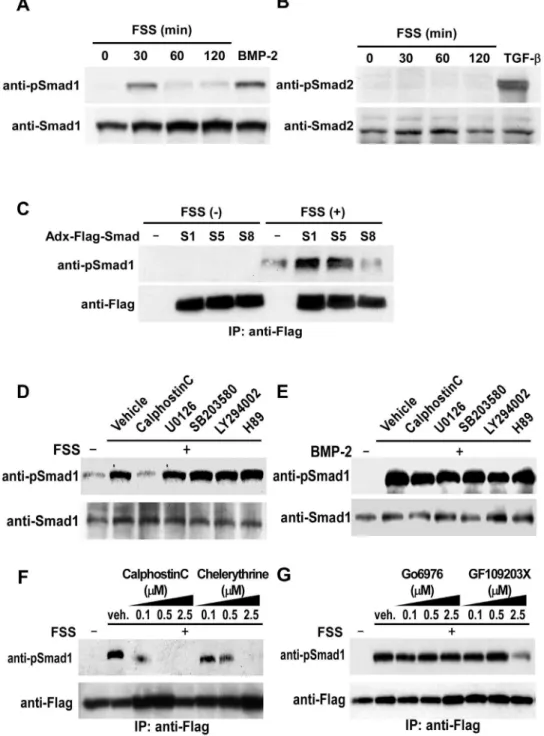
BMP-Smad pathway plays a central role in osteoblast differentiation and bone formation. In order to clarify the contribution of BMP-Smad signaling in osteoblasts in response to mechanical stress, we first investigated the effect of FSS on phosphorylation of Smads. FSS in vitro to primary osteoblasts derived from newborn mice calvariae enhanced phosphorylation of BR-Smads, Smad1/5/8, within 30 minutes using an antibody against phosphorylated Smad1 that cross-reacts with phosphory-lated Smad5/8 (Figure 1A). Because mechanical loading to osteoblasts does not alter the expression levels of BMP-2 (Figure S2), these results suggest that mechanical stress to osteoblasts enhances BR-Smads phosphorylation without stimulation by BMP. The effect of FSS on phosphorylation of transforming growth factor-b-specific R-Smads, Smad2/3, was also examined using an antibody against phosphorylated Smad2 that cross-reacts with phosphorylated Smad3. As shown in Figure 1B, FSS failed to induce phosphorylation of Smad2/3 in osteoblasts, showing that mechanical stress specifically phosphorylates BR-Smads in osteoblasts.
In order to clarify which BR-Smads were phosphorylated by FSS, we tested the effect of FSS on phosphorylation of exogenous Flag-Smad1, Flag-Smad5 or Flag-Smad8 in adenovirus vector infected into mPOBs. FSS induced phosphorylation of Smad1 and Smad5, but not of Smad8 in osteoblasts (Figure 1C). Because the antibody detects phosphorylated Ser463/465 of all BR-Smads and cannot differentiate among them, experiments using this antibody were interpreted as detecting phosphorylation of endogenous Smad1/5 in response to FSS.
Induction of BR-Smad phosphorylation by mechanical stress is dependent on PKC
In order to clarify intracellular signaling pathways that lead to phosphorylation of Smad1/5 by FSS, the effect of inhibitors of PKC, mitogen-activated protein kinase, phosphatidylinositol-3-OH kinase (PI3K), and protein kinase A (PKA) was examined using concentrations that can inhibit the activity of the respective enzymes. mPOBs were exposed to a PKC inhibitor calphostin C, a mitogen-activated protein kinase kinase inhibitor U0126, a p38 kinase inhibitor SB203580, a PI3K inhibitor LY294002, or a PKA inhibitor H89, and then subjected to FSS. Only a pan-specific PKC inhibitor, calphostin C, abolished phosphorylation of Smad1/5, whereas any of the other kinase inhibitors, U0126, SB203580, LY294002 or H89, failed to inhibit Smad1/5 phosphorylation by FSS (Figure 1D). Neither calphostin C nor other inhibitors affected BMP-induced phosphorylation of Smad1/5 (Figure 1E). These results are consistent with the notion that the mechanism of FSS-induced BR-Smad phosphorylation is different from that of BMP, and that PKC is involved in this process.
We next examined the effect of conventional PKC (cPKC) inhibitors with different dose-dependent inhibitory effects on other classes of PKC, chelerythrine, Go6976, and GF109203X, on FSS-induced phosphorylation of Smad1. High doses of chelerythrine and GF109203X inhibited Smad1 phosphorylation stimulated by FSS (Figure 1F and 1G), while Go6976 did not affect FSS-induced phosphorylation of Smad1 even at the highest dose (Figure 1G). Because chelerythrine and GF109203X at high doses are reported to act as pan-specific inhibitors for PKC [23,24], these observations suggest that PKC other than cPKC induces BR-Smad phosphorylation in response to FSS.
PKCd is activated by mechanical stress and phosphorylates BR-Smads
PKC family members are divided into three species; cPKC, novel PKC (nPKC) and atypical PKC (aPKC). Because cPKC and nPKC but not aPKC are activated by diacylglycerol, and because cPKC and nPKC can be down-regulated by a pretreatment with phorbol esters [25], the effect of a phorbol ester, 12-O-tetradecanoylphorbol 13-acetate (TPA), on Smad1 phosphorylation induced by BMP or FSS was examined. Pretreatment with TPA inhibited FSS-induced Smad1 phosphorylation, but did not inhibit BMP-induced Smad1 phosphorylation (Figure 2A), showing that mechanical stress induces phosphorylation of BR-Smads through an activation of nPKC.
We thus examined the effect of FSS on the activation of nPKCs, including PKCd, PKCe, PKCgand PKCh. When FSS was applied to mPOBs and serine/threonine or tyrosine phosphorylation of the respective nPKC was examined, only tyrosine phosphorylation of PKCdwas enhanced by FSS (Figure 2B). In contrast, none of the
other nPKCs, PKCe, PKCg or PKCh, showed FSS-dependent phosphorylation of serine/threonine or tyrosine. Tyrosine phos-phorylation at amino acid 311 (Y311) is critical for the activation of PKCd[26], and Western blot analysis using an antibody against phosphorylated Y311 of PKCdrevealed that Y311 of PKCdwas phosphorylated by FSS (Figure 2C). Furthermore, overexpression of mutant PKCd with modification of Y311 to F311 (Y311F) abrogated tyrosine phosphorylation of PKCdby FSS (Figure 2D). These results demonstrate that PKCd is specifically tyrosine-phosphorylated at Y311 by mechanical stress.
Activated PKCdby mechanical stress phosphorylates BR-Smads
To investigate whether phosphorylated PKCd by mechanical stress interacts with BR-Smads, we next performed Glutathione
Figure 1. Mechanical stress induces phosphorylation of BR-Smads via PKC.(A and B) Experiments were performed by culturing mPOBs for 48 h and then exposing them to FSS for the indicated times. Phosphorylation of endogenous BR-Smads using an antibody against phosphorylated Smad1 that reacts with phosphorylated Smad5/8 (A), and endogenous Smad2 using an antibody against phosphorylated Smad2 that cross-reacts with phosphorylated Smad3 (B) was analyzed by Western blot analysis. Proteins from 4 wells were analyzed in each time point, and the experiments were repeated for 3 times with similar results. Results from a representative experiment were presented. (C) Flag-Smad1, Flag-Smad5 or Flag-Smad8 in adenovirus vector was infected into mPOBs, and mPOBs were cultured as above. They were then exposed to FSS for 30 min and phosphorylation of exogenous Smad1/5/8 was analyzed by Western blot analysis using an antibody against phosphorylated Smad1 that cross-reacts with phosphorylated Smad5/8. Proteins from 4 wells were analyzed in each lane, and the experiments were repeated for 3 times with similar results. Results from a representative experiment were presented. (D and E) mPOBs were pretreated with vehicle, 10mM calphostin C, 10mM U0126, 10mM SB203580, 10mM LY294002, or 10mM H89 for 30 min and then exposed to FSS for 30 min (D). mPOBs were treated in the same way in the presence of 300 ng/ml BMP-2 (E). Phosphorylation of endogenous Smad1/5 was analyzed by Western blot analysis. Proteins from 4 wells were analyzed in each lane, and the experiments were repeated for 3 times with similar results. Results from a representative experiment were presented. (F and G) In order to examine dose-dependent suppression of BR-Smad phosphorylation by various PKC inhibitors, mPOBs were infected with an adenovirus expressing Flag-Smad1 for 48 hours. mPOBs were pretreated with the indicated doses of calphostin C or chelerythrine (F), or the indicated doses of Go6976 or GF109203X (G) for 30 min, and then exposed to FSS for 30 min. Cells were homogenized and Flag-Smad1 was immunoprecipitated using an anti-Flag antibody. Phosphorylation of exogenous Smad1 was analyzed by Western blot analysis. Proteins from 4 wells were analyzed in each lane, and the experiments were repeated for 2 times with similar results. Results from a representative experiment were presented.
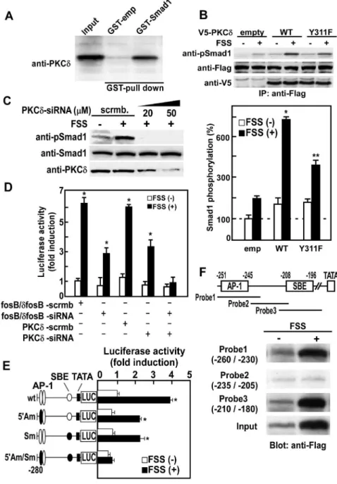
transferase (GST) pull-down assay. Western blot analysis using an anti-PKCdantibody revealed that PKCd binds to GST-Smad1 (Figure 3A), demonstrating that PKCd physically interacts with Smad1.
In order to clarify whether Y311 phosphorylation of PKCdcan in fact activate PKCd and phosphorylate BR-Smads, Smad1 phosphorylation by FSS was examined in mPOBs overexpressing wild-type or Y311F mutant PKCd. Smad1 phosphorylation was robustly enhanced by FSS in osteoblasts overexpressing wild-type PKCd, but was attenuated in osteoblasts overexpressing Y311F PKCd (Figure 3B). Moreover, knockdown of PKCd by small interfering RNA (siRNA) abrogated phosphorylation of endoge-nous Smad1/5 by FSS (Figure 3C). To further clarify whether Smad1/5 phosphorylation by PKCd stimulates IL-11 gene transcription in response to FSS, we examined the effect of PKCd
knockdown on IL-11 gene promoter activity. Knockdown of PKCdby siRNA markedly suppressed the enhancement ofIL-11
gene promoter activity by FSS, and FosB/DFosB knockdown to inhibit AP-1 signaling in combination with PKCd knockdown almost completely abolished the stimulation of IL-11 gene promoter activity by FSS (Figure 3D). Taken together, these observations demonstrate that mechanical stress induces phos-phorylation of BR-Smads by activation via Y311 phosphos-phorylation of PKCd, which in turn enhances the expression of IL-11 in cooperation with AP-1 signal in osteoblasts.
Phosphorylated BR-Smads by mechanical stress interact withDFosB/JunD on IL-11gene promoter
We have previously demonstrated that mechanical stress enhances IL-11 expression by an activation of IL-11 gene promoter via heterodimer formation of DFosB and JunD [13]. In order to clarify whether there is any interaction between
DFosB/JunD heterodimers and BR-Smads, we examined the effect of FSS on Smad1 binding toIL-11gene promoter as well as Figure 2. Mechanical stress activates PKCdby Y311 tyrosine phosphorylation.(A) mPOBs were infected with an adenovirus expressing Flag-Smad1 for 48 hours, and were pretreated with vehicle or 100 nM TPA for overnight, and then exposed to FSS for 30min or treated with 300 ng/ ml BMP-2. Phosphorylation of exogenous Smad1 was analyzed by Western blot analysis. Proteins from 4 wells were analyzed in each lane, and the experiments were repeated for 3 times with similar results. Results from a representative experiment were presented. (B) mPOBs were exposed to FSS for 30 min, and then PKCd, PKCe, PKCgand PKChwere immunoprecipitated from 500mg of total cell lysates obtained from 4 dishes using antibodies against PKCd, PKCe, PKCgand PKCh,. Western blot analysis was performed using anti-phosphorylated serine/threonine and tyrosine antibodies. The experiments were repeated for 3 times with similar results. (C) mPOBs were exposed to FSS for the indicated times, and then phosphorylation at S643, T505, and Y311 of PKCd was analyzed by Western blot analysis. (D) Mutant PKCdwith modification of Y311 to F311 (Y311F) was transiently transfected into mPOBs, and then cells were exposed to FSS for 30 min. In the upper panel, tyrosine phosphorylation was analyzed by Western blot analysis. Proteins from 4 wells were analyzed in each lane, and the experiments were repeated for 3 times with similar results. Results from a representative experiment were presented. In the lower panel, protein kinase activity was measured by kinase assay. The data were means6S.E.M. for three experiments, and difference among the groups was analyzed by one-way ANOVA. *p,0.05 compared with the wild type without FSS. doi:10.1371/journal.pone.0013090.g002
Figure 3. Mechanical stress-activated PKCdphosphorylates BR-Smads, and phosphorylated BR-Smads interact withDFosB/JunD on
interaction of BR-Smads with DFosB/JunD heterodimers. Anal-ysis of mouse IL-11 gene promoter activity with site-directed mutagenesis at a putative Smad-binding element (SBE) (2208 to
2196) and an upstream AP-1 binding site (2251 to2245) which confers IL-11 gene transcription by DFosB/JunD [13] revealed that both SBE and AP-1 sites were required for full activation of IL-11 gene promoter activity by FSS (Figure 3E). These observations can also explain why knockdown of PKCdby siRNA only partially suppressedIL-11gene promoter activity (Figure 3D), while Smad1 phosphorylation by FSS was almost completely abrogated by siRNA (Figure 3C).
In order to examine whether BR-Smads bind to the SBE on IL-11gene promoter, Flag-tagged Smad1 was transfected to mPOBs and DNA precipitation assay was performed. Because mechanical stress to mPOBs enhanced Smad1 phosphorylation by PKCd, transition of phosphorylated Smad1 into nucleus increased. As a result, the total amount of Smad1 in the nucleus shown as input in Figure 3F also increased by FSS. In addition to the increase in the total nuclear Smad1, not only DNA probe 3 (2210 to 2180) covering SBE but also probe 1 (2260 to2230) covering upstream AP-1 site bound to Flag-tagged Smad1 in an FSS-dependent manner (Figure 3F). Probe 2 (2235 to2205) that does not cover either SBE or AP-1 site did not bind Flag-Smad1 (Figure 3F). These results demonstrate that FSS-activated Smad1 is incorporated into a complex with DFosB/JunD heterodimer on the IL-11 gene promoter, and the complex is bound to both AP-1 site and SBE.
BR-Smads form complex with DFosB/JunD heterodimer by binding to JunD
In order to clarify the relationship between Smad1 andDFosB/ JunD heterodimer, V5-taggedDFosB, Flag-tagged JunD and Myc-tagged Smad1 were co-transfected into mPOBs and DNA precipitation assay was performed using upstream AP-1 probe (Probe 1) or SBE probe (Probe 3). V5-DFosB and Flag-JunD were bound to SBE when Smad1 was co-transfected, and Myc-Smad1 was bound to AP-1 when V5-DFosB and Flag-JunD were co-transfected (Figure 4A). These results demonstrate that Smad1 binds to SBE andDFosB/JunD heterodimer binds to AP-1 site on the IL-11 gene promoter, and that Smad1 and DFosB/JunD heterodimer form complex on the IL-11 gene promoter. In addition, a GST pull-down assay revealed that GST-Smad1 interacted with only Flag-JunD but not with V5-DFosB (Figure 4B), suggesting that Smad1 interacts with JunD to form a complex with
DFosB/JunD heterodimer. To further delineate the interaction between Smad1 andDFosB/JunD heterodimer, we next searched binding domain of JunD to Smad1 by creating full length Flag-JunD, as well as deletion mutants of Flag-JunD, Flag-JunD(1–344) and Flag-JunD(1–321). Full length and JunD(1–344) with intact Smad-binding domain similarly bound to Smad1, but JunD(1–321) that lacks C-terminal Smad-binding domain failed to bind to Smad1 (Figure 4C). Consistent with these observations, full length JunD and JunD(1–344), but not JunD(1–321), interacted with SBE in the presence of Smad1 (Figure 4D). Thus, Smad1 forms complex with
DFosB/JunD heterodimer by interacting with the C-terminal Smad-binding domain of JunD.
Discussion
In this paper, we report a new signal crosstalk between PKCd -Smads pathway and ERK-CREB-DFosB/JunD pathway in response to mechanical stress. Mechanical stress by FSS to osteoblasts enhances phosphorylation of BR-Smads via an activa-tion of PKCd. Phosphorylated BR-Smads form complex with
DFosB/JunD heterodimers via binding to the C-terminal region of JunD, and BR-Smad binds to SBE andDFosB/JunD heterodimer binds to AP-1 site of theIL-11gene promoter. Consequently, they cooperate to lead to a full activation of theIL-11gene transcription. In the present study, mechanical stress to osteoblasts transiently enhanced Smad1/5 phosphorylation without stimulation by BMP. Because BMP binding to its receptor phosphorylates BR-Smads by an activation of BMP receptor type I kinase, PKC inhibitors are not expected to inhibit BMP-induced phosphorylation of BR-Smads. However, in the present study, FSS-induced phosphory-lation of BR-Smads was almost completely blocked by an inhibition of PKCd, or transfection of Y311F mutant that cannot be phosphorylated by PKCd at amino acid 311, or silencing PKCdexpression by PKCdsiRNA. These observations indicate that mechanical stress-induced stimulation of BR-Smad signaling is independent of BMP-BMP receptor signaling pathway, but is dependent upon PKCdactivation. Stimulation of PKC activity by mechanical stress to osteoblastic cells has been reported using cyclic stretch load by Flexercell system in calvarial osteoblasts [27]. However, it has been unknown which class of PKC is activated by mechanical stress to osteoblastic cells. The present study revealed that activation of PKCd plays an important role for the phosphorylation and activation of BR-Smads by mechanical stress. One of the earliest responses after mechanical loading to bone is an increase in the expression of c-Fos. However, because overexpression of c-Fos develops osteosarcoma without an enhancement of bone formation [28], c-Fos appears to enhance transformation instead of differentiation of osteoblasts. We previously demonstrated that mechanical stress to osteoblasts rapidly enhances the transcription offosBgene with a similar time course to that ofc-fosgene, and that the enhancement offosBgene transcription is mediated via an activation of ERK-CREB signaling [14]. Activation of fosB gene transcription causes an increase in the expression of FosB and its truncated splice variant,
DFosB. Because DFosB overexpression in mice causes an enhancement of bone formation [29,30], the increase inDFosB in response to mechanical stress may play an important role in the stimulation of bone formation by mechanical stress. To this end, it is interesting to note thatDFosB forms heterodimer with JunD on the AP-1 site of theIL-11gene promoter, and that the binding of
DFosB/JunD heterodimer on theIL-11gene promoter enhances the promoter activity to increase the expression of IL-11 after mechanical stress [13]. However, AP-1 factors regulate the transcription of wide range of genes, and this signaling cascade in itself does not enhance a group of genes that are specific for osteoblast differentiation.
BMP regulates differentiation of various types of cells, but its main target is the regulation of osteoblast differentiation. Because Smads mediate canonical BMP signaling, activation of
BR-Values were expressed as the mean6S.E.M. for four experiments, and difference between FSS(2) and FSS(+) in each group was analyzed by Student’sttest. *p,0.01 compared with FSS (2) cells. (F) Flag-tagged Smad1 was transfected into mPOBs. After mPOBs were subjected to FSS for 30 min, nuclear lysates were prepared and analyzed for DNA binding to various DNA probes (probe 1–3) ofIL-11gene promoter by DNA precipitation assay. Flag-tagged Smad1 bound to DNA probes 1 to 3 was analyzed by Western blotting using an anti-Flag antibody. DNA probe 1 (2260 to2230) covers 59AP-1 site (2251 to2245), DNA probe 3 (2210 to2180) covers SBE (2208 to2196), and DNA probe 2 (2235 to2205) does not cover either AP-1 site or SBE (upper panel). Not only DNA probe 3 (2210 to2180) covering SBE but also probe 1 (2260 to2230) covering upstream AP-1 site bound to Flag-tagged Smad1 in an FSS-dependent manner, whereas probe 2 did not bind to Smad1 (lower panel).
doi:10.1371/journal.pone.0013090.g003
Smad signaling by mechanical stress via an activation of PKCdmay play an important role for the enhancement of osteoblastic differentiation by mechanical stress. In fact, inhibition of phosphor-ylation of BR-Smads by transfection of Y311F mutant of PKCdor knockdown of PKCd abrogated the stimulation of IL-11 gene promoter activity by mechanical stress. These results are consistent with the notion that BR-Smads andDFosB/JunD heterodimer act synergistically to enhance osteoblast differentiation, and that both the activation of BR-Smads and the formation of DFosB/JunD heterodimer are required for a full activation of IL-11 gene transcription in response to mechanical stress. Because IL-11 transgenic mice exhibit increased bone mass with enhanced bone formation without an increase in bone resorption [16], and because
an increase in IL-11 expression causes a down-regulation of inhibitors of canonical Wnt signaling [13], it is plausible to assume that the mechanical stress-induced increase in IL-11 expression constitutes an important signaling cascade mediating the mechan-ical stress signal to the enhancement of osteoblast differentiation.
The present study demonstrated that phosphorylated BR-Smads bind to the C-terminal region of JunD as well as to SBE of theIL-11 gene promoter. Our previous results demonstrated thatDFosB/JunD heterodimer binds to the AP-1 site on theIL-11 gene promoter [13]. These results suggest that phosphorylated BR-Smads form complex withDFosB/JunD heterodimer on the IL-11 promoter with binding of BR-Smads to the SBE and of
DFosB/JunD heterodimer to the AP-1 site, and that they form a Figure 4. BR-Smads form complex withDFosB/JunD heterodimer by binding to JunD.(A) V5-taggedDFosB, Flag-tagged JunD and Myc-tagged Smad1 were co-transfected into mPOBs. Nuclear lysates were prepared and analyzed for binding of nuclear proteins to DNA probe 1 or 3 by DNA precipitation assay followed by Western blotting using antibodies against V5, Flag and Myc. Nuclear lysates from 4 wells were analyzed in each lane, and the experiments were repeated for 3 times with similar results. Results from a representative experiment were presented. (B) GST-Smad1 fusion protein was bound to Glutathione-Sepharose beads, and Flag-JunD or V5-DFosB was incubated with GST-Smad1 fusion protein coupled to Glutathione-Sepharose. Complexes were then washed and bound proteins were eluted and separated on SDS-PAGE. JunD orDFosB bound to GST-Smad1 fusion protein was detected by Western blot analysis using an anti-Flag or an anti-V5 antibody. (C) GST-GST-Smad1 fusion protein was bound to Glutathione-Sepharose beads, and Flag-tagged WT or C-terminal truncated JunD was incubated with GST-Smad1 fusion protein coupled to Glutathione-Sepharose. Complexes were then washed and bound proteins were eluted and separated on SDS-PAGE. Flag-tagged WT or C-terminal truncated JunD bound to GST-Smad1 fusion protein was detected by Western blot analysis using an anti-Flag antibody (upper panel). Both JunD (WT) and JunD (1–344) (D1) had Smad-binding domain, but JunD (1–321) (D2) lacked C-terminal Smad-binding domain (lower panel). (D) V5-taggedDFosB and Flag-tagged JunD (WT), JunD (D1), or JunD (D2) along with Myc-tagged Smad1 were co-transfected into mPOBs. Nuclear lysates were prepared and analyzed for binding of nuclear proteins to DNA probe 1 or 3 by DNA precipitation assay, followed by Western blotting using antibodies against V5, Flag and Myc. Nuclear lysates from 4 wells were analyzed in each lane, and the experiments were repeated for 3 times with similar results. Results from a representative experiment were presented.
complex with other transcription co-factors to enhanceIL-11gene transcription. The mechanism whereby the formation of such transcription factor complex enhances IL-11 gene transcription remains to be clarified.
In conclusion, the present observations demonstrate that PKCd -Smad1 pathway plays an important role in the intracellular signaling in response to mechanical stress, and that a cross-talk between PKCd-Smad1 and ERK-CREB-DFosB/JunD pathways synergistically stimulates osteogenic program in response to mechanical stress in osteoblasts.
Materials and Methods
Ethics Statement
All experiments with animals were performed according to the guidelines for animal protection in the University of Tokushima, and approved by the Animal Experiments Committee, Tokushima University (#Toku-Animal06121) for animal protection.
Materials
Recombinant human transforming growth factor -b1 and recombinant human BMP-2 were purchased from Peprotech Inc. (Rocky Hill, NJ). Recombinant human PKCdwas purchased from Calbiochem (San Diego, CA). Calphostin C (PKC inhibitor), U0126 (mitogen-activated protein kinase kinase inhibitor), SB203580 (p38 kinase inhibitor), LY294002 (PI3K inhibitor), H89 (PKA inhibitor), Chelerythrine chloride (PKC inhibitor), Go6976 (PKC inhibitor) and Bisindolylmaleimide I (PKC inhibitor) were purchased from Calbiochem. TPA (PKC activator) was purchased from Sigma (St Louis, MO).
Antibodies against phospho-Smad1 which recognizes Ser463/ 465-phosphorylated Smad1/5/8, against phospho-Smad2 which recognizes Ser465/467-phosphorylated Smad2/3, and Smad2 were purchased from Cell Signaling Technology (Berverly, MA). Antibody against Smad1 was purchased from MILLIPORE Corp. (Bedford, MA). Antibody against b-actin was purchased from Sigma. Antibody against phospho-Serine/Threonine (pSer/Thr) was purchased from Invitrogen (Carlsbad, CA). Antibody against phospho-Tyrosine (pTyr), PKCd, PKCe, PKCgand PKChwere purchased from Santacruz Biotechnology (San Diego, CA). Antibody against Phospho-PKCd.. Tyr311
˙ was purchased from Stressgene (Victoria, Canada). Anti-Flag antibody was purchased from Sigma. Anti-V5 antibody was purchased from Invitrogen. Anti-Myc antibody was purchased from Santacruz Biotechnology. All chemicals for dual-luciferase assays were purchased from Promega (Madison, WI). The other chemical reagents were purchased from Sigma unless otherwise specified.
Cell culture andin vitromechanical loading by FSS Primary osteoblasts were prepared from calvaria of newborn mice by sequential digestion as previously described [14]. Isolated mPOBs were cultured inaminimum essential medium (aMEM; Invitrogen) supplemented with 10% fetal bovine serum (FBS; MBL Co., Ltd, Nagoya, Japan), penicillin/streptomycin (Invitro-gen) for 48 hours. Before experiments, 16105 cells/ml mPOBs were seeded on culture dishes, grown to 70–80% confluence, and serum-deprived inaMEM with 1% FBS for 24 hours. As reported previously, these mPOBs can undergo osteoblastic differentiation in vitroto become osteogenic cells [14].
For mechanical loadingin vitro, cells were exposed to FSS by placing 6-well culture dishes (35 mm diameter) on a horizontal rotating platform with rotating radius of 9 mm fixed inside the culture incubator and rotated at 120 rpm [14]. This horizontally rotating apparatus creates rotating fluid flow of culture medium in
culture dishes. In a cone viscometer, because the angle of cone is constant, the FSS to cells is almost constant in most part of culture dishes [31], whereas the fluid surface height is not proportional to the distance from the center in our system. In our apparatus, the cycles of rotator and the fluid were almost synchronized, and the fluid surface height at the edge is the highest where rotation of them is in the same direction while the surface height at the edge is the lowest where rotation of them is in the opposite direction. Therefore, the height of the fluid (z) ranged as follows:
1{c’=c
ð Þv2c22gSzSð1zc’=cÞv2c22g
Wherevis the angular velocity,ris the radius of culture dish,r9is
the radius of rotator, andgis the gravity forces (9.8 m/s2). Because r9is almost 1/2 ofr,zis approximately 1.4 mm at the lowest edge and 4.0 mm at the highest edge. The z values in our system correspond tortanain a cone viscometer. Therefore, hypothetical
fluid surface angles (a) on the line of the lowest ridge can be
calculated by the following formula:
aðdegreeÞ~tan{1ðz=cÞ:180=p
where zis the minimal value on the lowest ridge, and r is the distance from the center.
Based upon the calculated FSS (t) of 2.0 Pa from the Reynolds
number (R) and the following formulas in Reference 31:
t~ðgv=aÞf Rð Þ
f Rð Þ~1z2:58 R3=2 3:5zR
ð Þ
{0:86 R5=2 3:5zR
ð Þ
tcan be calculated as follows:
rðmmÞ v a R f Rð Þ t(Pa)
18 4p 4:1 3:0 2:7 0:33
4:5 4p 0:72 0:006 1:0 0:69
2:25 4p 0:25 v0:001 1:0 2:0
Thus, FSS in 6-well culture dish is expected to range from 0.33 to 2.0 Pa.
The above model may not be able to precisely estimate FSS at the very edge of the dish where the fluid contacts with the outer wall, and at the very center of the well. However, based on the calculations above, we expect that FSS in our system mostly remains within the physiological range of 2.0 Pa or lower [32].
Adenovirus Infection
All recombinant adenovirus used in this study (Flag-Smad1, Flag-Smad5, and Flag-Smad8) were reported previously [22], and high-titered stocks of recombinant adenoviruses were grown in HEK293 cells (Riken Bioresource Center, Ibaragi, Japan). For experiments, mPOBs passage two were infected with various recombinant adenoviruses at a multiplicity of infection (m.o.i) varying from 0 to 100. At 48 h after infection, cells were harvested and examined for expression or phosphorylation of Smads by Western blot analysis.
GST pull-down assay
GST-Smad1 plasmids were transformed into BL21 competent cells (Invitrogen) and the protein expression was induced by the addition of isopropyl-1-thio-b-D- galactopyranoside (IPTG) and then bacterial pellets were lysed by freeze-thaw cycles and
sonication. GST fusion protein then were mixed with Glutathione-Sepharose (GE Healthcare Life Sciences, Little Chalfont, Buck-inghamshire, UK) and incubated at 4Cu for 1 hour. The Sepharose beads were then washed three times and used for GST pull-down assays. mPOBs were lysed in lysis buffer (20 mM HEPES-KOH, pH7.5, 150 mM NaCl, 1% Triton-X100, 0.5 mM phenylmethylsulfonyl fluoride (PMSF), and 16protease inhibitor cocktail (SIGMA)) and aliquots of the lysate were then incubated under constant agitation for 1 hour at 4Cu with GST fusion protein coupled to Glutathione-Sepharose. Subsequently, com-plexes were washed three times and then bound proteins were eluted and separated on SDS-PAGE and detected by Western Blots using appropriate antibodies.
Protein analysis
Preparation of nuclear extracts or total cell lysates were prepared as described before [14]. For Western blot analysis, 30mg of protein was separated on a SDS-PAGE (10–20% gradient gel, ATTO, Tokyo, Japan) and transferred to PVDF membrane (MILLIPORE). The membrane was rinsed and blocked with 5% non-fat skim milk in Tris-buffered saline (TBS) with 0.1% Tween-20 for 1 hour at room temperature, then blotted sequentially with primary antibodies, then with a horseradish peroxidase-conjugated secondary antibody (Promega) for 1 hour, and the protein bands were visualized with an ECL-Plus detection system (GE Health-care Life Sciences).
Plasmid construction
Myc-tagged Smad1 used in this study was reported previously [22]. A cDNA encoded from mouse V5-tagged DFosB (full-length), Flag-tagged JunD (WT: full-length,D1: 1–344a.a.,D2: 1– 321a.a.), and V5-tagged PKCd(WT: full-length) were amplified by polymerase chain reaction (PCR) using PrimeSTAR (TaKaRa, Shiga, Japan) and subcloned into the mammalian expression vector using pcDNA3.1D Directional TOPO Expression Kit (Invitrogen) as referred to the manufacturer’s instructions. A PKCd mutant (PKCd Y311F) was generated by site-directed mutagenesis using the QuickChangeTMSite-Directed Mutagenesis Kit (STRATAGENE, La Jolla, CA) with mutagenic primers (sense: 59- CAGAGTCTGTCGGAATATTCCAGGGATTT-GAGAAGAA -39, antisense: 59- TTCTTCTCAAATCCCTG-GAATATTCCGACAGACTCTG-39). These expression plasmids were checked and verified sequencing (ABI model 377, Applied Biosystems) promoted by Support Center for Advanced Medical Societies, University of Tokushima.
MouseIL-11promoter construction
Fragments of mouse IL-11 gene (GeneBankTM number AY225468) upregulatory region from nucleotides 2269 to 26 (2280wt) [33] were cloned and subcloned into a luciferase reporter plasmid pGL3-basic (Promega), which lacks eukaryotic promoter and enhancer sequences (with the transcription start site being numbered+1). A series of mutant plasmids (22805’Am,2280Sm, and22805’Am/Sm) were generated by site-directed mutagenesis with mutagenic primers including 59- ACTCtca -39 (WT: 59 -tgactca -39, position at2251 to2245) for 59AP-1 mutation and 59 -TTTgcc -39(WT: 59- cccgccc -39, position at2201 to2196) for SBE mutation, respectively. Capital letters indicate substituted nucleotides that disrupt AP-1 and Smad binding sites.
Gene silencing
Duplexed siRNA was designed and synthesized by Hokkaido System Sciences (Sapporo, Japan). The siRNA sequences for
targeting mouse fosB/dfosB and mouse PKCd are as follows; mouse fosB/dfosB: 59- UCUCUUUACACACAGUGAAGUU-CAA -39and mouse PKCd: 59 -AGCUGAAGGGCAAAGAUAA-GUACUUAG-39. Scrambled siRNA was synthesized and used as a negative control. Transfection of the siRNA oligonucleotides was performed using Lipofectamine RNAiMAX (Invitrogen) in opti-MEM (Invitrogen) without serum. Gene silencing efficiency was evaluated by Western blotting using specific antibody for targeting gene products.
Measurement of transcriptional activity
mPOBs were seeded in 6-well culture plates at 50% confluence and co-transfected with 2mg of chimeric Firefly luciferase reporter plasmid combined with Renilla luciferase reporter plasmid (pRL-TK, Promega) as a control, and/or with mammalian expression plasmids using GenePORTER 2 Transfection Reagent (Genlantis, San Diego, CA) in opti-MEM (Invitrogen) supplemented with 1% serum. Sixteen hours after transfection, the medium was replaced by aMEM containing with 1% FBS and treated with various reagents and exposed to FSS by placing the culture plates on a shaking apparatus at 100 to 120 rpm. For dual luciferase assay, the cells were washed and lysed with passive cell lysis buffer (Promega) and both Firefly and Renilla luciferase activity was measured with luminometer (ATTO) by mixing 50ml of luciferase substrate solution (Promega) with 10ml of cleared cell lysates. Transcrip-tional activity was corrected for Renilla luciferase activity or protein concentrations.
Measurement of protein kinase activity
Cells were stimulated with different agents and then lysed with a lysis buffer for the kinase assay (20 mM Tris-HCl, pH 7.5, 137mM NaCl, 1 mM EDTA, 1 mM EGTA, 10% glycerol, 1% NP-40, 1 mM sodium orthovanadate (Na3VO4), 4.5 mM sodium
pyrophosphate, 47.6 mM sodium fluoride (NaF), 9.26 mM b -glycerophosphate, 0.5 mM dithiothreitol (DTT) and 16protease inhibitor cocktail). Lysates were then immunoprecipitated using appropriate antibodies. Kinase assays were performed in the immunoprecipitates using the PKC assay kit (Upstate Biotechnol-ogy, Lake Placid, NY) following the directions provided by the company with minor modifications. Briefly, Immunoprecipitaes bound to agarose beads were resuspended in assay dilution buffer containing phospatidylserine and diacylglycerol. The kinase reaction was initiated either the addition of a Mg2+
/adenosine triphosphate (ATP) mixtures (75 mM MgCl2 and 0.5 mM ATP)
containing 10 mCi of [c-32P]ATP (3,000 Ci/mmol) and the substrate (GST-Smad1 or histone H1 as a control). The reaction mixtures were briefly vortexed and then incubated at 30Cu for 30 min with occasional mixing. After incubation, the kinase reaction was terminated by adding 46sample buffer and boiling the samples. Samples were resolved using 4–20% SDS-PAGE gels and then dried and analyzed by autoradiography.
DNA precipitation assay
Biotinylated double-stranded oligonucleotide probes containing tandem AP-1 of the mouse IL-11 promoter sequences [33] were prepared by annealing complementary oligonucleotides followed by ethanol precipitation. Then annealed DNA probes were incubated with equal amounts of nuclear extracts and poly dI-dC (GE Healthcare LifeSciences) in reaction buffer (20 mM HEPES-KOH pH 7.4, 0.1 M NaCl, 0.5 mM EDTA, 0.5 mM EGTA, 0.5 mM DTT, 1 mM PMSF, 16 protease inhibitor cocktail, 20 mM NaF, and 1 mM Na3VO4) for 4Cuwith gentle
complexes bound to beads were collected by centrifugation and washed with ice-cold reaction buffer for 4 times. After the final wash, beads were collected and resuspended in 26sample buffer and boiled. DNA-bound proteins were separated on a SDS-PAGE and detected by Western blot analysis.
Statistical analysis
Values for parameters within a group are expressed as means6 S.E.M. For comparisons among groups, statistical significance was assessed using an unpaired two-tailed Student’sttest for two-group comparisons or one-way ANOVA, and the significance of each difference was determined by post-hoc testing using the Tukey-Kramer method. These analyses were performed on an Apple Macintosh computer with the use of Excel (Microsoft) and StatView statistical package (StatView 5.0; SAS Institute Inc.). A Pvalue of less than 0.05 was considered significant.
Supporting Information
Figure S1 IL-11 expression is enhanced by BMP-2. mPOBs were treated with 300 ng/ml BMP-2 for the indicated period of time, and quantitative real-time PCR analysis was performed using the SYBR Green chemistry. The reaction mixture (20ml) contained 200 nM PCR primers sets for IL-11 (forward: 59 -aaattcccagctgacggagatcac -39, reverse: 59- tacatgccggaggtaggacat-caa -39) and GAPDH (forward: 59- ggcaaattcaacggcacagtca -39,
reverse: 59- ggcaaattcaacggcacagtca -39), 10ml SYBR premix Ex Taq (TaKaRa, Shiga, Japan) and cDNA templates equivalent to 100 ng total RNA.
Found at: doi:10.1371/journal.pone.0013090.s001 (8.70 MB TIF)
Figure S2 Mechanical stress enhances IL-11 but not BMP-2 expression. mPOBs were exposed to FSS for the indicated period of time, and then quantitative real-time PCR analysis was performed using the SYBR Green chemistry. The reaction mixture (20ml) contained 200 nM PCR primers sets for IL-11 (forward: 59- aaattcccagctgacggagatcac -39, reverse: 59- tacatgccg-gaggtaggacatcaa -39), BMP-2 (forward: 59 cgcagcttccatcacgaaga -39, reverse: 59- tgttcccggaagatctggagtt -39), and GAPDH (forward: 59- ggcaaattcaacggcacagtca -39, reverse: 59- ggcaaattcaacggca-cagtca -39), 10ml SYBR premix Ex Taq (TaKaRa, Shiga, Japan) and cDNA templates equivalent to 100 ng total RNA. Data are means6S.E.M. for four experiments, and difference from non-treated cells was analyzed by one-way ANOVA. *P,0.05 compared with non-treated cells.
Found at: doi:10.1371/journal.pone.0013090.s002 (8.70 MB TIF)
Author Contributions
Conceived and designed the experiments: SK DI TI TM. Performed the experiments: SK YI. Analyzed the data: SK IE DI HT TI. Contributed reagents/materials/analysis tools: SK RKK YUM HT YI TI. Wrote the paper: SK RKK DI TI TM.
References
1. Morey ER, Baylink DJ (1978) Inhibition of bone formation during space flight. Science 201: 1138–1141.
2. Schneider V, Oganov V, LeBlanc A, Rakmonov A, Taggart L, et al. (1995) Bone and body mass changes during space flight. Acta Astronaut 36: 463–466. 3. Inoue M, Tanaka H, Moriwake T, Oka M, Sekiguchi C, et al. (2000) Altered
biochemical markers of bone turnover in humans during 120 days of bed rest. Bone 26: 281–286.
4. Watanabe Y, Ohshima H, Mizuno K, Sekiguchi C, Fukunaga M, et al. (2004) Intravenous pamidronate prevents femoral bone loss and renal stone formation during 90-day bed rest. J Bone Miner Res 19: 1771–1778.
5. Kodama Y, Nakayama K, Fuse H, Fukumoto S, Kawahara H, et al. (1997) Inhibition of bone resorption by pamidronate cannot restore normal gain in cortical bone mass and strength in tail-suspended rapidly growing rats. J Bone Miner Res 12: 1058–1067.
6. Burr DB, Robling AG, Turner CH (2002) Effects of biomechanical stress on bones in animals. Bone 30: 781–786.
7. Knothe Tate ML (2003) ‘‘Whither flows the fluid in bone?’’ An osteocyte’s perspective. J Biomech 36: 1409–1424.
8. Liu D, Genetos DC, Shao Y, Geist DJ, Li J, et al. (2008) Activation of extracellular-signal regulated kinase (ERK1/2) by fluid shear is Ca(2+)- and ATP-dependent in MC3T3-E1 osteoblasts. Bone 42: 644–652.
9. You J, Reilly GC, Zhen X, Yellowley CE, Chen Q, et al. (2001) Osteopontin gene regulation by oscillatory fluid flow via intracellular calcium mobilization and activation of mitogen-activated protein kinase in MC3T3-E1 osteoblasts. J Biol Chem 276: 13365–13371.
10. Hung CT, Allen FD, Pollack SR, Brighton CT (1996) Intracellular Ca2+stores and extracellular Ca2+are required in the real-time Ca2+response of bone cells experiencing fluid flow. J Biomech 29: 1411–1417.
11. Walker LM, Publicover SJ, Preston MR, Said Ahmed MA, El Haj AJ (2000) Calcium-channel activation and matrix protein upregulation in bone cells in response to mechanical strain. J Cell Biochem 79: 648–661.
12. Rawlinson SC, Pitsillides AA, Lanyon LE (1996) Involvement of different ion channels in osteoblasts’ and osteocytes’ early responses to mechanical strain. Bone 19: 609–614.
13. Kido S, Kuriwaka-Kido R, Imamura T, Ito Y, Inoue D, et al. (2009) Mechanical stress induces Interleukin-11 expression to stimulate osteoblast differentiation. Bone 45: 1125–1132.
14. Inoue D, Kido S, Matsumoto T (2004) Transcriptional induction of FosB/
DFosB gene by mechanical stress in osteoblasts. J Biol Chem 279: 49795–49803. 15. Kodama Y, Takeuchi Y, Suzawa M, Fukumoto S, Murayama H, et al. (1998) Reduced expression of interleukin-11 in bone marrow stromal cells of senescence-accelerated mice (SAMP6): relationship to osteopenia with enhanced adipogenesis. J Bone Miner Res 13: 1370–1377.
16. Takeuchi Y, Watanabe S, Ishii G, Takeda S, Nakayama K, et al. (2002) Interleukin-11 as a stimulatory factor for bone formation prevents bone loss with advancing age in mice. J Biol Chem 277: 49011–49018.
17. Urist MR (1965) Bone: formation by autoinduction. Science 150: 893–899. 18. Wozney JM, Rosen V, Celeste AJ, Mitsock LM, Whitters MJ, et al. (1988) Novel
regulators of bone formation: molecular clones and activities. Science 242: 1528–1534.
19. Afrakhte M, Moren A, Jossan S, Itoh S, Sampath K, et al. (1998) Induction of inhibitory Smad6 and Smad7 mRNA by TGF-bfamily members. Biochem Biophys Res Commun 249: 505–511.
20. Hollnagel A, Oehlmann V, Heymer J, Ruther U, Nordheim A (1999) Id genes are direct targets of bone morphogenetic protein induction in embryonic stem cells. J Biol Chem 274: 19838–19845.
21. Takase M, Imamura T, Sampath TK, Takeda K, Ichijo H, et al. (1998) Induction of Smad6 mRNA by bone morphogenetic proteins. Biochem Biophys Res Commun 244: 26–29.
22. Imamura T, Takase M, Nishihara A, Oeda E, Hanai J, et al. (1997) Smad6 inhibits signalling by the TGF-bsuperfamily. Nature 389: 622–626.
23. Hofmann J (1997) The potential for isoenzyme-selective modulation of protein kinase C. Faseb J 11: 649–669.
24. Jordan NJ, Watson ML, Yoshimura T, Westwick J (1996) Differential effects of protein kinase C inhibitors on chemokine production in human synovial fibroblasts. Br J Pharmacol 117: 1245–1253.
25. Jaken S (1996) Protein kinase C isozymes and substrates. Curr Opin Cell Biol 8: 168–173.
26. Konishi H, Yamauchi E, Taniguchi H, Yamamoto T, Matsuzaki H, et al. (2001) Phosphorylation sites of protein kinase C d in H2O2-treated cells and its activation by tyrosine kinase in vitro. Proc Natl Acad Sci U S A 98: 6587–6592. 27. Carvalho RS, Scott JE, Suga DM, Yen EH (1994) Stimulation of signal transduction pathways in osteoblasts by mechanical strain potentiated by parathyroid hormone. J Bone Miner Res 9: 999–1011.
28. Grigoriadis AE, Schellander K, Wang ZQ, Wagner EF (1993) Osteoblasts are target cells for transformation in c-fos transgenic mice. J Cell Biol 122: 685–701. 29. Sabatakos G, Sims NA, Chen J, Aoki K, Kelz MB, et al. (2000) Overexpression of DFosB transcription factor(s) increases bone formation and inhibits adipogenesis. Nat Med 6: 985–990.
30. Kveiborg M, Sabatakos G, Chiusaroli R, Wu M, Philbrick WM, et al. (2004)
DFosB induces osteosclerosis and decreases adipogenesis by two independent cell-autonomous mechanisms. Mol Cell Biol 24: 2820–2830.
31. Sakai K, Mohtai M, Shida J, Harimaya K, Benvenuti S, et al. (1999) Fluid shear stress increases interleukin-11 expression in human osteoblast-like cells: its role in osteoclast induction. J Bone Miner Res 14: 2089–2098.
32. Weinbaum S, Cowin SC, Zeng Y (1994) A model for the excitation of osteocytes by mechanical loading-induced bone fluid shear stresses. J Biomech 27: 339–360.
33. Tohjima E, Inoue D, Yamamoto N, Kido S, Ito Y, et al. (2003) Decreased AP-1 activity and interleukin-11 expression by bone marrow stromal cells may be associated with impaired bone formation in aged mice. J Bone Miner Res 18: 1461–1470.