Mouse Sphingosine Kinase 1a Is Negatively
Regulated through Conventional
PKC-Dependent Phosphorylation at S373 Residue
Yong-Seok Oh1*, Sun Sik Bae3¤a, Jong Bae Park2¤b, Sang Hoon Ha2, Sung Ho Ryu2,
Pann-Ghill Suh3*
1Department of Brain-Cognitive Science, Daegu-Gyeongbuk Institute of Science and Technology (DGIST), Hyeonpung-myeon, Dalseong-gun, Daegu, Republic of Korea,2Department of Life Science, Division of Molecular and Life Science, Pohang University of Science and Technology, Pohang, Republic of Korea,
3Department of Biological Sciences, Ulsan National Institute of Science and Technology (UNIST), Ulsan, Republic of Korea
¤a Current address: MRC for Ischemic Tissue Regeneration, Department of Pharmacology, Pusan National University, School of Medicine, Yangsan, Republic of Korea
¤b Current address: Specific Organs Cancer Branch, National Cancer Center, Goyang, Republic of Korea *[email protected](PGS);[email protected](YSO)
Abstract
Sphingosine kinase is a lipid kinase that converts sphingosine into sphingosine-1-phos-phate, an important signaling molecule with intracellular and extracellular functions. Although diverse extracellular stimuli influence cellular sphingosine kinase activity, the molecular mechanisms underlying its regulation remain to be clarified. In this study, we investigated the phosphorylation-dependent regulation of mouse sphingosine kinase (mSK) isoforms 1 and 2. mSK1a was robustly phosphorylated in response to extracellular stimuli such as phorbol ester, whereas mSK2 exhibited a high basal level of phosphoryla-tion in quiescent cells regardless of agonist stimulaphosphoryla-tion. Interestingly, phorbol ester-induced phosphorylation of mSK1a correlated with suppression of its activity. Chemical inhibition of conventional PKCs (cPKCs) abolished mSK1a phosphorylation, while overexpression of PKCα, a cPKC isoform, potentiated the phosphorylation, in response to phorbol ester. Fur-thermore, anin vitrokinase assay showed that PKCαdirectly phosphorylated mSK1a. In
addition, phosphopeptide mapping analysis determined that the S373 residue of mSK1a was the only site phosphorylated by cPKC. Interestingly, alanine substitution of S373 made mSK1a refractory to the inhibitory effect of phorbol esters, whereas glutamate substitution of the same residue resulted in a significant reduction in mSK1a activity, suggesting the sig-nificant role of this phosphorylation event. Taken together, we propose that mSK1a is nega-tively regulated through cPKC-dependent phosphorylation at S373 residue.
OPEN ACCESS
Citation:Oh Y-S, Bae SS, Park JB, Ha SH, Ryu SH, Suh P-G (2015) Mouse Sphingosine Kinase 1a Is Negatively Regulated through Conventional PKC-Dependent Phosphorylation at S373 Residue. PLoS ONE 10(12): e0143695. doi:10.1371/journal. pone.0143695
Editor:Qiming Jane Wang, University of Pittsburgh School of Medicine, UNITED STATES
Received:May 23, 2015
Accepted:November 8, 2015
Published:December 7, 2015
Copyright:© 2015 Oh et al. This is an open access article distributed under the terms of theCreative Commons Attribution License, which permits unrestricted use, distribution, and reproduction in any medium, provided the original author and source are credited.
Data Availability Statement:All relevant data are within the paper.
Introduction
Sphingolipids such as ceramide, sphingosine (SPH), and sphingosine-1-phosphate (S1P) are ubiquitous constituents of eukaryotic membranes that regulate cell growth, survival, apoptosis, differentiation, migration, and immune responses [1–4]. In contrast to ceramide and SPH, which are associated with apoptosis, S1P has been clearly established as a pro-survival molecule [5], as well as an important regulator of cellular trafficking, differentiation, angiogenesis, and inflammation [5]. S1P acts as both an intracellular second messenger and an extracellular ligand [1–4,6,7]. Inside cells, S1P is important for direct modulation of the activity of histone deacetylase [7], the ubiquitin ligase activity of TRAF2 [8], activation of MAP kinase [9], and Ca2+mobilization [10,11]. In another context, S1P functions as an extracellular ligand for a family of S1P-specific cell-surface G protein-coupled receptors (GPCRs) [5,12]. In addition, S1P is generated in and released from multiple types of cells [1]. Five S1P receptors (S1P1-5)
interact with S1P at the plasma membrane and then signal downstream via various G proteins including Gq, Gi/o, and G12/13, allowing for cell type-specific responses [1,5,12].
Sphingosine kinase (SK) is a lipid kinase that converts SPH into S1P by ATP-dependent phosphorylation [3]. The level of S1P in the cell is regulated in response to extracellular stimuli, probably by adjusting the balance between SK-mediated synthesis and degradation by SPP lyase or phosphatase [1]. To date, it is not clear that the activity of S1P lyase or phosphatase is transiently regulated; by contrast, many studies have established that the activity of cellular SK is regulated dynamically in the context of cellular physiology [3]. Indeed, SK is activated by multiple stimuli, including as PDGF [13], serum [13,14], TNFα[15], NGF [16], VEGF [17], acetylcholine [18,19], phorbol ester [20], forskolin [21], and FcgRII ligation [22], and formyl peptide [23]. On the other hand, SK activity could be negatively regulated in response to extra-cellular stimuli. For example, HDL profoundly inhibits TNF-stimulated sphingosine kinase activity in endothelial cells, resulting in decreased S1P production [24]. Despite extensive stud-ies about the physiological roles of SK and its product S1P, the molecular mechanisms underly-ing SK regulation have remained largely unclear.
The mouse genes,mSK1andmSK2, encode the mSK1 and mSK2 proteins, respectively, each of which has multiple splice variants that differ only at the N-terminus [1,5]. Just like the human isoforms, mSK1 and mSK2 exhibit different tissue distributions and subcellular location patterns [25]. Genetic knockout of each SK isoform has confirmed that the two isoforms may have dis-tinct physiological functions [26–29], in addition to some functional redundancy [30,31]. Except for the catalytic domains, the two SK isoforms differ significantly in protein sequence and molec-ular structure [1,32,33]. mSK1 harbors multiple Ca2+/calmodulin-binding sequences [32] and both isoforms have a proline-rich motif that might be involved in the interaction with SH3 domain-containing protein(s) [33], suggesting that the two isoforms might be differentially regu-lated through both specific and common signaling pathways. However, the possibility that each mSK isoform is regulated by distinct phosphorylation event has not been explored yet.
In this study, we analyzed the phosphorylation pattern of mSK isoforms in response to extracellular stimuli. Based on the initial observation that mSK1a is robustly phosphorylated in response to phorbol ester, we characterized the molecular pathway leading to mSK1a phos-phorylation and elucidated its influence on SK activity.
Material and Methods
Materials
S1P and SPH were from Biomol (Enzo Life Sciences, US). [32P]ATP was purchased from Perki-nElmer NEN Products (Boston, MA, USA). Phorbol 12-myristate 13-acetate (PMA), 4-deoxy Competing Interests:The authors have declared
pyridoxine, GF109204X, Go6976, and RO-31-8220 were from Merck Millipore (San Diego, USA). Silica TLC G60 plate was from Merck Millipore. Serum and medium were obtained from HyClone (Logan, UT), and other chemicals were from Sigma-Aldrich (St. Louis, USA).
Cell culture
COS-7 cells (from ATCC) were maintained in Dulbecco's modified Eagle's medium supple-mented with 10% FBS. Cells were grown at 37°C in humidified atmosphere containing 5% CO2.
Immunoblot Analysis
Cell lysates (30μg) were resolved by 12% SDS-PAGE and transferred to NC membrane (Schlei-cher & Schuell Bioscience). Membranes were blocked by incubation for 30 min at room tem-perature with T-TBS (TBS and 0.1% (v/v) Tween 20) containing 5% (w/v) nonfat milk. Primary antibodies were incubated overnight at 4°C in the blocking buffer at the appropriate dilutions. The bound primary IgGs were detected by incubation with secondary antibodies conjugated to HRP and developed using the ECL system (GE Healthcare Life Sciences).
Plasmid construction and mutagenesis
IMAGE clones 4221357 (mSK1a) and 2650442 (mSK2) were obtained from the Human Genome Mapping Project Resource Centre in UK and verified by direct sequencing with appropriate primers. Full-length cDNAs were amplified with PCR using primer pairs for
mSK1a (5’-gc gaa ttc atg gaa cca gaa tgc cct cga-3’/5’-gc gga tcc tta tgg
ttc ttc tgg agg tgg- 3’) and mSK2 (5’-ccc aag ctt atg gcc cca cca cca cta ctg-3’/5’-ccg gaa ttc tca ggc ttg tgg ctt ttg acc-3’). The PCR products were digested withEcoRI/BamHI (for mSK1a) orHindIII/EcoRI (for mSK2), and subsequently ligated into pCMV2-FLAG.
Site-directed mutagenesis was carried out using the splice-overlap extension method. For the substitution of S373 with alanine, PCR was carried out with wild-type (WT) mSK1a as the
template DNA, the forward primer5'- ggc cgg gac gcc cgg cgg ggg- 3’, and the
reverse primer5’- ccc ccg ccg ggc gtc ccg gcc-3’. From the PCR product, the
EcoRI/BamHI fragment was cloned into pCMV2-FLAG. The strategy was used to generate the other mutations using the following oligonucleotides: forward primer,5’-aga ccc gcc gcc aca ctg gtg-3’/reverse primer,5’-cac cag tgt ggc ggc ggg tct-3’(S225A);
for-ward primer,5’-gag ccc agg gcc cag agg ggc-3’/reverse primer,5’-gcc cct ctg
ggc cct ggg ctc-3’(S332A); forward primer,5’-ggc cgg gac gcg cgg cgg
ggg-3’/reverse primer,5’-ccc ccg ccg cgc gtc ccg gcc-3’(S373A); and forward primer,
5’-ggc cgg gac gag cgg cgg ggg-3’/reverse primer,5’-ccc ccg ccg ctc gtc ccg gcc-3’(S373E). All mutations were verified by direct sequencing of the entire ORFs while confirming the absence of undesired mutations.
Transfection
COS-7 cells were plated on 35 mm or 10 cm culture dishes at a density of 1.5 × 10⁵or 7 × 10⁵ cells/dish, respectively. The next day, 1–4μg of plasmid DNAs (pCMV2-control,
pCMV2-mSK2, pCMV2-mSK1a WT, pCMV2-mSK1a S373A, pCMV2-mSK1a S373E,
pCMV2-mSK1a S225A, pcDNA3.1, pcDNA3.1 PKCα, or pcDNA3.1 PKCδ) were transfected
Measurement of SK Activity
In vitroSK assay. As described previously with minor modifications [34], the cells were
washed with ice-cold PBS and scraped in SK assay buffer (20 mM Tris buffer [pH 7.2], 10 mM MgCl2, 20% glycerol, 1 mM dithiothreitol, 1 mM EDTA, 1 mM Na3VO4, 15 mM NaF, 10μg/
ml leupeptin and aprotinin, 1 mM PMSF, and 0.5 mM 4-deoxypyridoxine). For cell lysis, cells were ruptured by sonication (Branson Sonifier, output control 3) in SK assay buffer supple-mented with 0.25% Triton X-100. Cell homogenates were centrifuged at 15,000 rpm to remove the insoluble fraction. SK activity in cell extracts was measured by incubation in SK assay buffer with 50μM SPH, solubilized in 0.25% Triton X-100 and 1 mM [32P] ATP for 20 min at 37°C. The labeled lipids were extracted and resolved by TLC in the solvent of 1-butanol/ethanol/ace-tic acid/water (8:2:1:2). The formation of S1P was visualized and quantitated on a PhosphoI-mager system (Fuji Film, Tokyo).
Measurement of [3H] S1P formation. COS-7 cells, transfected with either control or mSK1a constructs, were serum-deprived for 24 hrs. The transfected cells were equilibrated in DMEM containing 1 mg/ml fatty acid-free BSA for 30 min, and then the medium was replaced with serum-free DMEM containing [3H]SPH (5 × 10⁵cpm, final concentration ~15 nM) pre-loaded onto 1 mg/ml fatty acid-free BSA, together with PMA (100 nM) or vehicle. After incu-bation for 5 min, the assay medium was replaced with 1 ml of pre-chilled methanol. The lipids were extracted, dried, and resolved by TLC in 1-butanol:acetic acid:water (3:1:1) as described previously [18]. SPH (Rf 0.67) and S1P (Rf 0.51) spots were visualized by ninhydrin and molybdenum blue staining. Each spot was scraped off, and radioactivity was measured by liq-uid scintillation counter.
Immuno-affinity Purification of mSK1a
After transfection with mSK1a, COS-7 cells were lysed and sonicated in lysis buffer A (20 mM Tris, pH 8.0, 1 mM EDTA/EGTA, 0.1 mM DTT) supplemented with 0.25% Triton X-100 and protease inhibitors (1 mM PMSF, 10μg/ml leupeptin, and 10μg/ml aprotinin). The lysates were centrifuged at 15,000 rpm for 20 min at 4°C. The supernatants were incubated at 4°C overnight with constant agitation with 50μl ofα-FLAG (M2 clone) Affi-Gel (Sigma-Aldrich, USA). The resin was washed four times with 1 ml of lysis buffer A. Bound protein was eluted with lysis buffer A containing 4μg/ml 3×FLAG peptide (Sigma-Aldrich).
Expression and Purification of PKC
α
Recombinant baculovirus expressing PKCαwas a generous gift from Dr. W. Cho (University of Illinois). PKCαwas expressed in baculovirus-infected Sf9 cells and purified as described with some modifications [35]. Monolayers of Sf9 cells (2 × 10⁷cells/150 mm dish, ten dishes) were infected at a multiplicity of infection of 10. PKCαwas purified using Hitrap-Q FPLC, fol-lowed by Phenyl-Superose FPLC (GE Healthcare Life Sciences). The purity of PKCαwas assessed higher than 90% by SDS-PAGE and Coomassie stain.
In vivo
and
in vitro
32P-labeling mSK1a and Purification
Forin vivophosphorylation, COS-7 cells in 60 mm dishes were transfected with mSK1a plas-mid and metabolically labeled with [32P] inorganic phosphate (0.4 mCi) in 2 ml of phosphate-free DMEM for 3 hrs. After PMA (100 nM) treatment for 10 min, cells were washed twice with ice-cold PBS, lysed in 500μl of lysis buffer (20 mM Tris [pH 7.4], 1 mM EDTA, 1 mM EGTA,
15 mM NaF, 1 mM Na3VO4, 30 mM sodium pyrophosphate, 10 mMβ-glycerophosphate, 1%
min at 4°C. mSK1a was isolated withα-FLAG Affi-Gel, separated with SDS-PAGE, electro-blotted onto NC membrane, and analyzed by autoradiography.
In vitrophosphorylation by PKC was carried out under standard kinase assay conditions. Briefly, recombinant mSK1a (50 ng) was incubated with purified PKCα(10 ng) in the presence or absence of 1μM PMA in a total volume of 50μl of PKC assay buffer (50 mM MES [pH 6.5], 1.25 mM EDTA, 12.5 mM MgCl2) containing 2.5μCi of [32P]-ATP and 10μM ATP for the
times specified at 30°C. Reactions were terminated by adding 12.5μl of 5×SDS sample buffer and boiling for 5 min. Proteins were separated by SDS-PAGE and electro-blotted onto NC membrane. The phosphorylated protein bands were localized by autoradiography to Phos-phoImager or photographic film.
Phosphopeptide Mapping and Phospho-amino Acid Analysis
In vivoimmunoprecipitated orin vitro32P-labeled mSK1a was resolved by 10% SDS-PAGE and electro-blotted onto NC membrane. Two-dimensional phosphopeptide mapping of phos-phorylated mSK1a and phosphoamino acid analysis were carried out on a Hunter thin layer electrophoresis system (CBS Scientific Company, Del Mar, CA, USA) as described previously [36]. Briefly, phosphorylated mSK1a proteins were separated by SDS-PAGE. mSK1a protein was washed and digested with trypsin for 12 hrs at 37°C before the addition of another 2μg of trypsin; the total incubation time was 24 hrs. The phosphopeptides were separated by two-dimensional chromatography.
Phosphoamino acid analysis was performed essentially as described previously [36]. Phos-phopeptides localized by autoradiography were scraped and eluted from the cellulose matrix with 200μl of pH 1.9 buffer. Acid-hydrolyzed phosphopeptides were separated by two-dimen-sional electrophoresis using the HTLE 7000 electrophoresis system. Standard amino acids (ser-ine, threon(ser-ine, and tyrosine) were visualized with ninhydrin, and the phosphoamino acid content of mSK1a was analyzed by autoradiography.
Phosphate-release assays were performed utilizing Edman degradation chemistry. Aliquots of purified phosphopeptides were covalently linked to a Sequelon AA filter using the Sequelon AA reagent kit (Millipore), and phosphate was extracted from each cycle with three 0.5 ml ali-quots of 90% methanol/0.015% phosphoric acid as the solvent. Fractions from each cycle were collected and counted in a liquid scintillation counter.
Two-dimensional Gel Electrophoresis of mSK1a
A ready-to-use immobilized pH gradient strip (GE Healthcare Life Sciences) was rehydrated for 12 hrs in 2D lysis buffer (7 M urea, 2 M thiourea, 4% CHAPS, 2% DTT, protease inhibitors, and phosphatase inhibitors). mSK1a-containing immune complex was solubilized by 2D lysis buffer with gentle vortexing for 1 hr at room temperature. Sample was applied to the rehy-drated strips and run on a horizontal MultiPhorII IEF unit. Second-dimension gel electropho-resis was performed as specified by the manufacturer (GE Healthcare Life Sciences).
Immunoblotting was performed as described elsewhere.
Calmodulin-affinity Binding Analysis
buffer and separated by SDS-PAGE and electro-blotted onto NC membrane. The blot was probed withα-FLAG antibody. Sepharose CL-4B (GE Healthcare Life Sciences) was used as a negative control to monitor the level of nonspecific binding of mSK1a to the beads themselves.
Results
PMA-induced phosphorylation of mSK1a
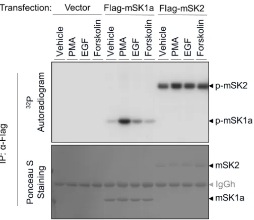
To investigate the mechanism of post-translational regulation of the mouse SK (mSK) iso-forms, mSK1a and mSK2, we first examined whether these proteins were phosphorylated in response to external stimuli. After overexpression of either mSK1a or mSK2 and32P metabolic labeling, we treated COS-7 cells with various types of extracellular stimuli, including PMA, for-skolin, and epidermal growth factor (EGF), that have been previously identified as activators of human SK isoforms [13,20,21,37,38]. Consistent with a previous study of hSK isoforms [21,
37,38], phosphorylation of mSK1a is strongly induced in response to PMA, and to a lesser extent by EGF. By contrast, the basal level of mSK2 phosphorylation remained high, even in quiescent cells subjected to serum withdrawal, and agonist stimulation did not alter mSK2 phosphorylation markedly as shown in mSK1a (Fig 1). These results revealed that the phos-phorylation levels of mSK1a and mSK2 are differentially regulated in response to extracellular stimuli. In particular, mSK1a is robustly phosphorylated by PMA stimulation, suggesting a possibility that mSK1a is regulated by this phosphorylation event.
Fig 1. Phosphorylation of mSK isoforms in response to extracellular stimuli.COS-7 cells were transfected with control vector, FLAG-tagged mSK1a, or FLAG-tagged mSK2 constructs. After serum deprivation and metabolic labeling with [32P] inorganic phosphate, the cells were treated for 10 min with
various agonists, as indicated: PMA (100 nM), EGF (100 ng/ml), or forskolin (20μM). mSK isoforms were immunoprecipitated usingα-FLAG Affi-Gel, transferred on nitrocellulose membrane, and exposed to X-ray photographic film (top panel) after Ponceau S staining (bottom). This result is representative of three independent experiments.
Inverse correlation between mSK1a phosphorylation and its activity
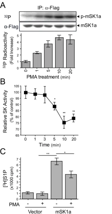
Next, we examined the correlation between mSK1a phosphorylation and enzyme activity. When measured over a time course, mSK1a phosphorylation progressively increased in response to PMA treatment up to 4.5-fold (Fig 2A), whereas itsin vitroactivity was suppressed (Fig 2B). Furthermore, we observed that PMA treatment resulted in the marked reduction of cellular S1P formation in the mSK1a-transfected cells, by inhibiting its cellular activity (Fig 2C). These results strongly suggested that mSK1a is negatively regulated by PMA treatment and that phosphorylation of mSK1a is closely related to the suppression of its activity.
Conventional PKC (cPKC)-dependent phosphorylation of mSK1a
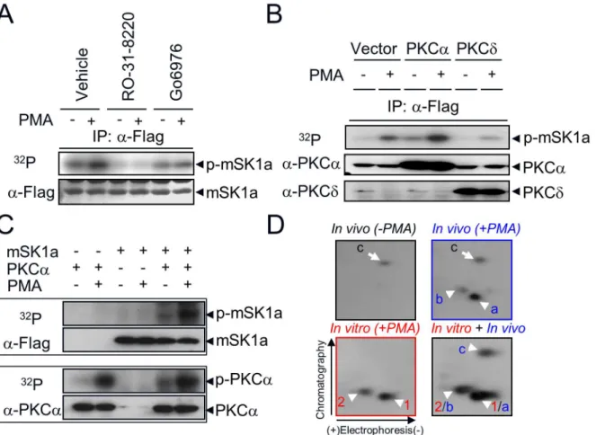
PMA is a potent activator that stimulates multiple subclasses of PKC isoforms, including both conventional (α,βI,βII,γ) and novel (δ,ε,η,θ) isozymes in various cell types [39]. To deter-mine which PKC isozyme mediates PMA-induced mSK1a phosphorylation, we used two inde-pendent approaches. First, we examined the effect of pharmacological PKC inhibitors, such as PKC-nonselective RO-31-8220 [40], and PKCα/β1-selective Go6976 [41], on mSK1a phos-phorylation. PMA-induced phosphorylation of mSK1a was completely abolished by pretreat-ment with RO-31-8220 or Go6976 (Fig 3A). Second, we examined the effect of overexpression of each isozyme on mSK1a phosphorylation. mSK1a phosphorylation was significantly increased by overexpression of PKCα, a cPKC isoform, but not PKCδ, a novel PKC isoform (Fig 3B), which suggests the specific PKC pathway mediates mSK1a phosphorylation. These results suggested that the PKCαor possibly other cPKC isozymes mediates PMA-induced phosphorylation of mSK1a in COS-7 cells.
After the functional role of cPKC upstream of mSK1a phosphorylation was established, it remained unclear whether mSK1a is the direct substrate of cPKC. To address this question, we conductedin vitrokinase assays by reconstituting purified mSK1a and PKCαin the presence or absence of PMA. mSK1a was directly phosphorylated by PKCα, a cPKC isoform,in vitro, as it isin vivo(Fig 3C). Furthermore, we compared the 2D-phosphopeptide maps derived from mSK1a proteins phosphorylatedin vitroandin vivo(Fig 3D). PMA treatment of cells overex-pressing mSK1a led to the generation of twoin vivophosphopeptides (designated a and b). In addition, one basal phosphopeptide (designated as c) was present irrespective of PMA treat-ment. Notably,in vitrophosphorylation of mSK1a generated two phosphopeptides (designated as 1 and 2) that completely overlapped within vivophosphopeptides a and b, respectively, as revealed by analysis of the mixture (in vivo+in vitro, designated 1/a and 2/b). By contrast,in vivopeptide c, which was not responsive to PMA treatment, was not present in mSK1a phos-phorylated by PKCαin vitro. These data suggested that PKCαdirectly phosphorylates mSK1a, at the same residue bothin vivoandin vitro.
Determination of
in vivo
phosphorylation sites of mSK1a
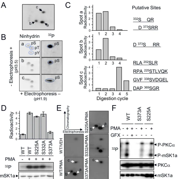
To determine the PKC-dependent (a and b) and -independent (c) phosphorylation sites of mSK1a, we extracted32P-labeled phosphopeptides (Fig 4A). Purified phosphopeptides were divided into two fractions; one was subjected to phosphoamino analysis and the other was sub-jected to32P release assay. Phosphoamino analysis revealed that all three phosphopeptides (a, b, and c) exclusively harbor phosphoserine residues within their sequences (Fig 4B). Moreover,
32P release assay after Edman degradation revealed that the phosphoserine residues were
Fig 2. PMA-induced phosphorylation and inhibition of mSK1a.(A) Time-dependent phosphorylation of mSK1a in response to PMA treatment. COS-7 cells were transfected with mSK1a cDNA. After metabolic labeling with [32P]ortho-phosphate, the cells were treated with 100 nM PMA for indicated periods of time.
mSK1a was immunoprecipitated using the FLAG epitope. The autoradiogram and immunoblot images using α-FLAG antibody are shown (top and middle panels), and relative radioactivity was quantitated in duplicate (bottom). Data represent the means±SE. (B) Time course of PMA-induced suppression of mSK1a activity. COS-7 cells, transfected with mSK1a cDNA, were stimulated with 100 nM PMA for the indicated periods of time. Data are from triplicate determinations from independent cultures, and are expressed as percentages relative to untreated controls. Data represent the means±SEM. (One-way ANOVA test,*p<0.05, **p<0.01). (C) PMA-induced suppression of [3H]S1P formation by mSK1a. Formation of [3H]S1P was
determined in COS-7 cells that are transfected with either control vector or mSK1a cDNA, and further stimulated with 100 nM PMA for 10 min prior to [3H]SPH labeling. Data represent the means±SEM. (t-test,
*p<0.05,**p<0.01).
one of the others (S140, S225, S338, S369) was the putative site in phosphopeptide c. To deter-mine the exact phosphorylation sites of mSK1a, we performed alanine mutagenesis at each of these residues. Ala mutation at S373 (S373A) resulted in a marked reduction in the level of mSK1a phosphorylation in response to PMA treatment (Fig 4D). Furthermore, comparison of phosphopeptide maps revealed that the reduction of32P-incorporation into the S373A mutant was due to the disappearance of two PMA-responsive phosphopeptides, a and b (indicated by arrowheads in the WT inFig 4E). By contrast, mutation of S332, the other candidate, had no effect on the level of32P-incorporation. These data suggest that S373 of mSK1a is the only phosphorylation site by PKCα. Because partial digestion occurs frequently in tandem Arg or Lys residues in the protein sequence [42], the two phosphopeptides arising in response to PMA Fig 3. Direct phosphorylation of mSK1a by cPKC.(A) Effect of PKC inhibition on mSK1a phosphorylation. COS-7 cells were transfected with FLAG-tagged mSK1a and then labeled with [32P]ortho-phosphate. As indicated, the cells were treated with PKC inhibitors (10μM Ro-31-8220, or 0.5μM Go6976)
for 15 min prior to PMA treatment (100 nM, 10 min). mSK1a immobilized onto nitrocellulose membrane (NC) was exposed to a photographic film (top). The same NC membrane was immunoblotted with anti-FLAG antibody (bottom). (B) Effect of PKC overexpression on PMA-induced mSK1a phosphorylation. mSK1a cDNA were co-transfected with control vector or cDNAs encoding PKCαor PKCδas indicated. Relative radioactivity of immunoprecipitated mSK1a was visualized as described above (top panel), and the overexpression of each PKC isozyme was confirmed by immunoblot analysis of total homogenates using antibodies specific for each isozyme (PKCαor PKCδ). (C) Direct phosphorylation of mSK1a by purified PKCα. One hundred nanograms of mSK1a and/or 10 ng of PKCαwere incubated in 25μl of phosphorylation buffer containing [32P]-ATP in the presence or absence of 1μM PMA for 20 min as
indicated. Incorporation of radioactivity was determined after separation of the reaction mixtures by gel electrophoresis and autoradiography. The upper panel is an autoradiogram of NC membrane corresponding to mSK1a and PKCα, respectively. The lower panel is an immunoblot of the same NC membrane showing that equal amounts of proteins were used in the phosphorylation reactions. (D) Comparison of mSK1a phosphopeptides phosphorylatedin vitroand
in vivo. mSK1a phosphorylatedin vivowas obtained from COS-7 cells after they were treated with PMA (In vivo+PMA) or not treated (In vivo-PMA). mSK1a (in vitro+PMA) phosphorylated by PKCαin vitrowas digested with TPCK-trypsin. The tryptic digests ofin vivoandin vitrophosphorylated mSK1a, as well as
a mixture of the two (in vivo + in vitro), were subjected two-dimensional phosphopeptide mapping analysis consisting of electrophoresis and subsequent
chromatography. The arrowheads indicate two overlapping phosphopeptides (1/a, 2/b) and the arrow is for non-overlapping peptide (c).
Fig 4. mSK1a S373 is the cPKC-dependent phosphorylation site.(A) PKC-dependent (spots a and b: arrowheads) and -independent (spot c: arrow) phosphopeptide(s) are indicated. Each peptide was eluted from the TLC plate and divided into two fractions, which were subjected to two-dimensional phosphoamino acid analysis or32P-release assay. (B) Phosphoamino acid analysis of three phosphopeptides. Acid-hydrolyzed peptides were subjected to 2-dimensional phosphoamino acid analysis. Dotted circles indicate the migration positions of phosphoserine (pS), phosphothreonine (pT), and
phosphotyrosine (pY) standards (left panel) and the image from autoradiography (right panel). (C)32P-release assay of three radiolabeled peptides.
Radiolabeled peptides loaded onto the sequencing membrane (Millipore) were digested sequentially from the N-terminus by the Edman degradation method. Putative candidate sites for each phosphopeptide were predicted based on the relative position from the tryptic cleavage site, as shown on the right. (D) Site-directed mutagenesis of candidate sites. COS-7 cells were transfected with FLAG-tagged mSK1a constructs including wild-type (WT) and its mutant (S225A, S332A, S373A) and treated with PMA (100 nM). The relative phosphorylation level of each mSK1a form was assessed in duplicate. Data represent the means±SE. (E) Two-dimensional phosphopeptide analysis of WT mSK1a and its S/A mutants (S225A, S332A, S373A). cPKC-dependent spots (a and b) and -independent spot (c) are indicated by arrowheads and arrow, respectively. (F)In vitrophosphorylation of WT mSK1a and its S/A mutants by PKCα. Wild-type mSK1a and S/A mutants (S373A, S225A) were expressed in COS-7 cells and purified usingα-FLAG Affi-Gel. Each protein (100 ng) was incubated with PKCα(10 ng) in 1× phosphorylation buffer in the presence or absence of PMA and GF109204X (GFX). Incorporation of radioactivity was measured, and the transferred membrane was immunoblotted to monitor the input amount of each protein, as indicated.
treatment are likely to be generated from partial tryptic digestion of tandem Arg residues in Asp-phosphoSer-Arg-Arg-Gly into two similar peptides, Asp-phosphoSer-Argand Asp-phos-phoSer-Arg-Arg. Thein vitroPKC assay revealed that S373 is the only PKCαphosphorylation
site in mSK1a. PKCαcan phosphorylate WT mSK1a and the S225A mutant, but not the S373A
mutant (Fig 4F). These data clearly showed that the S373 residue, located in the C-terminus of mSK1a, is the direct phosphorylation site of PKCα.
In addition, Ala mutation at S225 (S225A) resulted in a marginal reduction in the level of mSK1a phosphorylation in response to PMA treatment (Fig 4D). Furthermore, comparison of phosphopeptide maps revealed that the slight decrease of32P-incorporation into the S225A mutant was due to the disappearance of phosphopeptide c (indicated by arrows in the WT in
Fig 4E), confirming that S225 is a basal phosphorylation site that is not responsive to PMA treatment. Furthermore, we examined the relationship between different phosphorylation sites. Phosphorylation at S373 was not affected by mutation at S225 (Fig 4F). Conversely, muta-tion at S373 had no effect on the phosphorylamuta-tion level at S225. Thus, phosphorylamuta-tion of S373 and S225 occur under independent control.
PMA-induced mSK1a inhibition is abolished by S373A mutation
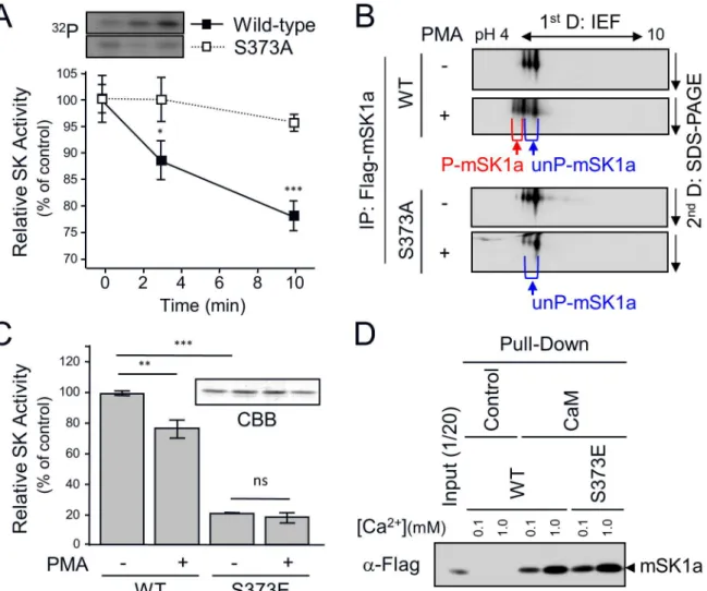
We next examined whether S373 phosphorylation of mSK1a results in an alteration in its activ-ity. Wild-type (WT) mSK1a was phosphorylated and inhibited in a time-dependent manner, whereas the S373A mutant, which was defective in phosphorylation, was refractory to inhibi-tion by PMA treatment over all time periods examined (Fig 5A). Furthermore, we tried to esti-mate the level of mSK1a phosphorylation by separating phosphorylated and unphosphorylated mSK1a by 2-D gel electrophoresis. In WT transfected cells, but not S373A-transfected cells, PMA treatment shifted mSK1a toward acidic field concomitant with the appearance of addi-tional mSK1a spots, which may be attributable to the generation of a phosphorylation-depen-dent negative charge in mSK1a (Fig 5B). The phosphorylation level could be roughly estimated at around 20–30% of total mSK1a, roughly correlated with the level of mSK1 inhibition in response to PMA treatment. To determine whether S373 phosphorylation is sufficient to inhibit mSK1a activity, we analyzed the effects of S373E mutagenesis, mimicking the phos-phorylated state of S373. The activity of WT mSK1a was inhibited after PMA treatment, whereas the S373E mutant was constitutively inactive irrespective of PMA treatment (Fig 5C), suggesting that S373 phosphorylation plays a significant role in inhibiting mSK1a activity.
Next, we investigated whether alteration in SK activity as a result of the S373E mutation is a result of the aberrant folding of the protein itself. Previous work showed that correctly folded hSK1 protein interacts with Ca2+-bound calmodulin [43], which provides an indicator for the proper protein folding of SK1a. Hence, we conducted calmodulin-binding analysis with WT mSK1a and the S373E mutant at two different concentrations of Ca2+ion. We observed that mSK1 interacted with calmodulin in a Ca2+concentration-dependent manner. Importantly, the S373E mutation did not alter its calmodulin interaction under any conditions tested (Fig 5D), suggesting that altered activity of mSK1a S373 mutant is unlikely to aberrant protein fold-ing durfold-ing protein synthesis. We have further examined whether the S373 mutation of mSK1a influences on protein-protein interactions of mSK1a. While multiple proteins were found co-precipitated with mSK1a, we couldn’t detect any clear difference between WT mSK1a and the S373 mutant (S1 Fig).
Discussion
regulation remain incompletely understood. In this study, we explored the phosphorylation-dependent regulation of mSK1a. We observed that S373 of mSK1a is directly phosphorylated by cPKC in response to PMA treatment. Furthermore, this phosphorylation results in the inhi-bition of mSK1a activity.
Although previous work showed that mSK1a contains several consensus motifs for phos-phorylation by protein kinases such as PKA, casein kinase II, and PKC [32], the physiological relevance of putative phosphorylation events in mSK1a have not been rigorously tested. Our results demonstrate that phosphorylation of mSK1a is strongly elicited by PMA, a potent Fig 5. S373 phosphorylation-dependent Inhibition of mSK1a.(A) The effect of mSK1a S373A mutation on PMA-induced inhibition of mSK1a activity. Wild-type (WT) mSK1a or the S373A mutant was transfected into COS-7 cells. One day after transfection, the cells were serum-deprived for 24 hr and treated with 100 nM PMA for the indicated periods of time. Protein extracts from COS-7 cells were assayed for SK activityin vitro. SK activity is expressed as the percentage of activity relative to the non-treated control. These data represent the means±SEM. Inset: time-dependent phosphorylation of WT mSK1a and the S373A mutant (S373A). (B) Approximate stoichiometry of mSK1a phosphorylation. After PMA stimulation, WT mSK1a and its S373A mutant were immunoprecipitated and subjected to 2-D gel electrophoresis. mSK1a was detected using immunoblot analysis usingα-FLAG Ab. S373-phosphorylated (P-mSK1a, red) and unphosphorylated (un-P-(P-mSK1a, blue) proteins are indicated below the images. (C) Effect of mSK1a S373E mutation on PMA-induced inhibition of mSK1a activity. Wild-type mSK1a and the S373E mutant were purified from transfected COS-7 cells usingα-FLAG Affi-Gel. The same amounts (5 ng) of purified WT mSK1a and S373E mutant were assayed for SK activity. The data are the means±SEM (t- test,*p<0.05,**p<0.01). Inset: The amounts of purified mSK1a and S373E mutant were confirmed by SDS-PAGE followed by Coomassie Blue (CBB) Staining. (D) Calmodulin interaction of WT mSK1a and the S373 mutant. Identical concentrations of mSK1a and the S373E mutant were incubated with either calmodulin-agarose beads or control beads at two different Ca2+concentrations, as indicated. mSK1a bound to beads were assessed by immunoblotting usingα-FLAG antibody. See alsoS1 Fig
for additional data about the protein interactions of WT mSK1a and the S373E mutant.
activator of PKC, but not by forskolin, an activator of PKA, indicating that this phosphorylation of mSK1a is kinase-specific. Our results also showed that cPKC including PKCα, phosphory-lates mSK1a bothin vivoandin vitro. Mutation at S373 of mSK1a completely abolishes mSK1a phosphorylation in response to PMA treatment, confirming that S373 is the sole site phosphor-ylated by cPKC. Notably, peptide sequences surrounding S373 residue of mSK1a (-pS-R-R-G-) are consistent with the representative consensus motif for cPKC phosphorylation, -[pS/pT]-[_/F/K/V/L/Q/V]-[K/R]-[K/R/G]- [46], further confirming mSK1a as a novel substrate of acti-vated cPKC within cells. cPKC isoforms including PKCα, -β, and -γphosphorylate a number of common substrates including GSK-3β, GAP-43, and EGFR [47]. Thus, another cPKC isoform (s) could also be involved in S373 phosphorylation of mSK1a, either alone or together with PKCα, although this was not demonstrated in the present study. Still, the expression and distri-bution of cPKC isoforms vary between cell types [48]. PKCαare found widely expressed in vari-ous cell types whereas others seem to be more restricted in their distributions. Presumably, the relative dominance of PKCαover other cPKC isoforms for the phosphorylation of mSK1a might be influenced by the expression level of each isoform in the given cell type.
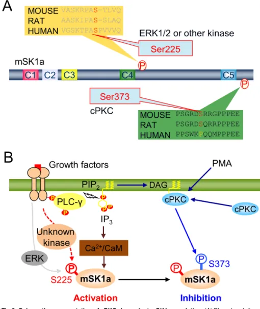
of the plasma membrane without altering the total mSK1a level in cells. In this regard, it would be interesting to examine if mSK1a S373 phosphorylation can influence the export of mSK1a protein and its extracellular activity on the surface of the plasma membrane in future studies. We found that mSK1a is phosphorylated in two distinct sites, S225 and S373. Our results identify S373 of mSK1a as the cPKC-dependent phosphorylation site, as well as S225 as the PKC-independent phosphorylation site. A previous report showed that S225 phosphorylation of hSK1a in response to PMA treatment is mediated by ERK1/2, which plays a key role in PMA-induced activation of mSK1a by facilitating both membrane translocation and enzymatic activation of hSK1a [37]. Our result confirmed that phosphorylation at the S225 residue of SK1 is conserved between species. However, it was noteworthy that S225 phosphorylation of mSK1a is barely sensitive to PMA treatment. In fact, sequence alignment between human and Fig 6. Schematic representation of cPKC-dependent mSK1a regulation.(A) Phosphorylation-dependent regulation of mSK1a. (B) Schematic model for mSK1a regulation in response to extracellular stimuli. Abbreviations: mSK1a (mouse sphingosine kinase 1a), PIP2(phosphatidylinositol 4,5-bisphosphate), DAG
(diacylglycerol), cPKC (conventional protein kinase C), PMA (phorbol 12-myristate 13-acetate), ERK (extracellular signal-regulated kinase), CaM (calmodulin), IP3, (inositol trisphosphate), PLC-γ(phospholipase
C-gamma), P (phosphate group).
mouse mSK1a revealed a meaningful difference in the proline residue immediately adjacent to the phosphorylated S225 residue (Fig 6A). This proline residue is an essential component of the consensus motif for ERK1/2 phosphorylation [46]. Presumably, the absence of P226 resi-due in mSK1a makes the S225 resiresi-due of mSK1a less optimal for the phosphorylation by ERK, which may account for no significant change in S225 phosphorylation by ERK in the cells. In this regard, S225 phosphorylation of mSK1a is unlikely mediated by ERK1/2, but rather by another kinase that is insensitive to PMA treatment, suggesting the significant difference in the signaling pathway to SK1a activation. On the other hand, our current study clearly demon-strated that mSK1a is inhibited through S373 phosphorylation in response to PMA stimulus, which is not consistent with the previous report using hSK1a[37]. Of note, we found that S373 of mSK1a is conserved in mouse and rat, but not in human (Fig 6A), indicating that this phos-phorylation and inhibition of mSK1a happens in species-specific manner. Collectively, distinct phosphorylation pattern at two mSK1a phosphorylation sites, S225 and S373 may account for the apparent discrepancy between species regarding PMA-induced SK1a regulation [37]. In particular, our current observation may contribute to better understanding the molecular basis underlying the interspecies differences in sphingolipid metabolismin vivo.
In summary, our results demonstrate that mSK1a is inhibited by cPKC-dependent phos-phorylation, and that mSK1a phosphorylation is mediated specifically and directly by cPKC. We also demonstrated that phosphorylation at S373, located in the C-terminal regulatory region, plays a pivotal role in the negative regulation of mSK1a. Nonetheless, we have to admit that our study was conduced mostly with cell stimulation with PMA, a type of phorbol esters which does not exist endogenously in mammals, but mimic the action of diacyl glycerol (DAG), potent activator of PKC [52]. In this regards, the physiological relevance of our obser-vation may need to be further validated in the research models of mSK1a-dependent cellular processes including cell proliferation, cell survival, apoptosis, and angiogenesis [2,45].
In the context of cellular signaling, mSK1a is activated through S225 phosphorylation by unknown kinase probably downstream of growth factor receptor stimulation (Fig 6B). In paral-lel, phospholipase C (PLC)-dependent cleavage of PIP2generates IP3and diacyl glycerol
(DAG), leading to elevation of intracellular Ca2+and cPKC activation. The increase in cellular Ca2+may result in Ca2+-calmodulin-dependent activation of mSK1a via direct interaction, whereas cPKC activation may lead to S373 phosphorylation, which results in subsequent inac-tivation of mSK1a (Fig 6B). This schematic model illustrates the highly dynamic temporal reg-ulation of mSK1a through distinct phosphorylation events triggered by extracellular stimuli.
Supporting Information
S1 Fig. Effect of mSK1a phosphorylation on its molecular interaction.COS-7 cells were transfected with control vector, FLAG-tagged mSK1a wild-type (WT), or its S373E mutant (S373E). After serum deprivation, the cells were treated for 10 min with PMA (100 nM). The cell lysates were incubated withα-FLAG Affi-Gel. The protein complexes co-immunoprecipitated with mSK1a were analyzed with SDS-PAGE followed by Coomassie Brilliant Blue (CBB) stain-ing. Abbreviations: IgGh; Immunoblobulin G heavy chain, IgGl; immunoglobulin G light chain. (TIF)
Acknowledgments
Author Contributions
Conceived and designed the experiments: YSO PGS. Performed the experiments: YSO SSB JBP SHH. Analyzed the data: YSO SHH SHR PGS. Contributed reagents/materials/analysis tools: YSO PGS SHR. Wrote the paper: YSO PGS.
References
1. Pitson SM. Regulation of sphingosine kinase and sphingolipid signaling. Trends in biochemical sci-ences. 2011; 36(2):97–107. doi:10.1016/j.tibs.2010.08.001PMID:20870412.
2. Pyne NJ, Tonelli F, Lim KG, Long J, Edwards J, Pyne S. Targeting sphingosine kinase 1 in cancer. Advances in biological regulation. 2012; 52(1):31–8. doi:10.1016/j.advenzreg.2011.07.001PMID: 21791223.
3. Spiegel S, Milstien S. Functions of the multifaceted family of sphingosine kinases and some close rela-tives. The Journal of biological chemistry. 2007; 282(4):2125–9. doi:10.1074/jbc.R600028200PMID: 17135245.
4. Hannun YA, Obeid LM. Principles of bioactive lipid signalling: lessons from sphingolipids. Nature reviews Molecular cell biology. 2008; 9(2):139–50. doi:10.1038/nrm2329PMID:18216770.
5. Pyne NJ, Pyne S. Sphingosine 1-phosphate and cancer. Nature reviews Cancer. 2010; 10(7):489–503. doi:10.1038/nrc2875PMID:20555359.
6. Shen H, Giordano F, Wu Y, Chan J, Zhu C, Milosevic I, et al. Coupling between endocytosis and sphin-gosine kinase 1 recruitment. Nature cell biology. 2014; 16(7):652–62. doi:10.1038/ncb2987PMID: 24929359.
7. Hait NC, Allegood J, Maceyka M, Strub GM, Harikumar KB, Singh SK, et al. Regulation of histone acet-ylation in the nucleus by sphingosine-1-phosphate. Science. 2009; 325(5945):1254–7. doi:10.1126/ science.1176709PMID:19729656; PubMed Central PMCID: PMC2850596.
8. Alvarez SE, Harikumar KB, Hait NC, Allegood J, Strub GM, Kim EY, et al. Sphingosine-1-phosphate is a missing cofactor for the E3 ubiquitin ligase TRAF2. Nature. 2010; 465(7301):1084–8. doi:10.1038/ nature09128PMID:20577214; PubMed Central PMCID: PMC2946785.
9. Pyne S, Chapman J, Steele L, Pyne NJ. Sphingomyelin-derived lipids differentially regulate the extra-cellular signal-regulated kinase 2 (ERK-2) and c-Jun N-terminal kinase (JNK) signal cascades in airway smooth muscle. European journal of biochemistry / FEBS. 1996; 237(3):819–26. PMID:8647130.
10. Choi OH, Kim JH, Kinet JP. Calcium mobilization via sphingosine kinase in signalling by the Fc epsilon RI antigen receptor. Nature. 1996; 380(6575):634–6. doi:10.1038/380634a0PMID:8602265.
11. Olivera A, Zhang H, Carlson RO, Mattie ME, Schmidt RR, Spiegel S. Stereospecificity of sphingosine-induced intracellular calcium mobilization and cellular proliferation. The Journal of biological chemistry. 1994; 269(27):17924–30. PMID:8027049.
12. Lee MJ, Van Brocklyn JR, Thangada S, Liu CH, Hand AR, Menzeleev R, et al. Sphingosine-1-phos-phate as a ligand for the G protein-coupled receptor EDG-1. Science. 1998; 279(5356):1552–5. PMID: 9488656.
13. Olivera A, Edsall L, Poulton S, Kazlauskas A, Spiegel S. Platelet-derived growth factor-induced activa-tion of sphingosine kinase requires phosphorylaactiva-tion of the PDGF receptor tyrosine residue responsible for binding of PLCgamma. FASEB journal: official publication of the Federation of American Societies for Experimental Biology. 1999; 13(12):1593–600. PMID:10463951.
14. Olivera A, Spiegel S. Sphingosine-1-phosphate as second messenger in cell proliferation induced by PDGF and FCS mitogens. Nature. 1993; 365(6446):557–60. doi:10.1038/365557a0PMID:8413613.
15. Xia P, Gamble JR, Rye KA, Wang L, Hii CS, Cockerill P, et al. Tumor necrosis factor-alpha induces adhesion molecule expression through the sphingosine kinase pathway. Proceedings of the National Academy of Sciences of the United States of America. 1998; 95(24):14196–201. PMID:9826677; PubMed Central PMCID: PMC24350.
16. Rius RA, Edsall LC, Spiegel S. Activation of sphingosine kinase in pheochromocytoma PC12 neuronal cells in response to trophic factors. FEBS letters. 1997; 417(2):173–6. PMID:9395290.
17. Shu X, Wu W, Mosteller RD, Broek D. Sphingosine kinase mediates vascular endothelial growth factor-induced activation of ras and mitogen-activated protein kinases. Molecular and cellular biology. 2002; 22(22):7758–68. PMID:12391145; PubMed Central PMCID: PMC134718.
19. van Koppen CJ, Meyer zu Heringdorf D, Alemany R, Jakobs KH. Sphingosine kinase-mediated calcium signaling by muscarinic acetylcholine receptors. Life sciences. 2001; 68(22–23):2535–40. PMID: 11392623.
20. Buehrer BM, Bardes ES, Bell RM. Protein kinase C-dependent regulation of human erythroleukemia (HEL) cell sphingosine kinase activity. Biochimica et biophysica acta. 1996; 1303(3):233–42. PMID: 8908158.
21. Machwate M, Rodan SB, Rodan GA, Harada SI. Sphingosine kinase mediates cyclic AMP suppression of apoptosis in rat periosteal cells. Molecular pharmacology. 1998; 54(1):70–7. PMID:9658191
22. Melendez A, Floto RA, Gillooly DJ, Harnett MM, Allen JM. FcgammaRI coupling to phospholipase D ini-tiates sphingosine kinase-mediated calcium mobilization and vesicular trafficking. The Journal of bio-logical chemistry. 1998; 273(16):9393–402. PMID:9545263.
23. Alemany R, Meyer zu Heringdorf D, van Koppen CJ, Jakobs KH. Formyl peptide receptor signaling in HL-60 cells through sphingosine kinase. The Journal of biological chemistry. 1999; 274(7):3994–9. PMID:9933590.
24. Xia P, Vadas MA, Rye KA, Barter PJ, Gamble JR. High density lipoproteins (HDL) interrupt the sphingo-sine kinase signaling pathway. A possible mechanism for protection against atherosclerosis by HDL. The Journal of biological chemistry. 1999; 274(46):33143–7. PMID:10551885.
25. Taha TA, Hannun YA, Obeid LM. Sphingosine kinase: biochemical and cellular regulation and role in disease. Journal of biochemistry and molecular biology. 2006; 39(2):113–31. PMID:16584625.
26. Jo SK, Bajwa A, Ye H, Vergis AL, Awad AS, Kharel Y, et al. Divergent roles of sphingosine kinases in kidney ischemia-reperfusion injury. Kidney international. 2009; 75(2):167–75. doi:10.1038/ki.2008.400 PMID:18971925; PubMed Central PMCID: PMC2646633.
27. Lai WQ, Irwan AW, Goh HH, Melendez AJ, McInnes IB, Leung BP. Distinct roles of sphingosine kinase 1 and 2 in murine collagen-induced arthritis. Journal of immunology. 2009; 183(3):2097–103. doi:10. 4049/jimmunol.0804376PMID:19596980.
28. Oskeritzian CA, Alvarez SE, Hait NC, Price MM, Milstien S, Spiegel S. Distinct roles of sphingosine kinases 1 and 2 in human mast-cell functions. Blood. 2008; 111(8):4193–200. doi: 10.1182/blood-2007-09-115451PMID:18178871; PubMed Central PMCID: PMC2971746.
29. Wadgaonkar R, Patel V, Grinkina N, Romano C, Liu J, Zhao Y, et al. Differential regulation of sphingo-sine kinases 1 and 2 in lung injury. American journal of physiology Lung cellular and molecular physiol-ogy. 2009; 296(4):L603–13. doi:10.1152/ajplung.90357.2008PMID:19168577; PubMed Central PMCID: PMC2670763.
30. Allende ML, Sasaki T, Kawai H, Olivera A, Mi Y, van Echten-Deckert G, et al. Mice deficient in sphingo-sine kinase 1 are rendered lymphopenic by FTY720. The Journal of biological chemistry. 2004; 279 (50):52487–92. doi:10.1074/jbc.M406512200PMID:15459201.
31. Mizugishi K, Yamashita T, Olivera A, Miller GF, Spiegel S, Proia RL. Essential role for sphingosine kinases in neural and vascular development. Molecular and cellular biology. 2005; 25(24):11113–21. doi:10.1128/MCB.25.24.11113-11121.2005PMID:16314531; PubMed Central PMCID:
PMC1316977.
32. Kohama T, Olivera A, Edsall L, Nagiec MM, Dickson R, Spiegel S. Molecular cloning and functional characterization of murine sphingosine kinase. The Journal of biological chemistry. 1998; 273 (37):23722–8. PMID:9726979.
33. Liu H, Sugiura M, Nava VE, Edsall LC, Kono K, Poulton S, et al. Molecular cloning and functional char-acterization of a novel mammalian sphingosine kinase type 2 isoform. The Journal of biological chemis-try. 2000; 275(26):19513–20. doi:10.1074/jbc.M002759200PMID:10751414.
34. Olivera A, Rosenthal J, Spiegel S. Sphingosine kinase from Swiss 3T3 fibroblasts: a convenient assay for the measurement of intracellular levels of free sphingoid bases. Analytical biochemistry. 1994; 223 (2):306–12. doi:10.1006/abio.1994.1589PMID:7887476.
35. Medkova M, Cho W. Mutagenesis of the C2 domain of protein kinase C-alpha. Differential roles of Ca2 + ligands and membrane binding residues. The Journal of biological chemistry. 1998; 273(28):17544– 52. PMID:9651347.
36. Boyle WJ, van der Geer P, Hunter T. Phosphopeptide mapping and phosphoamino acid analysis by two-dimensional separation on thin-layer cellulose plates. Methods in enzymology. 1991; 201:110–49. PMID:1943760.
38. Hait NC, Bellamy A, Milstien S, Kordula T, Spiegel S. Sphingosine kinase type 2 activation by ERK-mediated phosphorylation. The Journal of biological chemistry. 2007; 282(16):12058–65. doi:10.1074/ jbc.M609559200PMID:17311928.
39. Mochly-Rosen D, Das K, Grimes KV. Protein kinase C, an elusive therapeutic target? Nature reviews Drug discovery. 2012; 11(12):937–57. doi:10.1038/nrd3871PMID:23197040; PubMed Central PMCID: PMC3760692.
40. Nixon JS, Bishop J, Bradshaw D, Davis PD, Hill CH, Elliott LH, et al. Novel, potent and selective inhibi-tors of protein kinase C show oral anti-inflammatory activity. Drugs under experimental and clinical research. 1991; 17(8):389–93. PMID:1822831.
41. Martiny-Baron G, Kazanietz MG, Mischak H, Blumberg PM, Kochs G, Hug H, et al. Selective inhibition of protein kinase C isozymes by the indolocarbazole Go 6976. The Journal of biological chemistry. 1993; 268(13):9194–7. PMID:8486620.
42. Jensen ON, Vorm O, Mann M. Sequence patterns produced by incomplete enzymatic digestion or one-step Edman degradation of peptide mixtures as probes for protein database searches. Electrophoresis. 1996; 17(5):938–44. doi:10.1002/elps.1150170516PMID:8783020.
43. Pitson SM, D'Andrea R J, Vandeleur L, Moretti PA, Xia P, Gamble JR, et al. Human sphingosine kinase: purification, molecular cloning and characterization of the native and recombinant enzymes. The Bio-chemical journal. 2000; 350 Pt 2:429–41. PMID:10947957; PubMed Central PMCID: PMC1221270.
44. Ader I, Malavaud B, Cuvillier O. When the Sphingosine Kinase 1/Sphingosine 1-Phosphate Pathway Meets Hypoxia Signaling: New Targets for Cancer Therapy. Cancer Res. 2009; 69(9):3723–6. doi:10. 1158/0008-5472.CAN-09-0389WOS:000265761900001. PMID:19383898
45. Karliner JS. Sphingosine kinase and sphingosine 1-phosphate in the heart: a decade of progress. Bio-chimica et biophysica acta. 2013; 1831(1):203–12. doi:10.1016/j.bbalip.2012.06.006PMID:
22735359; PubMed Central PMCID: PMC3479372.
46. Yaffe MB, Leparc GG, Lai J, Obata T, Volinia S, Cantley LC. A motif-based profile scanning approach for genome-wide prediction of signaling pathways. Nature biotechnology. 2001; 19(4):348–53. doi:10. 1038/86737PMID:11283593.
47. Dekker LV, Parker PJ. Protein kinase C—a question of specificity. Trends in biochemical sciences. 1994; 19(2):73–7. doi:10.1016/0968-0004(94)90038-8PMID:8160269.
48. Nishizuka Y. The molecular heterogeneity of protein kinase C and its implications for cellular regulation. Nature. 1988; 334(6184):661–5. doi:10.1038/334661a0PMID:3045562.
49. Wang Z, Min X, Xiao SH, Johnstone S, Romanow W, Meininger D, et al. Molecular basis of sphingosine kinase 1 substrate recognition and catalysis. Structure. 2013; 21(5):798–809. doi:10.1016/j.str.2013. 02.025PMID:23602659.
50. Ancellin N, Colmont C, Su J, Li Q, Mittereder N, Chae SS, et al. Extracellular export of sphingosine kinase-1 enzyme. Sphingosine 1-phosphate generation and the induction of angiogenic vascular matu-ration. The Journal of biological chemistry. 2002; 277(8):6667–75. doi:10.1074/jbc.M102841200 PMID:11741921.
51. Rigogliuso S, Donati C, Cassara D, Taverna S, Salamone M, Bruni P, et al. An active form of sphingo-sine kinase-1 is released in the extracellular medium as component of membrane vesicles shed by two human tumor cell lines. J Oncol. 2010; 2010:509329. doi:10.1155/2010/509329PMID:20508814; PubMed Central PMCID: PMCPMC2875746.