Dina Raquel da Silva Coelho
Dissertation presented to obtain the Ph.D degree in Biology
Instituto de Tecnologia Química e Biológica António Xavier | Universidade Nova de LisboaOeiras,
The Role of the Endoplasmic
Reticulum Stress Transducer Ire1
during Photoreceptor
Dina Raquel da Silva Coelho
Dissertation presented to obtain the Ph.D degree in Biology
Instituto de Tecnologia Química e Biológica António Xavier | Universidade Nova de LisboaOeiras, December, 2013
The Role of the Endoplasmic
Reticulum Stress Transducer Ire1
during Photoreceptor
Dina Raquel da Silva Coelho The Role of the Endoplasmic Reticulum Stress Transducer Ire1
during Photoreceptor Differentiation in Drosophila Oeiras, December,
Acknowledgments
First and foremost I would like to thank my PhD advisor, Pedro Domingos, who took me in the laboratory as an inexperienced master’s student and gave me conditions to grow and evolve as a scientist. It was a long journey since Pedro introduced me to the basics in the laboratory until we finally got our paper accepted in a high standard journal. Thank you for pushing me and giving me the motivation that I needed. I feel that my work was completely rewarded and most of the times rewarding, as well. Thank you for supporting me and caring for everybody in the lab.
I want to thank everyone who has helped in my scientific education, but mainly to people in the lab. Fátima Cairrão who showed me molecular biology techniques and Vanya Rasheva who explained me some classical genetics tricks. I appreciate the companionship from girl members of the lab, Nadine, Rita and Tânia.
I am grateful to people who contributed to my paper and provided important results for this thesis: Elisabete Pires, Ana Varela Coelho, Hyung Don Ryoo, Xiaomei Zeng and Fátima Cairrão.
I want also to acknowledgment ITQB, IGC and CEDOC for giving me access to equipment and facilities. In particular I want to acknowledgment Jaime Mota Lab and Cristina Silva Pereira Lab for always sparing reagents and consumables with us and for giving me some precious protocol advices.
I want to acknowledge Gulbenkian Foundation for paying my PhD fellowship and Fundação para a Ciência e Tecnologia for supporting the project through grants PEst-OE/EQB/LA0004/2011, PTDC/BIA-BCM/105217/2008, PTDC/SAU-OBD/104399/2008, and PTDC/ BEX-BCM/1217/2012.
Table of contents
List of Figures ... vii
Abbreviations ... x
Summary ... xiv
Sumário ... xv
Chapter I – Introduction 1.The Endoplasmic Reticulum ... 2
1.1 Folding and Post-translational Modifications ... 2
1.2 ER-Associated Degradation ... 6
1.3 ER Stress and Unfolded Protein Response ... 9
1.4 Ire1 Signaling ... 11
1.5 ATF6 Signaling ... 22
1.6 Perk Signaling ... 24
1.7 The UPR in Disease and Development ... 27
2. Morphogenesis of the Drosophila Eye ... 32
2.1 Apical/Basolateral Specification ... 35
2.2 Formation of the Interrhabdomeral Space ... 38
2.3 Morphogenesis of the Rhabdomere ... 40
2.4 Targeted Vesicular Trafficking to the Rhabdomere ... 43
2.5 Rhodopsin1 Biogenesis and Retinal Degeneration ... 44
3. Aims of the work ... 45
Chapter II – Ire1 signaling is required for rhabdomere morphogenesis and photoreceptor differentiation in Drosophila Summary ... 48
Materials and Methods ... 51
Results ... 55
Ire1 signaling is active during photoreceptors differentiation in the pupa ... 55
Ire1 is not required for photoreceptors specification and maintenance of the apical/basolateral polarity... 58
Ire1 is necessary for secretion of Spacemaker into the interrhabdomeral space and Rhodopsin1 localization to the rhabdomere ... 61
ER markers are down-regulated in PBac{WH}Ire1f02170 mutant photoreceptors ... 65
Discussion... 66
Acknowledgments and author contribution ... 69
References ... 69
Chapter III – Generation and characterization of Xbp1 deficiencies Summary ... 76
Introduction ... 76
Materials and Methods ... 78
Results ... 84
P{lacW}Xbp1k13803 does not have a phenotype for photoreceptor morphology ... 84
Characterization of P{lacW}Xbp1k13803 excisions ... 86
Characterization of P{SUPor-P}CG9418KG05183excisions ... 88
Characterization of P{GSV3}GS6093 excisions ... 92
P{SUPor-P}CG9418KG05183excisionspresent photoreceptors with a normal morphology ... 96
P[PTT-GB]Xbp1CB02061 excisions show a normal morphogenesis of the rhabdomere ... 97
Xbp1spliced is sufficient but not necessary for ninaA expression ... 101
Ectopic expression of ninaA does not rescue Rhodopsin1 localization in
PBac{WH}Ire1f02170clones ... 101
Discussion... 103
Acknowledgments and author contribution ... 105
References ... 105
Chapter IV – Regulation of Fatp by RIDD is critical for rhabdomere morphogenesis Summary ... 110
Introduction ... 110
Materials and Methods ... 113
Results ... 116
Regulation of Fatp by RIDD is critical for rhabdomere morphogenesis ... 116
Rab11 positive vesicles accumulate in the cytoplasm of PBac{WH}Ire1f02170 mutant photoreceptors ... 120
Increased levels of fatty acids in PBac{WH}Ire1f02170 retinas disrupt rhabdomere morphogenesis ... 121
RNase activity of Ire1 is essential for rhabdomere morphogenesis .... 123
Atf4 is up-regulated in PBac{WH}Ire1f02170 clones ... 125
Discussion... 126
Acknowledgments and author contribution ... 129
References ... 129
Chapter V – General discussion ... 135
List of Figures
Figure 1.1 The endoplasmic reticulum. P.2
Figure 1.2. The endoplasmic reticulum is the entry site for the secretory pathway. P.4
Figure 1.3 The events and components of ERAD. P.9
Figure 1.4 Ire1 Signaling. P.12
Figure 1.5 Structure of Ire1. P.16
Figure 1.6 Recruitment of Xbp1 mRNA to the ER membrane in mammals. P.18
Figure 1.7 ATF6 and CREBH signaling. P.23
Figure 1.8. PERK Signaling. P.26
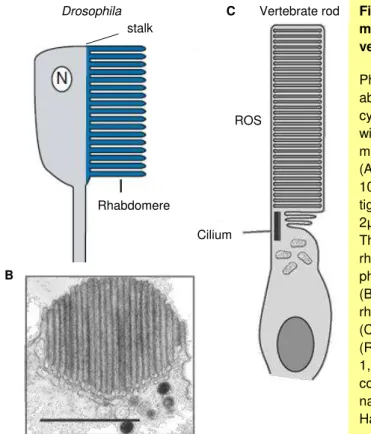
Figure 1.9 The structure of the Drosophila compound eye. P.33
Figure 1.10. The photoreceptive membranes in Drosophila and vertebrates. P.34
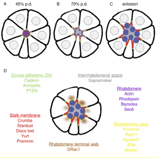
Figure 1.11 Differentiation of photoreceptors in the pupa and markers of cell polarity P.37
Figure 1.12 Formation of the interrhadomeral space and morphogenesis of the rhabdomere in the pupa. P.39
Figure 1.13. Rhabdomere morphogenesis and Rhodopsin1 distribution. P.42
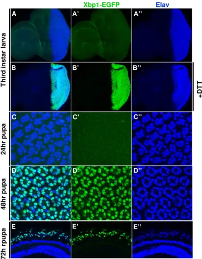
Figure 2.1 The Xbp1-EGFP reporter is activated in the photoreceptors during pupal stages. P.56
Figure 2.2. Xbp1-EGFP expression persists in some cells of the adult eye. P.57
Figure 2.3 Ire1 is required for Xbp1-EGFP activation in the Drosophila Eye. P.59
Figure 2.4. Ire1 is not required for photoreceptor specification and maintenance of apical/basolateral polarity. P.60
Figure 2.5 Ire1 is required for Spacemaker (Spam) secretion and formation of the interrhabdomeral space. P.62
Figure2.7 PBac(WH)Ire1f02170 photoreceptors degenerate early in the adult. P.65
Figure 2.8 ER markers are down-regulated in PBac{WH}Ire1f02170 mutants. P.67
Figure 3.1 P(lacW)Xbp1k13803 show a normal morphology of the photoreceptors in the adult. P.85
Figure 3.2 Genetic scheme followed to generate P(lacW)Xbp1k13803 excisions. P.85
Figure 3.3 Characterization of P(lacW)Xbp1k13803 excisions by PCR. P.87
Figure 3.4. Characterization of P{SUPor-P}CG9418KG05183 excisions by PCR. P.89
Figure 3.5 Excision30 and Excision101 originate truncated Xbp1 mRNAs that are spliced by Ire1. P.91
Figure 3.6. Determination of ExcisionF211 genomic limits by PCR. P.93
Figure 3.7 P{GSV3}GS6093 transposon continues inserted in the original site in ExcisionF211. P.94
Figure 3.8. Rescue of ExcisionF211 clones viability. P.95
Figure 3.9 Xbp1exc30, Xbp1exc101 and Xbp1250 show a normal morphology of the photoreceptors in 72hr pupa. P.96
Figure 3.10 Xbp1exc79 and Xbp1exc81 have a normal morphology of photoreceptors at 62hr pupation. P.98
Figure 3.11. Xbp1exc79 and Xbp1exc81 photoreceptors present mild defects in the delivery of Rh1 to the rhabdomere. P.99
Figure 3.12 ER markers are down-regulated in Xbp1exc79 and Xbp1exc81 mutant photoreceptors. P.100
Figure 3.13. Xbp1spliced induces NinaA expression. P.102
Figure 4.1. Ire1 regulates mRNA decay of Fatp in Drosophila eye. P.118
Figure 4.2. Drosophila Xbp1 and Fatp mRNAs are cleaved by purified human Ire1. P.119
Figure 4.3 Increased levels of Fatp deregulate the delivery of Rh1 to the rhabdomere. P.120
Figure 4.5 Levels of phosphatidic acid are increased in PBac{WH}Ire1f02170 retinas. P.124
Figure 4.6 Ire1 RNase mutant (Ire1H890A) and Ire1 kinase mutant (Ire1K576A) fail to rescue Xbp1-EGFP expression and Rh1 localization in the rhabdomeres of PBac(WH)Ire1f02170 homozygous photoreceptors. P.125
Abbreviations
ADP adenosine diphosphate
aPKC: atypical protein kinase C
ARF1: ADP-ribosylation factor 1
Arm: armadillo
Asn: asparagine
ASK1: apoptosis signal-regulating kinase 1
ATF4: activating transcription factor 4
ATF6: activating transcription factor 6
ATP: adenosine triphosphate
BBS: bip-binding site ,
BCL2: B-cell CLL/lymphoma 2
BE: bipartite element
Bip binding immunoglobulin protein
Bp: base pairs
BSA: bovine serum albumin
bZIP: basic leucine zipper domain
cDNA: complementary DNA
CHOP: C/EBP homologous protein
CL: cell lethal
Cnx: calnexin
CRB2: crumbs homolog 2
CRE: cAMP response element
CREBH: cyclic AMP response element binding protein hepatocyte
CTR: c-terminal domain
Dlt: discs-lost
DSHB: developmental studies hybridoma bank
DSS: dextran sodium sulfate
DTT: dithiothreitol
EDAM2: ER degradation enhancer, mannosidase alpha-like 2
EGFP: enhanced green fluorescent protein
EGFR: epidermal growth factor receptor
EGUF: eyeless-Gal4 UAS-Flipase
eiF2: eukaryotic translation initiation factor 2
Elav: embryonic lethal abnormal vision
ER: endoplasmic reticulum
Ero1: ER oxidoreductin 1
FATP: fatty acid transporter protein
Flp: flipase recognition target
FRT: flipase recombination target
GADD34: growth arrest and DNA-damage inducible protein-34
GMR: glass multimer reporter
GRP78: 78 kDa glucose-regulated protein
GRP94: 94 kDa glucose-regulated protein
HAC1: homologous to ATF/CREB1
Herp: homocysteine-induced endoplasmic reticulum protein
HR2: hydrophobic region2
HRD1: HMG-CoA reductase degradation protein 1
Hsc3: heat shock cognate 3
HSP40: heat shock protein 40
HSP70: heat shock protein 70
HSP90: heat shock protein90 IKK: inhibitor kβ kinase
Ire1: inositol-requiring enzyme 1
JNK: cJun-N terminal kinase
Kb: kilobase
KEN: kinase extension nuclease
LC-MS: liquid chromatography-mass spectroscopy
MHC: major histocompatibility complex
MyoV: Myosin V
Neo: neomycin
NF-κB: factor nuclear kappa B
ninaA: neither inactivation nor afterpotential A
ninaE: neither inactivation nor afterpotential E (Rhodopsin1 gene)
Nt: nucleotide
PA: phosphatidic acid
Par-3: partitioning defective 3 homolog
pd: pupal development
Pdi: protein disulfide isomerase
Perk: protein kinase (PKR)-like ER kinase
PLD: phosphatidylcholine-hydrolyzing phospholipase D
PP1: protein phosphatase 1
PTEN: Phosphatase and tensin homolog
R1-8: photoreceptor 1-8
Rh1: rhodopsin 1
RIDD: regulated Ire1 dependent decay
RNC ribosome-nascent chain
RNAi: RNA interference
RTW: rhabdomere terminal web
S1P: site 1 protease
S2P: site 2 protease
Ser: serine
SD: standard deviation
SOD1: Super Oxide Dismutase 1
SRP: signal recognition particle
SUMO: small ubiquitin-like modifier
Thr: threonine
TPRs: tetratricopeptide repeats
TRAF2: TNFR-associated factor 2
Trl1: tRNA ligase 1
UAS: upstream activating sequence
UGT: UDP glucose-glycoprotein glucosyl transferase
uORF: upstream open reading frames
UPR: unfolded protein response
UPRE: unfolded protein response element
UTR: untranslated region
USP14: ubiquitin specific protease 14
VCP: Valosin-containing protein (VCP)
Xbp1: X-box binding protein 1
Summary
The accumulation of misfolded proteins in the lumen of the endoplasmic
reticulum (ER) causes ER stress and activates a homeostatic mechanism
termed the Unfolded Protein Response (UPR). The most conserved arm of the
UPR is mediated by the ER transmembrane protein Ire1 that removes an
unconventional intron from Xbp1 mRNA upon ER stress. Xbp1spliced is an
effective transcription factor that up-regulates ER chaperones and enzymes.
This project started with the observation that the ER stress reporter
Xbp1-EGFP is activated in the photoreceptors of Drosophila eye at mid and late pupal
stages, when the morphogenesis of the light sensing organelle (rhabdomere)
occurs. I generated Xbp1 null mutations, which I used to investigate the function
of the Ire1/Xbp1 signaling during photoreceptor differentiation. I found that Ire1
is required for photoreceptor differentiation and rhabdomere morphogenesis,
including delivery of Rhodopsin1 to the rhabdomere and secretion of
Spacemaker into the interrhabdomeral space. Xbp1 null mutations do not
present the same phenotypes observed in Ire1 mutants. I identified an
alternative mechanism for these Xbp1-independent signaling events
downstream of Ire1. I found that mRNAs targeted for Regulated Ire1-Dependent
Decay (RIDD) are up-regulated in Ire1 mutant eyes, including the mRNA of
Fatty acid transport protein (Fatp), a regulator of Rhodopsin1 protein levels.
RIDD dependent regulation of Fatp is critical for rhabdomere morphogenesis,
since the defects observed in Ire1 mutant photoreceptors are rescued by
down-regulation of Fatp by RNA interference and over-expression of Fatp results in
phenotypes similar to Ire1 mutants. In addition, I have shown that dysregulation
of Fatp in Ire1 mutants results in higher levels of phosphatidic acid and
decreasing the levels of phosphatidic acid in Ire1 mutants by expression of
Lazaro, an enzyme that converts phosphatidic acid to diacylglycerol, rescues
photoreceptor phenotype. Thus, Ire1 regulates the rhabdomere morphogenesis
Sumário
A acumulação de proteínas misfolded no lúmen do retículo endoplasmático
(RE) causa stress celular e ativa um mecanismo homeostático desigando
Unfolded Protein Response (UPR). A via de sinalização mais conservada do
UPR é mediada por Ire1,uma proteína transmembranar do RE que remove um
intrão não conventional do mRNA de Xbp1 em condições de stress no RE.
Xbp1spliced é um factor de transcrição eficiente que activa chaperones e
enzimas do RE.
Este projecto começou com a observação que o repórter de stress no RE
Xbp1-EGFP é ativado nos fotoreceptores do olho de Drosophila em fases
intermédias e avançadas do desenvolvimento na pupa, aquando da
morfogénese do organelo sensível à luz (rabdómero). Após gerar mutantes
nulos de Xbp1, investiguei a função da sinalização mediada por Ire1/Xbp1
durante a diferenciação dos photoreceptores. Descobri que Ire1 é necessária
para a diferenciação dos fotoreceptores e morfogénse do rabdómero,
nomeadamente para o transporte de Rodopsina1 para o rabdómero e para a
secreção de Spacemaker para o espaço interrabdomérico. Os mutantes nulos
de Xbp1 não apresentam os mesmos fenótipos observados nos mutantes de
Ire1. Identifiquei um mecanismo alternativo de sinalização a jusante de Ire1,
independente de Xbp1. Descobri que a expressão de mRNAs alvo de
Regulated Ire1-Dependent Decay (RIDD) está aumentada em photoreceptores
mutantes para Ire1, incluindo o mRNA de Fatp (Fatty acid transport protein), um
regulador dos níveis de Rodopsina1. A regulação de Fatp através de RIDD é
crítica para a morfogénese do rabdómero já que defeitos observados em
photoreceptores mutantes para Ire1 são recuperados através da sob-regulação
de Fatp por RNA de interferência e a sobre-expressão de Fatp resulta em
fenótipos semelhantes aos observados em mutantes de Ire1. Mostrei ainda que
a desregulação de Fatp em olhos mutantes para Ire1 leva a níveis aumentados
de ácido fosfatídico e a expressão de Lazaro, uma enzima que converte ácido
selvagem. Portanto, Ire1 regula a morfogénese do rabdómero através da sua
Introduction
1. The Endoplasmic Reticulum
The endoplasmic reticulum or ER is an organelle present in all eukaryotic
cells. The ER is organized in an extensive network of flattened sac and branching
tubules and its membrane constitutes more than half of the total membrane in the
cell, forming a continuum with the nuclear envelope (Figure1.1) (Cooper, 2000).
The ER is the main cellular storage of intracellular calcium and works as a factory
for the synthesis of membrane lipids like phospholipids and cholesterol. The ER
is also the entry site for the secretory pathway: all proteins targeted for the
plasma membrane, extracellular space and some organelles (the ER itself, the
Golgi apparatus, the lysosomes, the endosomes) are imported into the ER from
the cytosol, where they are folded and modified (Cooper, 2000).
1.1 Folding and Post-translational Modifications
Proteins can be translocated into the ER after translation by free ribosomes
in the cytosol (post-translationally) or during translation by ER membrane bound
Figure 1.1 The endoplasmic reticulum.
ribosomes (co-translationally) (Wickner and Schekman, 2005). In both cases, the
unfolded polypeptide transverse the ER membrane through a pore formed by the
Sec61 complex (Robson and Collinson, 2006). Co-translational translocation is
initiated when a signal recognition particle (SRP) detects a hydrophobic signal
sequence in the nascent polypeptide chain and directs it to the ER membrane
(Lütcke, 1995). The process of protein synthesis drives the translocation of
growing polypeptide chains. Post-translational translocation does not require
SRP and the signal sequence is recognized by receptor proteins of the Sec62/63
complex associated with Sec61 in the ER membrane (Ng et al., 1996). In the
luminal face of the ER membrane, the chaperone GRP78 binds the polypeptide
and pulls it into the ER lumen.
In the ER lumen, polypeptide chains fold into their correct three-dimensional
conformation and polypeptides are assembled into multi-subunit proteins
(Figure1.2). For any protein, the number of possible conformations corresponds
to the possible interactions of its amino acid residues, which is determined by the
characteristics of its amino acid sequence. During folding, the proteins undergo
transient conformational changes until they achieve the most stable conformation
and with less free energy, that is called native conformation (Anfinsen and
Scheraga, 1975).
To form secondary and tertiary structures, in which residues far apart in the
polypeptide chain interact, such as β-sheets or disulfide bonds, the polypeptide must be maintained in a folding competent state. Resident molecular chaperones
of the ER bind folding intermediates and facilitate their folding by shielding
hydrophobic patches at their surface and avoiding improper interactions with
other proteins in the lumen of the ER (Blond-Elguindi et al., 1993; Flynn et al.,
1991). Interaction with chaperones consumes ATP and the folding of many
secretory proteins is inhibited by depletion of ATP (Dorner et al., 1990). ER
chaperones and co-chaperones are classified into several groups according to
their cytosolic counterparts: HSP70 class includes Bip/GRP78 (Haas and Wabl,
al., 1984), DNA-J-like/HSP40 class (Chevalier et al., 2000; Cunnea et al., 2003;
Schlenstedt et al., 1995) and GrpE class (Chung et al., 2002). Foldases catalyze
protein folding steps to increase their rates, for instance, cis-trans peptidyl-propyl
isomerases catalyze the cis-trans isomerization of peptidyl-propyl bonds ( Price
et al., 1991; Spik et al., 1991).
Figure 1.2. The endoplasmic reticulum is the entry site for the secretory pathway.
Proteins are translocated into the ER lumen, where chaperones and folding enzymes assist in the folding of the nascent polypeptide. Correctly folded proteins are transported to the Golgi and
proceed in the secretory pathway en route to the plasma membrane or the extracellular space.
The ER is also the site where proteins undergo covalent modifications, such
as disulfide bond formation, lipidation, initial stages of glycosylation, and addition
of glycolipid anchors to plasma membrane proteins. Disulfide bond formation
between the side chains of cysteine residues (S—S) has an important role in the
structure of secreted and membrane surface proteins. Pdi (Protein disulfide
isomerase) catalyses the formation of disulfide bond and Ero-1 (ER oxidoreductin
1) reconverts Pdi into its reduced form (Freedman, 1989; Frand and Kaiser,
1998).
Nascent polypeptides are glycosylated (N-linked glicosilation) while they are
translocated into the ER. An oligosaccharide with 14 sugars is transferred to the
side chain of an asparagine residue in the consensus sequence Asn-x-Ser/Thr by
a membrane bound oligosaccharyl transferase (Hubbard and Ivatt, 1981;
Kornfeld and Kornfeld, 1985). Some proteins require N-linked glycosylation for
proper folding in the ER. One arm of the ER quality control, the
calnexin/calreticulin machinery, monitors the folding status by interaction with
sugar residues in folding intermediates (Ellgaard and Helenius, 2003). After
addition of the core oligosaccharide, two glucoses are sequentially removed by
glucosidase I and glucosidase II. The monoglycosilated glycan of unfolded
proteins interact with the lectins calnexin and calreticulin, retaining them in the
ER. When a third glucose is removed from the glycan, the protein dissociates
from calnexin/calreticulin and can leave the ER. However, if the protein is still
unfolded, UGT (UDP glucose-glycoprotein glucosyl transferase) adds another
glucose residue and the unfolded protein goes through another cycle of
interaction with calnexin/calreticulin. An unfolded protein undergoes continuous
cycles of de- and re-glucosylation, and maintains an affinity for calnexin and
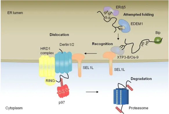
1.2 ER-Associated Degradation
Proteins that fail to fold into their native conformation are targeted to the ER
associated degradation (ERAD) pathway (Figure1.3). Proteins marked as
terminally misfolded are dislocated to the cytoplasm, where they are degraded by
the ubiquitin-proteosome system (Claessen et al., 2012; Smith et al., 2011).
ERAD machinery includes chaperones that extract misfolded proteins from the
pro-folding machinery, a complex in the ER membrane which coordinates and
drives protein dislocation across the membrane, and
ubiquitination/deubiquitination proteins and proteosome in the cytoplasm.
Damaged ER compartments and aggregated misfolded proteins might also be
eliminated by autophagy (Bernales et al., 2006). Autophagy is a survival
response linked to adaptation to starvation that involves the sequestration of
substrates in autophagosomes and fusion with lysosome containing hydrolases.
ERAD and autophagy might be coordinated processes, with autophagy being
up-regulated upon proteasome impairment (Nedelsky et al., 2008).
Depending on the topology of the substrate, i. e., misfolded domains in the
ER lumen, membrane, or cytosolic compartments of the protein, a distinct ERAD
pathway is engaged. Studies in yeast and metazoan have revealed a great
complexity and diversity of key ERAD components and adaptor proteins for some
model substrates. The transmembrane complexes associated with E3 ubiquitin
ligases have a central role and are common to all pathways (Carvalho et al.,
2006; Denic et al., 2006; Gauss et al., 2006a). E3 ubiquitin ligases have a
variable number of transmembrane domains, a cytoplasmic RING domain and
catalyze substrate ubiquitylation (Gardner et al., 2000). E3 ligase complexes
organize the machinery that coordinates events on both sides of the ER
membrane. The prototipycal E3 ligase is Hrd1 (HMG-CoA reductase degradation
protein 1) in yeast or HRD1, its mammalian homolog. Hrd1 over-expression is
sufficient to promote the degradation of ER luminal proteins (Carvalho et al.,
Usa1. However Usa1 is not required for the degradation of some subtracts and a
mammalian homolog has not be identified (Carvalho et al., 2006).
Adaptor proteins in the E3 ligase bound complex determine substrate
specificity by recruiting misfolded proteins to the membrane. The most well
characterized adaptor protein is Hrd3 in yeast and its counterpart SEL1 in
mammals. Hrd3 binds misfolded proteins, a interaction that is facilitated by a
large luminal domain with multiple tetratricopeptide repeats (TPRs) (Gauss et al.,
2006b). Housekeeping chaperones such as Bip can also work as adaptor
proteins and delivery proteins to the E3 ligase complexes.
The E3 complex members Derlins have a function which is not fully
understood. Yeast Der1p is a multi-pass transmembrane protein that interacts
with Hrd1p and Hrd3p as well as with substrates (Gauss et al., 2006a; Carvalho
et al., 2006) and are likely protein adaptors. In mammals, Derlin1, Derlin2 and
Derlin3 may form homo- and heterodimers, giving rise to the hypothesis that they
form (part of) a protein conducting channel (Bagola et al., 2011). Derlins are
inactive members of the rhomboid family of proteases that conserve a
membrane-embedded hydrophilic domain and can interact with translocation
machinery via its C-terminus (Greenblatt et al., 2011). Derlins recruits p97/VCP to
functional ERAD complexes for the extraction of misfolded secreted proteins from
the cytoplasmic face of the ER membrane (Ye et al., 2004).
The modification of a substrate with ubiquitin can recruit either one of two
multiprotein complexes that extract the protein from the ER membrane: the
ATPase Cdc48 (p97/VCP in metazoans) or the 19S lid complex of the
proteasome (Kalies et al., 2005; Stolz et al., 2011). The core of each complex
consists of a ring-shaped, hexameric ATPase of the AAA family and ATP
hydrolysis generates the energy necessary for protein dislocation across the
membrane and unfolding. 19C docks to the Sec61 channel at the ER membrane
(Kalies et al., 2005; Ng et al., 2007). Proteins might be retro-translocated via this
channel and delivered directly to the proteasome, which pulls the protein from the
In the cytoplasm, p97 recruits peptide glycanase (PNGase) to remove
N-linked oligossacharides moiety from substrates before they enter the proteosome
(Hirsch et al., 2003). P97 also recruits a de-ubiquitinase YOD1.
The mechanism of ERAD is best understood for the case of luminal
glycoproteins. As glycoproteins attempt to achieve their native conformation
entering the calnexin/calreticulin cycle, mannosidases mediate competing
reactions that remove mannoses residues from the glycan moiety (Ruddock and
Molinari, 2006). Removal of a α1,2-linked mannose from the A chain, the only
acceptor for UGT-catalyzed protein re-glucosylation, abrogates interaction with
calnexin/calreticulin and thus prevents defective folding glycoproteins from
entering futile cycles in the calnexin/calreticulin cycle. The kinetics of the
de-mannosidase reaction is slow thus only proteins that fail to mature in a timely manner are marked as ‘misfolded proteins’, a mechanism known as the mannose timer (Helenius, 1994).
In mammals, over-expression of EDEM1 (ER degradation enhancing
mannosidase-like Protein 1) enhances the rate of ERAD by accelerating the
release of defective folding polypeptides from calnexin (Molinari et al., 2003; Oda
et al., 2003). Possibly EDEM1 recognizes proteins marked as misfolded and
deliver them to the retro-translocation complex in the membrane. The core
component of the E3 complex SEL1L (Hrd3) forms a complex with glycan-binding
lectins Yos9p in yeast and OS-9 and XTP3-B in mammals to recruit misfolded
proteins ((Bernasconi et al., 2008; Christianson et al., 2008; Denic et al., 2006;
Gauss et al., 2006)). EDEM3 (or yeast Htm1p) functions as a exomannosidase that exposes a terminal α(1,6)-bonded mannose in glycoproteins, a commitment step required for effect degradation, which is recognized by Yos9p/OS-9/XTP3-B
((Clerc et al., 2009; Hosokawa et al., 2009; Quan et al., 2008)). To facilitate
retro-translocation ER enzymes help processing ERAD substrates, for example ERdj5
can reduce disulfide bonds of aberrantly linked proteins and alleviate compact
1.3 ER stress and Unfolded Protein Response
A number of cellular stress conditions such as low ATP levels, redox stress
or abnormal ER calcium content may perturb protein maturation in the ER and
interfere with ER folding capacity. Mutations in the amino acid sequence may
render some proteins incompetent to fold and lead to their constitutive retention
in the ER (Malhotra and Kaufman, 2007). Many physiological processes may
challenge the ER by representing a constant or fast source of large amounts of
proteins in the ER (Moore and Hollien, 2012). This is the case of differentiation of
highly secretory cells, such as the pancreatic β-cells, that produce insulin in
Figure 1.3 The events and components of ERAD.
The key steps of ERAD for a luminal glycoprotein in metazoans. See text for details. Adapted
response to sucrose stimulation. The imbalance between the ER folding capacity
and the burden of incoming proteins into the ER leads to the accumulation of
misfolded/unfolded proteins in the lumen of the organelle and causes ER stress.
Adaptation to ER stress is mediated by the engagement of the Unfolded Protein
Response or UPR (Hetz, 2012; Ron and Walter, 2007). The UPR terms a
collection of integrated signaling pathways triggered by ER-localized
transmembrane receptors, with luminal portions that sense stress in the ER and
cytoplasmatic effector portions that interact with the transcriptional or translational
apparatus. The UPR changes expression of proteins related to nearly every
aspect of the secretory pathway.
The UPR was first described in yeast, where a single ER transmembrane
protein, Ire1 (inositol-requiring enzyme 1), mediates one linear pathway (Mori et
al., 1992). In higher eukaryotes the UPR gained complexity and is mediated by
three ER transmembranar sensors: pancreatic ER kinase (PKR)-like ER kinase
(Perk), activating transcription factor 6 (ATF6) and Ire1 (Harding et al., 2002) The
outcomes of the three UPR pathways are temporally coordinated: first the
general rate of translation is attenuated to reduce the load of protein synthesis
into the ER and prevent further accumulation of unfolded proteins; second, genes
encoding ERAD components are up-regulated to increase the retro-translocation
and degradation of misfolded proteins from the ER; and third, genes encoding ER
chaperones and enzymes are up-regulation to enhance the ER folding capacity.
If UPR mechanisms are insufficient to overcome ER stress and restore ER
homeostasis, cells undergo apoptosis (Rasheva and Domingos, 2009). Chronic
ER stress and deleterious effects of the UPR have been involved in the pathology
of human diseases, such as cancer, diabetes, neurodegenerative disorders and
inflammation (Wang and Kaufman, 2012). Therefore there has been increasing
interest in controlling ER stress pathways and discover new therapeutic targets to
1.4 Ire1 Signaling
Ire1 is the most evolutionarily conserved arm of the UPR, with homologs in
yeast, plants, worms, fliesand vertebrates (Cox et al., 1993; Koizumi et al., 2001;
Mori et al., 1993a; Shen et al., 2001; Wang et al., 1998). Ire1 is a type I
ER-resident transmembrane protein with an ER luminal dimerization domain and a
cytoplasmic domain with Ser/Thr kinase and endoribonuclease activities (Shamu
and Walter, 1996; Tirasophon et al., 1998; Liu et al., 2002).
In the budding yeast the only known substrate of Ire1 is the mRNA of the
bZIP transcription factor Hac1 (Figure1.4) (Cox and Walter, 1996; Mori et al.,
1996; Nikawa et al., 1996). Under normal conditions, Hac1 mRNA is not
translated due to a translational attenuation exerted by the base pairing between
the 5’UTR and a 262bp intron (Rüegsegger et al., 2001). In case of ER stress,
Ire1 oligomerizes and activates its RNase domain by autophosphorylation (Liu et
al., 2000; Papa et al., 2003; Shamu and Walter, 1996; Welihinda and Kaufman,
1996). Activated Ire1 recognizes a double stem loop in the intron of Hac1 mRNA
and cleaves it twice, leading to the excision of the 252bp intron (Sidrauski and
Walter, 1997) A tRNAse ligase, Trl1, joins both ends of the exons (Sidrauski et
al., 1996). This unconventional splicing has two consequences: release of the
translational repression of Hac1 mRNA and introduction of a new C-terminus
during translation that transforms Hac1spliced into an effective transcription
factor (Chapman and Walter, 1997; Cox and Walter, 1996). The functional
homolog of Hac1 in mammals, worms and flies is Xbp1 (Calfon et al., 2002;
Yoshida et al., 2001a); Ryoo et al., 2007; Souid et al., 2007). The intron spliced
by Ire1 in Xbp1 mRNA has only 26bp in mammals (or 23bp in Drosophila) and
does not exert a translational repression. Xbp1 unspliced mRNA is translated
under normal conditions but originates a inhibitor of the UPR, which is rapidly
degraded (Calfon et al., 2002; Yoshida et al., 2006). Xbp1 spliced protein
comprises the original N-terminal DNA binding domain plus an alternative
introduced upon a frame-shit during translation of the spliced Xbp1 mRNA. So
far, the ligase that joins the ends of Xbp1 spliced mRNA was not identified.
Figure 1.4 Ire1 signaling.
Spliced Hac1 binds the unfolded protein response element (UPRE)
containing the consensus sequence CAGCGTG and Xbp1spliced binds
promoters containing CRE (cAMP response element)-like elements
[GATGACGTG(T/G)NNN(A/T)T] (Mori et al., 1992, 1998; Clauss et al., 1996).
Genetic profiling and genetic analyses reveled that Hac1/Xbp1 control the
expression of genes related to the UPR including chaperone induction,
up-regulation of ERAD machinery, membrane biogenesis and ER quality control
(Lee et al., 2003; Shaffer et al., 2004). In mammals, Xbp1 coordinates the
expression of unexpected cell type specific targets linked to cell differentiation,
signaling and DNA damage (Acosta-Alvear et al., 2007).
Mechanism of ER stress sensing by Ire1
The mechanism by which the ER luminal domain of Ire1 senses misfolded
proteins remains somewhat enigmatic. It has been known that the ER chaperone
Bip binds to Ire1 and maintains it in an inactive monomeric state under normal
conditions (Bertolotti et al., 2000; Okamura et al., 2000). The model suggested
that Bip is a negative regulator of Ire1 activity; and under stress conditions, Bip is
mobilized to misfolded proteins allowing Ire1 to multimerize and
autophosphorylate on its cytosolic domain. However, mutagenesis analysis
showed that variants of Ire1 that do not bind Bip respond normally to stress,
leading to the proposal that Bip binding is more important for preventing
inappropriate activation of Ire1 rather than eliciting activation by ER stress
(Kimata et al., 2003).
Recently the resolution of the crystal structure of the ER luminal domain of
yeast and human Ire1 protein brought new insights about the mechanism of Ire1
activation (Credle et al., 2005; Zhou et al., 2006). The luminal domain of yeast
Ire1 displays a MHC-like groove and mutations in the amino acids of the groove
or in the dimerization interface, abrogate the ability of Ire1 to engage the UPR
(Credle et al., 2005). This observation led to the speculation that misfolded
oligomerization. In vitro studies show that yeast Ire1 binds to a constitutively
misfolded form of carboxypeptidase Y through its luminal groove and peptide
binding causes Ire1 oligomerization (Gardner and Walter, 2011). A more direct
model of activation has now gained strength: first Bip dissociates from Ire1
leading to its dimerization and then direct binding of unfolded proteins to Ire1
luminal domain may contribute to cluster formation and engage full ribonuclease
activity (Kimata et al., 2007).
Additional studies with mammalian Ire1 suggest that this mechanism of ER
stress sensing might not be conserved. The MHC-like groove of mammalian Ire1
might be too narrow to accommodate an unfolded peptide, as indicated by the
crystal structure (Zhou et al., 2006). Furthermore, Ire1 mutants for Bip binding
become active even under unstressed conditions and the luminal domain of
mammalian Ire1 did not interact with unfolded proteins in an in vitro assay
(Oikawa et al., 2009). It may be that regulation of mammalian Ire1 depends on
Bip dissociation and not on the ligation of misfolded proteins, contrary to the
yeast counterpart.
Activation and Regulation of Ire1 RNase activity
The sequential steps of Ire1 activation - oligomerization, autophosphorylation
and RNase activitation - have been known for some time. The recent crystal
structure models of Ire1 cytosolic domain elucidated the molecular mechanism of
its activation, in particularly explaining the coupling of Kinase and RNase
activities.
The cytosolic domain of yeast Ire1 was crystallized in an activated state with
bound ADP-Mg and phospharylated at the kinase activation loop (Figure 1.5).
The crystal structure revealed a typical bilobed protein kinase domain, with the
ATP-binding site in the cleft between the N-lobe and C-lobe, and an atypical
RNase domain formed exclusively by alpha helices, named the KEN (kinase
extension nuclease) domain. The KEN domain is contiguous and fused to the
symmetric back-to-back dimers, creates a cleft with the RNase catalytic active
site. Dimers may assemble into rodshaped oligomers with a helical symmetry. In
oligomers, the phosporyilated activation loop is ordered, and contributes to a
stable active conformation by forming salt bridges with residues in cis(Korennykh
et al., 2009).
The crystal structure of a cytoplasmatic portion of dephosphorylated human
Ire1α bound to ADP further supported the previous data from yeast Ire1 and
added new details. The model for human Ire1 predicted a face-to-face dimer with
the ATP binding pockets facing each other and the activation loops juxtaposing,
in a position favoring trans-autophosphorylation (Ali et al., 2011). The
face-to-face dimer is predicted to be an intermediate state that precedes the back to back
activated state described in yeast.
Association into high order oligomers reinforces RNase activity, as
suggested by cooperative activation of the Ire1 RNase in vitro and stabilization of
the RNase active site in crystallized oligomers (Li et al., 2010). The RNA binding
cavity created by two RNase domains accommodates only one RNA stem loop
and the docking is probably asymmetric. Both Ire1 monomers participate in the
recognition of the stem loop but only one active site, with the catalytic residues
H1061 and Y1043 is used (Korennykh et al., 2011a).
Cofactors bind the ATP-binding pocket of the Ire1 kinase and modulate the
RNase activity and oligomerization of Ire1. Following dimerization, the alpha
C-helix moves into an active conformation, enabling productive ATP docking and
phosphorylation (Korennykh et al., 2009, 2011b). ADP, which is the natural
co-factor, modulates the position of the alpha C-helix. ADP and synthetic molecules
as APY29 may induce a 200-fold increase in the RNase activity (Korennykh et
al., 2011b). A screen to identify new Ire1 modulators identified flavonoids as
activators of the RNase activity. The most potent flavonoid activator of RNase
activity was quercitin, which binds to a deep buried hydrophobic pocket at the
Besides promoting stronger Ire1 self association, autophosphorylation may
have a role in shutting down Ire1 after ER adaptation. Yeast cells with kinase
inactive Ire1 have defects in the deactivation of Ire1, causing chronic ER stress
(Rubio et al., 2011). Phosphorylation may serve as a timer to count the duration
of the Ire1 signaling and hiper-phosphorylation may help to the disassembly
of Ire1 signaling complexes. The existence of this timer is observed in human
Figure 1.5 Structure of Ire1.
(A) A schematic representation of the yeast Ire1 structure indicating the kinase and RNase domains, BiP-binding site (BBS), the MHC-like domain, linker region and transmembrane region (TM).
Ire1, where oligomers disassemble concomitantly with Ire1 dephosphorylation,
even if the source of stress has not been mitigated (Li et al., 2010). After
inactivation, Ire1 enters a refractive state in which Ire1 no longer responds to ER
stress.
Targeting of Hac1/Xbp1 mRNA to Ire1
To be unconventionally spliced by Ire1, Hac1/Xbp1 mRNA must reach the
ER surface where Ire1 is localized. The mechanism of recruitment of the
Hac1/Xbp1 mRNA to the ER membrane seems to differ considerably between
yeast and mammals. Under non-stressed conditions, unspliced Hac1 mRNA is
found mostly in the cytoplasm, in association with stalled ribosome. Upon ER
stress, Hac1 mRNA is recruited to Ire1 clusters in the ER membrane, in a
process that depends on translational repression of Hac1 mRNA and on a
bipartite element (BE) at its 3’ UTR (Aragón et al., 2009). Disruption of Ire1 dimerization interfaces, prevented cluster assemble of Ire1 and impaired Hac1
mRNA targeting to the ER. Robust Ire1 oligomerization and Hac1 mRNA
targeting serves to concentrate both key UPR components into foci and ensure
efficient RNA processing.
In mammals, Xbp1 mRNA localization to the ER membrane is independent
of Ire1 and does not require secondary structures on unspliced Xbp1 mRNA
(Yanagitani et al., 2009). The unspliced Xbp1 mRNA is translated under normal
conditions and originates a polypeptide that associates with membranes through
a hydrophobic region at its C-terminus (the hydrophobic region2 - HR2) (Figure
1.6). The HR2 is a conserved region predicted to form a α-helice that has
propensity to interact with membrane lipid bilayers (Yanagitani et al., 2009).
Presumably, upon unspliced Xbp1 mRNA translation the HR2 on the nascent
polypeptide associates with the ER membrane and brings the Xbp1
mRNA-ribosome-nascent chain (RNC) complex to the vicinity of Ire1, facilitating
The HR2 is only 53 amino acid distant from the STOP codon. Before the
Xbp1 unspliced polypeptide is released from the RNC, a translational pause
stabilizes the complex and gives time for its recruitment to the ER membrane
(Yanagitani et al., 2011). The C-terminal region (CTR) in the Xbp1 unspliced -
with conserved Leu and Trp at positions 246 and 256,respectively, in humans – is
essential for elongation pausing when the HR2 portion of the Xbp1 unspliced
polypeptide protrudes from the ribosome exit tunnel(Yanagitani et al., 2011).
The mechanism underlying Xbp1 mRNA targeting to the ER was not yet
addressed in Drosophila. VISTA analysis for comparative genome analysis
shows that the 3’UTR of Xbp1 mRNA is conserved in four Drosophila species,
raising the possibility that the 3’UTR has affinity for Ire1. The Kyte and Doolittle
Figure 1.6 Recruitment of Xbp1 mRNA to the ER membrane in mammals.
(A) Structure of Xbp1 unspliced and Xbp1 spliced. ZIP, Leu zipper. TP, Translational pausing domain. HR2, hydrophobic region 2.
hydrophobicity scale predicts a region similar to mammalian HR2 in the
Drosophila unspliced Xbp1 protein. In addition the conserved Leu and Trp of the
CTR are also present. These Xbp1 features may have an important physiologic
role, taking into account that addition of yeast 3′UTR and mammalian CTR and
HR2 coding regions to a Drosophila ER reporter increased greatly its sensitivity
(Sone et al., 2013).
Xbp1-independent Ire1 functions
In contrast to budding yeast, where Ire1 and Hac1 are in a linear pathway, in
metazoans Ire1 has several signaling functions besides nucleolytic cleavage of
Xbp1 mRNA. Non-overlapping defects in Ire1 or Xbp1 mutants in C.elegans first
evidenced the existence of Ire1 alternative roles (Shen et al., 2005). Ire1 is
thought to regulate apoptosis, autophagy, and ERAD through interaction with
cytoplasmic partners and independently of its RNase activity (Hetz and Glimcher,
2009). However other targets of Ire1 kinase activity besides Ire1 itself are
unknown until date (Korennykh and Walter, 2012).
The cytosolic domain of Ire1 interacts with Traf2 (TNFR-associated factor 2),
and activates ASK1 (Apoptosis signal-regulating kinase1), triggering the JNK
(cJun-N terminal kinase) pathway (Urano et al., 2000; Nishitoh et al., 2002). This
Ire1/Traf2 interaction may lead to the activation of apoptosis under irreversible
ER stress (Mauro et al., 2006). Activated Ire1 may also instigate cell death by sequestrating IKK (inhibitor kβ kinase) in a complex with Traf2 (Hu et al., 2006). IKK is a negative regulator of the NF-kβ (factor nuclear kappa B) pathway,
associated with inflammation. Ire1 and NF-kβ induce TNF expression and
promote cell death in a caspase-8-dependent manner (Hu et al., 2006).
Ire1 may also control levels of autophagy under ER stress through activation
of the JNK pathway (Ogata et al., 2006). The phosphorylation of the
anti-apoptotic BCL2 (B-cell CLL/lymphoma 2) at the ER by JNK stimulates autophagy,
by modulating the activity of Beclin1. BCL2 forms a complex with Beclin1 and
yeast two-hybrid screen, Ire1 physically interacts with the ubiquitin specific
protease 14 (USP14), and this association is inhibited by ER stress (Nagai et al.,
2009). Moreover, inactive Ire1 interacts with ERAD components, such as Derlin,
SEL1 and HRD1. USP14, which is recruited to the ERAD machinery via
interaction with inactive Ire1, inhibits ERAD through deubiquitination-independent
mechanism (Nagai et al., 2009). Inactive Ire1 may form a macromolecular
complex that sequestrates/inactivates ERAD under non-stressed conditions.
Regulated Ire1-dependent decay (RIDD)
A genome-wide analysis in the budding yeast to find specific mRNA
substrates cleaved in vitro by Ire1 RNase or degraded in the absence of the Trl1
tRNA ligase only retrieved Hac1, supporting that Hac1 is an exclusive target of
Ire1 RNase (Niwa et al., 2005). Computational simulations in mammals searching
for mRNAs with structural and regulatory elements similar to Xbp1 (overlapping
long open reading frames and Ire1 cleavage sites flanking a short intron) did not
identify other mRNAs demonstrating the singularity of the pathway (Nekrutenko
and He, 2006). However a report using Drosophila S2 cells uncovered a novel
function for Ire1 RNase with a broad range of mRNA substrates and independent
of Xbp1. The group of Jonathan Weissman found through gene profiling that a
subset of mRNAs are degraded during ER stress by a mechanism that is
dependent of Ire1 but not Xbp1 (Hollien and Weissman, 2006). The degraded
mRNAs encoded mostly proteins with signal peptide/transmembrane domains
that would represent an additional challenge to the ER folding machinery under
ER stress. This mechanism was named RIDD, for regulated Ire1 dependent
decay, and was later described also in mammalian cells and in the fission yeast
S. pombe, which lacks any Hac1/Xbp1 homologue (Cross et al., 2012, Hollien et
al., 2009; Kimmig et al., 2012). While in Drosophila cells RIDD down-regulates
many RNAs by 5-10 fold, in mammals the magnitudes of expression changes
were small, twofold or less for many targets (Hollien and Weissman, 2006;
The mechanism of targeting mRNAs to RIDD seems to have diverged
throughout evolution (Hollien, 2013). In Drosophila, RIDD has a broad scope of
targets and there is a strong correlation between interaction of a mRNA with the
ER membrane and the extension of its degradation by RIDD (Gaddam et al.,
2013; Hollien and Weissman, 2006). In fact, deletion of the signal peptide from a
known RIDD target prevents its degradation and conversely, addition of a signal
peptide to GFP is sufficient to promote its degradation by RIDD. One interesting
exception is the mRNA of PlexinA, which although is strongly associated with ER
membrane is protected from RIDD and is continuously translated even during ER
stress (Gaddam et al., 2013). PlexinA mRNA has regulatory upstream ORFs, in
similarity with Gadd34, which are necessary for its protection from RIDD.
In mammalian cells, RIDD targets are enriched for mRNAs containing a
cleavage site with a consensus sequence (CTGCAG) and a predicted secondary
structure very similar to the conserved Ire1 recognition loop in Xbp1 mRNA (Han
et al., 2009; Hur et al., 2012; Oikawa et al., 2010). Deletion of the stem loop or
mutagenesis of the conserved bases abrogated RIDD (Oikawa et al., 2010). The
presence of Ire1 recognition sites in many RIDD substrates suggests that
mammals have evolved sequence specificity to better control the transcripts that
are subjected to degradation. In Drosophila, the mRNA encoding Smt3, a
homologue of SUMO (small ubiquitin-like modifier) is cleaved by RIDD on a stem
loop structure despite not being stably associated to the ER membrane (Moore et
al., 2013).
Under conditions of overwhelming ER stress or chronic low level stress (e.g.
expression of a mutant folding variant or treatment with 10nM thapsigargin),
mRNAs encoding secretory cargo proteins and resident ER proteins and other
secretory organelle proteins start to decay (Han et al., 2009). ER chaperone Bip
and Golgi localized glycosylating enzyme Gyltl1b are targets of RIDD.
Unmitigated ER stress may deplete important cell surface proteins or secretory
pathway proteins by continuous decay and promote apoptosis. In fact, expression
expression of Ire1 kinase active/RNase dead mutants do not induce apoptosis,
arguing that an active RNase is required to induce pro-apoptotic signals (Han et
al., 2009).
The two Ire1 functions, RIDD and Xbp1 splicing, can be uncoupled in vitro,
allowing better understanding of the physiological output of each pathway.
Ire1I642G mutant has an enlarged kinase pocket that prevents autophosphorylation and activation of the RNase. The need for ATP binding can be bypassed by
1NM-PP1, an ATP analog that binds specifically to the designed pocket of
Ire1I642G and activates the RNase by an allosteric mechanism (Papa et al., 2003). Addition of the 1NM-PP1 is sufficient to induce Xbp1 splicing when Ire1I642G is overexpressed, even in the absence of ER stress. However RIDD function can
only be engaged by 1NM-PP1 in the presence of ER stress, suggesting different
activation modes of Ire1 (Han et al., 2009; Hollien et al., 2009). Other
compounds, known as KIRAs (kinase inhibiting RNase attenuators) can bypass
the need for autophosphorylation to activate wild-type Ire1, stimulating Xbp1
splicing and tempering RIDD (Han et al., 2009).
ATF6 Signaling
The second arm of the UPR in metazoans is ATF6, an ER localized type II
transmembrane protein, whose cytosolic domain encodes a transcription factor
(Figure 1.7) (Haze et al., 1999; Wang et al., 2000; Yoshida et al., 2000) ATF6 is
retained in the ER by interaction with Bip and calnexin, that bind to its luminal
domain (Shen et al., 2002; Hong, 2004). Upon ER stress, underglycosylation of
ATF6 prevents interaction with calnexin and dissociation of Bip uncovers Golgi
localization signals on ATF6. ATF6 is then translocated to the Golgi, where is
cleaved sequentially by resident proteases S1P (site 1 protease) and S2P (site 2
protease) (Ye et al., 2000). The cytosolic fragment is released and moves to the
nucleus, where activates gene expression through a bZip domain.
ATF6 is more sensitive to perturbations of redox equilibrium in the ER than
ATF6 oligomers and induces its dissociation. In the monomeric form, ATF6 can
more easily be translocated to the Golgi (Nadanaka et al., 2007).
ATF6 binds to the ATF/CRE element and the ER stress response elements I
and II (ERSE), to induce the transcription of many UPR genes related to ERAD
Figure 1.7 ATF6 and CREBH signaling.
and folding, for instance bip, xbp1, herp, among others (Yoshida et al., 2003;
Kokame, 2000; Yoshida, 1998). ATF6 can form heterodimers with Xbp1, which
have higher affinity for UPR elements than either homodimer to promote maximal
expression of ER chaperones and degradation machinery (Yamamoto et al.,
2007).
Newly identified ATF6 homologs OASIS, CREBH, LUMAN/CREB3, CREB4,
BBF2H7 are controlled by ER stress in specific tissues (Bailey and O’Hare,
2007). All are ER-anchored bZip transcription factors that are processed in the
Golgi the same way as ATF6 but whose physiological role is poorly
characterized. CREBH (cyclic AMP response element binding protein
hepatocyte) expressed in hepatocytes induces the expression of hepcidin, both
basally and in response to ER stress and inflammation (Vecchi et al., 2009).
CREBH links ER stress to the production of acute phase proteins (serum proteins
that are associated with inflammation) (Zhang et al., 2006).
Perk Signaling
The third ER transducer, PERK, is an ER associated type I transmembrane
protein with a structure similar to Ire1: a luminal stress sensing domain and a
cytoplasmic portion with a protein kinase (Figure 1.8). The luminal domains of
Ire1 and PERK are phylogenetically related and can be experimentally
interchangeable in S. cerevisae, where Perk can splice Hac1 (Liu et al., 2000).
Like Ire1, Perk binds Bip under non stressed conditions (Bertolotti et al., 2000;
Ma et al., 2002). Upon ER stress, Bip dissociates and PERK is activated by
trans-autophosphorylation after oligomerization and phosphorylates the α-subunit
of the eukaryotic translation initiation factor 2 (eiF2) at Serine 51 (Harding et al.,
1999). Phosphorylation of eiF2 inhibits its recycling to an active GTP-bound form.
Limiting availability of active eiF2 for translation initiation attenuates global protein
synthesis and decreases protein influx into the ER lumen.
Expression profiling in ER stressed PERK knock-out cells or with a Ser51Ala
of UPR genes (Harding et al., 2003; Scheuner et al., 2001) Certain mRNAs
escape translational attenuation during ER stress due to the presence of
regulatory upstream open reading frames (uORFs) in their 5’ UTRs. Conditions
that limit eIF2 activity lead to ribosome skipping the inhibitory uORFs so that
downstream ORFs can be translated. ATF4 or its yeast homolog Gcn4 are
translationally induced by phosphorylation of eiF2α (Lu et al., 2004; Vattem and
Wek, 2004). ATF4 is a transcription factor responsible for activating genes
involved in amino acid metabolism and protection from oxidative stress, including
CHOP (C/EBP homologous protein) and Gadd34 (growth arrest and
DNA-damage inducible protein-34), besides other targets overlapping with ATF6 and
Xbp1 (Harding et al., 2000; Jiang et al., 2004; Ma et al., 2002b). Other stress
signaling pathways triggered by amino acid starvation, double-stranded RNA
accumulation or haem depletion also converge on eif2α phosphorylation,
activating the so called integrated stress response, which share common targets
(Harding et al., 2003).
The levels of phosphorylated eIF2α must be tightly regulated to avoid
permanent damage to the cell. Dephosphorylation of eIF2α is mediated by CReP
(constitutive repressor of eiF2 phosphorylation) and Gadd34 (Jousse et al., 2003;
Connor et al., 2001; Novoa et al., 2001). CReP is constitutive expressed and
encodes the substrate subunit of Protein Phosphatase 1 (PP1). GADD34 also promotes eIF2α dephosphorylation in complex with PP1, and works in a negative feedback loop to downregulate PERK signaling (Brush et al., 2003; Ma and
Hendershot, 2003; Novoa et al., 2003).
Activation of PERK occurs in a reversible manner and PERK is rapidly
dephosphoryilated after ER homeostasis is restored, allowing the fast recovery
from translational attenuation (Bertolotti et al., 2000; Jousse et al., 2003). The
HSP40 co-chaperone p58IPK is up-regulated by ATF6 during late phases of the
UPR to inhibit PERK activity by binding to its kinase domain (van Huizen, 2003;
The UPR in Disease and Development
Figure 1.8. PERK signaling.
In response to endoplasmic reticulum (ER) stress, Bip dissociates from PERK luminal domain, allowing PERK to dimerize and be activated by trans-autophosphorylation. PERK
phosphorylates the α subunit of eukaryotic translation initiation factor-2 (eIF2) on Ser51,
attenuating global protein translation by inhibition of eIF2B complex. Lower global protein synthesis reduces ER unfolded protein load but also affects gene transcription. eiF2 phosphorylation selectively increases the translation of ATF4, which under normal conditions is inhibited by conserved upstream open reading frames. The ATF4 protein up-regulates Xbp1, the proapoptotic transcription factor (CHOP) and genes encoding amino acid transporters and genes that protect against oxidative stress. The target genes of CHOP include GADD34 that
dephosphorylates eIF2α and ER oxidase-1 (ERO1) that promote protein folding within the ER
lumen. GADD34 associates with protein phosphatase I (PPI) to dephosphorylate eIF2α and
relieve PERK dependent translation attenuation in a negative-feedback circuit. A constitutive
Ire1α or xbp1 knock-out mice embryos present growth retardation and fetal
liver hipoplasia and die after 12.5 days of gestation (Reimold et al., 2000; Zhang
et al., 2005). Because the liver becomes the main hematopoietic organ in the
fetus, Ire1 or Xbp1 knock-out mice have reduced numbers of red blood cells but
can still differentiate erythroid and myeloid lineage cells. Ire1α is required in a
later step of the lymphoid differentiation for the immunoglobulin gene
rearrangement and formation of the B cell receptor in pro-B and pre-B cells
(Zhang et al., 2005). In the adult, splicing of Xbp1 by Ire1 is necessary for the
terminal differentiation of mature B-cell into antibody secreting plasmocytes
(Iwakoshi et al., 2003; Zhang et al., 2005). Introduction of Xbp1 into B cell
lineages is sufficient to induce plasma-cell differentiation and Xbp1 deficient B
cells are defective in secreting antibodies in vivo upon antigenic stimulation
(Reimold et al., 2001).
The targeted expression of Xbp1 in the liver of xbp1-/- embryos rescues
viability, but mice present severe defects in exocrine pancreas and salivary
glands that compromised secretion of digestive enzymes (Lee et al., 2003).
Pancreatic acinar cells of the xbp1-/-; liverxbp1 mice have a poorly developed ER
with few, disorganized cisternae, and immature zymogen granule precursors
located in the ER lumen. The failure in the ER expansion of pancreatic exocrine
cells during the embryonic development is consistent with Xbp1 role in
membrane biogenesis and phospholipid synthesis (Shaffer et al., 2004; Sriburi et
al., 2004). The liver is a secretory organ that produces the lipoprotein particles
that transport lipids to other tissues and Xbp1 is necessary for hepatic
lipogenesis. A Xbp1 conditional knock-out in the liver of adult mice reduces
plasma cholesterol and triglycerides due to a down-regulation of lipogenic genes
(So et al., 2012).
Deletion of xbp1 or ire1β in intestinal epithelial cells causes increased ER stress and exacerbated colitis induced by dextran sodium sulfate (DSS), a toxin
that disrupts mucosal barrier function (Bertolotti et al., 2001; Kaser et al., 2008).
infection, as a result of Paneth cells dysfunction and apoptosis and lack of
bactericidal killing activity (Kaser et al., 2008). Single-nucleotide polymorphisms
within the xbp1 gene locus on chromosome 22q12.1 are a risk factor for human inflammatory bowel diseases, Crohn’s disease and ulcerative colitis (Kaser et al., 2008).
Ire1α and xbp1 knock-outs mice do not have phenotype in the endocrine
pancreas (Iwawaki et al., 2010; Lee et al., 2005). However, several experiments
in vivo and in vitro point for a role of the Ire1/Xbp1 pathway during insulin
maturation in the ER. Pancreatic β-cells deficient for Xbp1 are inefficient to
process and secrete insulin (Lee et al., 2011). Analysis of the morphology of
Islets of Langerhans lacking Xbp1 reveals a highly disorganizated structure and
abnormal β–cells with fewer insulin granules and distended ER (Lee et al., 2011).
These observations are in agreement with Xbp1 being required for expression of
ER chaperone genes such as bip (Hspa5), ERdj4 (Dnajb9), and p58IPK (Dnajc3).
Perk deficient mice are viable but present growth retardation and have
defects in the pancreas and skeleton formation (Zhang et al., 2002). Pancreatic
acinar cells of perk-/- mice have a distended and fragmented ER, and undergo
increased apoptosis by the fourth post-natal week, a phenotype similar with
xbp1-/-; liverxbp1 mice. The inappropriate production of digestive enzymes causes
maldigestion of lipids in Perk-/- mice (Harding et al., 2001; Zhang et al., 2002).
perk-/- mice show deficient mineralization and impaired formation of skeleton due
mostly to osteoblasts defects. Compact bone is formed by type I collagen and
other components of the extracellular matrix, which are secreted by osteoblasts.
Perk-/- osteoblasts have distended and fragmented ER filled with an electron-dense material that corresponds to unprocessed procollagen (Zhang et al.,
2002).
Deletion of Perk leads to progressive degeneration of pancreatic β-cells, deregulation of glucose metabolism and early onset diabetes (Harding et al.,
2001; Zhang et al., 2002). In humans, Perk loss-of-function mutations are