Vo l. 9(5), p p . 132-139, 3 Fe b rua ry, 2015 DO I: 10.5897/ JMPR2014.5141
Artic le Numb e r: 8DC DE7550675 ISSN 1996-0875
C o p yrig ht © 2015
Autho r(s) re ta in the c o p yrig ht o f this a rtic le http :/ / www.a c a d e mic jo urna ls.o rg / JMPR
Journal of Medicinal Plants Research
Full Length Research Paper
In vitro and in vivo evaluation of quinones from
Auxemma oncocalyx Taub. on Leishmania braziliensis
Sayonara de Melo Viana
1, Maria Augusta D. Ferreira
2, Priscila Valera Guerra
1,
Glauce S. Barros Viana
3and Maria Jania Teixeira
1*
1
Departamento de Patologia e Medicina Legal, Faculdade de Medicina, Universidade Federal do Ceará (UFC), Rua Alexandre Baraúna, 949, 60.430-160 Fortaleza, CE, Brazil.
2
Departamento de Farmácia, Faculdade de Farmácia, Odontologia e Enfermagem, UFC, Rua Capitão Francisco Pedro 1210, 60430-370 Fortaleza, CE, Brazil.
3
Departamento de Fisiologia e Farmacologia, Faculdade de Medicina, UFC, Av. Cel. Nunes de Melo, 1127, 60430-270 Fortaleza, CE, Brazil.
Received 25 June, 2013; Accepted 3 February, 2015
The effect of a quinone fraction (QF) isolated from the heartwood of Auxemma oncocalyx Taub. was
investigated in vitro and in vivo on Leishmania braziliensis. Although QF (1-10 μg/mL) showing marked
in vitro anti-Leishmania activity (81 to 94%), the oral treatment with the compound did not protect
hamsters against progressive L. braziliensis infection. When QF was administered intraperitoneally (20
mg/kg) the lesion size was reduced by 65%; however, it was not able to promote parasite eradication, as evidenced by the high number of parasites in draining lymph nodes. Quinones are highly redox active molecules and with their semiquinones radicals can lead to formation of reactive oxygen species (ROS).
The generation of ROS could probably explain the in vitro leishmanicidal activity of the quinones, since
promastigotes are susceptible to H2O2 lethal effect in vitro. In conclusion, although quinones seem to be
effective against Leishmania parasites in vitro, they do not demonstrate a therapeutic effect in
experimental leishmaniasis. In addition, it can be hypothesized that QF in vivo might possibly be
converted into non-active metabolite(s) or be inactivated either by reduction or by interaction with serum proteins, losing its leishmanicidal activity.
Key words:Leishmania braziliensis, Auxemma oncocalyx,quinone, hamster, cutaneous leishmaniasis.
INTRODUCTION
Leishmaniasis are protozoan diseases which represent a risk for 350 million people worldwide, and 2 million new cases occur yearly (World Health Organization [WHO], 2010). Etiologic agents are intracellular parasites of the genus Leishmania that display a spectrum of a manifestation which goes from cutaneous involvement ith
late destruction of mucous membranes to generalized systemic visceral disease with fatal outcome, if not trea-ted (Pearson et al., 2000; Desjeux, 2004). There is still no effective vaccine to control the wide range of disease caused by different species of Leishmania (Oliveira et al., 2009).Thediseasemayregressspontaneouslyor evolve,
*C o rre sp o nd ing a utho r. E-ma il: mjte ixe ira 601@ g ma il.c o m. Te l: +55-85-33668311. Fa x: +55-85-33668316.
thus requiring treatment (Piscopo and Mallia, 2006). The antileishmanial first-line drugs are the pentavalent antimonials, meglumine antimoniate (Glucantime) and sodium stibogluconate (Pentostam). They are generally effective during the acute infection stages but not against the late stages, and produce significant side effects due to high toxicity and tissue drug accumulation, which includes myalgias, nausea, vomiting, cardiac arrhythmia, hepatitis, or pancreatitis (Croft et al., 2006; Palumbo, 2009). Amphotericin B and pentamidine are better tolerated drugs, but require long courses of parenteral administration (Amato et al., 2008). Miltefosine and fluconazole have recently showed effectiveness against cutaneous leishmaniasis (CL) caused by Leishmania braziliensis (Machado et al., 2010; Sousa et al., 2011), but despite the lower toxicity, these second line drugs are not useful against other forms of leishmaniasis (Palumbo, 2009). Furthermore, the continuous use of ineffective drugs has led to the development of resistance to their compounds (Escobar et al., 2001), which have stirred an urgent need for novel, effective, and safe drugs for treatment of leishmaniasis.
Since plant derivatives are among the most active agents against infections (Delorenzi et al., 2001), many researchers have been looking for better effects and less toxicity to treat leishmaniasis in these substances, especially those already used by people living in endemic areas (Weniger et al., 2001). Auxemma oncocalyx Taub. belongs to the Boraginaceae family, it is known as “pau branco” (white wood), and is easily available by rural settlers in Northeastern Brazil. The stem bark of the tree is astringent and popularly used in the treatment of wounds (Braga, 1976).
Previous pharmacological studies reported that the hydroalcoholic extract of the stem presents antioxidant, analgesic and anti-inflammatory properties (Ferreira et al., 2004, 2008). Alantoin and -sitosterol glycoside have been isolated from A. oncocalyx and are responsible for some of its pharmacological properties (Pessoa and De Lemos, 1997). Besides these compounds, at least six quinones were successfully isolated and oncocalyxone A, obtained in significant amounts, seems to be partially involved in the bioactivity of the plant (Leyva et al., 2000). Several quinones present antileishmanial (Sauvain et al., 1993; Sittie et al., 1999; Teixeira et al., 2001), antitumoral (Morello et al., 1995; Itoigawa et al., 2003), antifungal (Perry et al., 1991; Gafner et al., 1996), or antimalarial activity (Figueiredo et al., 1998), either in vitro or in vivo.
The aim of the present work was to evaluate the in vitro
and in vivo activity of the quinone fraction from A. oncocalyx Taub. against L. braziliensis.
MATERIALS AND METHODS
Plant extraction and purification of the quinone fraction
A. oncocalyx Taub. was collected in the city of Pentecoste, Ceará
State, Northeastern Brazil, and identified by Prof. A. G. Fernandes from the Biological Sciences Department. A voucher specimen has been deposited under the number 18459 at the Prisco Bezerra Herbarium of the Universidade Federal do Ceará, Brazil. The quinone fraction (QF) was prepared from grinded heartwood ethanolic extract through exhaustive aqueous extraction followed by lyophilization. Hydrosoluble components contained around 80% of oncocalyxone A, according to a previous characterization (Pessoa et al., 1993).
Parasites and animals
Three to four months adult female and male golden hamsters (Mesocricetus auratus), weighing 80 to 90 g, obtained from the central animal facility of Departamento de Patologia e Medicina Legal of Universidade Federal do Ceará (DPML/UFC), and housed in groups of six to eight per cage with free access to water and food. The Animal Care and Utilization Committee from UFC approved all experimental procedures (process n° 65/08). The L. braziliensis (MHOM/BR/94/H-3227) was originally isolated from skin lesions of a patient with CL from Ceará State, and previously typed using isoenzymes electrophoresis and monoclonal antibodies (De Oliveira et al., 2004).
The parasites, stored in liquid nitrogen, were thawed and cultured as promastigotes at 26°C in Schneider’s insect medium (Sigma-Aldrich, Chemical Co., St. Louis, USA) supplemented with 10% heat-inactivated fetal calf serum (Sigma), 2% sterile normal human urine, 2 mM L-glutamine (Gibco BRL, Grand Island, NY), and antibiotics [100 U/ml penicillin, 100 µg/ml streptomycin sulfate (Sigma-Aldrich)]. Subcultures were made in the stationary phase of growth and parasites were used at no later than the fourth passage. Prior to infection, promastigotes were harvested from culture, washed in sterile saline, counted in Neubauer’s chamber and adjusted to the appropriate concentration.
Anti-promastigote activity
Infection, treatment and lesion development
Hamsters were infected subcutaneously in the right hind footpad with 106 stationary phase L. braziliensis promastigotes in 20 µl of sterile saline. Treatment was initiated two weeks after inoculation when lesions were well defined. Animals were randomly divided into groups of six to eight and the drugs administered daily for 28 consecutive days. QF was dissolved in distilled water containing 0.1% of Tween 80 and 0.5% of carboxymethylcellulose (CMC) and administered using the following routes: (a) oral (p.o.), with 10 or 20 mg/kg by intragastric intubation; (b) intraperitoneal (i.p.) with 20 mg/kg. Alternatively, Glucantime (Sanofi-Aventis Farmacêutica, São Paulo, Brazil) was injected at the dose of 60 mg/kg/day intramuscularly (i.m.). Control groups received p.o. or i.p. drugless vehicle (Tween 80 + CMC + H2O) at equivalent volumes. Before
treatment was conducted, a toxicity test using various concentrations of QF that demonstrated concentrations of 10 and 20 mg/kg did not kill the animals. Lesion sizes were measured weekly with a dial gauge caliper (Mitutoyo, 0.01 mm sensitivity) and expressed as the difference between the thicknesses (mm) of the infected and contralateral uninfected footpads.
Treatment outcome
The number of parasites in the popliteal lymph was quantified by the limiting dilution technique as previously described (Lima et al., 1997). Briefly, after the treatment, the animals were euthanized by inhalation of halothane (Sigma-Aldrich) and submerged in 3% iodized alcohol up to 3 min to allow decontamination. The lymph nodes were removed aseptically and macerated in a Petri dish with 2 ml of Schneider medium. After removal of debris by sedimentation for 5 min, the homogenates were serially diluted (1:10) in Schneider’s medium supplemented with 100 U/ml of penicillin/ml, 100 μg/ml of streptomycin/ml, 10% fetal calf serum and 2% sterile human urine. One hundred microliters of these dilutions was distributed into 96-well flat bottom plates containing agar-blood in 6 replicates per concentration. The plates were incubated at 25°C and observed under an inverted microscope (Nikkon, Japan) every 3 days, up to a maximum of 30 days, to record the dilutions containing promastigotes. The final number of parasites per tissue was determined using the ELIDA software, version 12c for window (Taswell, 1984).
Statistical analysis
The data are presented as mean ± standard error of the mean. The significance of the results relating to the parasite load was calculated by Mann-Whitney test. The anti-promastigota activity and lesion sizes from treated and untreated animals were analyzed by the one-way analysis of variance (ANOVA) and complemented by the Bonferroni test for multiple comparisons. All analysis and graphs were performed using GraphPad Prism version 5.0 (GraphPad Software, San Diego, USA). Values of P < 0.05 were considered significant.
RESULTS
QF displayed marked in vitro anti-promastigote activity at concentration of 1 and 10 μg/mL (80.5 ± 2.80 and 91.0 ± 0.65%, respectively) against L. braziliensis, being similar to Amphotericin B (Figure 1). To determine the effect of QF on L. braziliensis infection in hamsters, the animals days via oral. The results showed that hamsters treated with QF (10 or 20 mg/kg, p.o.) did not present significant
reduction of the lesion thickness as compared to untreated animals. Also, doses of 20 and 10 mg/kg (p.o.), did not produce significant reduction of parasite load in lymph nodes as compared to untreated group (Figure 2A and B). As expected, animals treated with glucantime showed antimonial drug effectively brought footpad sizes down to normal within 2 weeks of treatment and also suppressed parasite growth in lymph nodes (Figure 2A and B).
To investigate if a different administration route of the drug could be more effective on controlling L. braziliensis
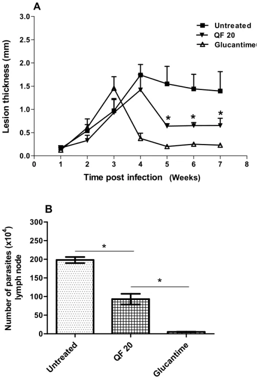
infection, hamsters were treated with 20 mg/kg QF intraperitoneally for 28 days (the highest dose, but yet not toxic to animals). The results showed that the lesion size of hamsters treated with QF i.p. decreased by 65% (P < 0.05), and parasite burden in the lymph nodes was significantly lower (9.3 ± 3.5 × 10⁵) when compared with untreated controls (1.98 ± 2.0 × 10⁶) (Figure 3A and B).
DISCUSSION
Antileishmanial activity has been reported in several compounds extracted from medicinal plants belonging to diverse chemical groups, including quinones (Chan-Bacab et al., 2001; Rocha et al., 2005). Other studies have found simple quinones isolated from dried trunks of
Jacaranda copaia to present significant anti-promastigote and anti-amastigote activities in vitro against L. amazonensis, but only weak activity when tested against
L. amazonensis-induced lesion in mice (Sauvain et al., 1993). Benzoquinones were found to be active in vitro
against trypanosomes, but this has not been confirmed in vivo (Grady et al., 1984; Pahn et al., 1988).
In murine model, Leishmania major- and Leishmania donovani-infected BALB/c treated with buparvaquone formulation (BPQ), showed parasite burden decrease in lesions and liver, smaller and not ulcerated lesions, in comparison with untreated control (Garnier et al., 2007). Recently, a study using the molecular hybridization of a naphthoquinone core with a pterocarpan moiety (LGB-118) led to significant reduction in skin lesions development, swelling, ulceration and parasite burden of BALB/c L. amazonensis-infected (Da Cunha-Júnior et al., 2011). Also, acetylisolapachol, a hydroxyquinone deriva-tive, showed in vitro activity against L. braziliensis, and in vitro and in vivo against L. amazonensis (Lima et al., 2004).
The leishmanicidal activity of a drug may be selective and direct against the parasite, or it may act indirectly by activating macrophage microbicidal mechanisms for instance. According to in vitro model systems, the macrophage microbicidal response to Leishmania
infection can follow two distinct pathways. Upon infection, promastigotes elicit a respiratory burst with the generation of reactive oxygen intermediates such as hydrogen peroxide (H2O2),
•
OH radical, superoxide (•O2)
Me
di
um
DM
SO
A
m
ph
ot
er
ic
in
10
ug
Am
pho
te
ric
in
0
,1u
g
Q
F 10
ug
QF
1ug
Q
F
0.1
ug
QF 0
.01
ug
0
20
40
60
80
100
In
h
ibi
ti
on
of
g
row
th
(
%
)
***
Figure 1. Antileishmanial activity in vitro of a quinone fraction (QF) isolated from the heartwood of Auxemma oncocalyx Taub. against promastigores of L. braziliensis.The inhibition of growth was expressed as the percent decrease of radioactive incorporation in treated parasites when compared with untreated control. ***P < 0.05 (test ANOVA).
and peroxynitrate as part of an oxygen dependent mechanism to kill promastigotes, however, a small percentage of phagocytosed organisms can survive (Beaman and Beaman, 1984). Second, murine or human macrophages can be activated to kill intracellular amastigotes, the form present during established infection, by previous exposure to cytokines such as
IFN- and TNF-, which activate both oxidative and non-H2O2-associated microbicidal mechanisms (Bogdan et
al., 1990; Liew, 1992; Assreuy et al., 1994; McSorley et al., 1996; Panaro et al., 1999). Studies have demonstrated that both H2O2-associated and non-H2O2
-associated pathways contribute to Leishmania killing and that their relative degree of importance may differ during initial promastigote invasion versus established amastigote infection (Chang, 1983; Murray and Nathan, 1999; Erel et al., 1999).
Quinones are highly redox active molecules and with their semiquinones radicals can lead to formation of reactive oxygen species (ROS), including •O2, H2O2, and
ultimately to hydroxyl radicals (Bolton et al., 2000). The formation of ROS could probably explain the in vitro
leishmanicidal activity of the quinones in this study, since
promastigotes are readily susceptible to killing by H2O2in
vitro (Murray, 1981; Zarley et al., 1991). It has been shown that Leishmania chagasi promastigotes in vitro are susceptible to killing by both H2O2 and the redox-cycling
compound menadione, a quinone that causes the gene-ration of •O2 in the presence of promastigotes (Wilson et
al., 1994). However, this source of quinone free radicals seems to be often more readily apparent in vitro than in vivo. Menadione providing an excellent example of this phenomenon, although presents anticancer activity in vitro in combination with other chemotherapeutic agents. However, menadione does not demonstrate the same activity in vivo even at high doses (Nestor et al., 1991; Djuric et al., 1995). Besides, in a BALB/c mouse model of leishmaniasis, sublethal concentrations of menadione caused L. chagasi promastigotes to become more virulent (Wilson et al., 1994).
0 1 2 3 4 5 6 7 8 0.0
0.5 1.0 1.5 2.0 2.5
3.0 Untreat
Glucant QF 20 QF 10
*
*
*
A
Time post infection (weaks)
L
esi
o
n
t
h
ickn
ess
(m
m
)
(Weeks)
U
nt
re
at
ed
QF
10
QF
2
0
G
lu
ca
nt
im
e
0 50 100 150 200 250 300
B
*
*
N
u
m
b
e
r of
pa
ra
s
it
e
s
(
x
1
0
4
)
l
y
m
p
h
no
de
Figure 2. Effect of oral treatment with QF (20 or 10 mg/kg). (A) Lesion growth, and (B) Parasite load in lymph node. Hamsters were infected with 106 L.
braziliensis promastigotes (8 per group). Animals were left untreated or were treated with daily dose of 20 mg/kg QF or with 10 mg/kg or 60 mg/kg/i.m. glucantime. Treatment started three weeks post infection. Lesion thickness was measured weekly using a dial gauge caliper (mean SE, n = 8). Parasite load was evaluated after the treatment. *P < 0.05 (A, Glucantime versus untreated or QF treatment; B, Glucantime versus QF treatment).
mean a decrease in the parasite load, but only a decrease of the local inflammatory reaction. QF of A. oncocalyx has a wide range of biological effects including anti-parasitic, antitumoral and antiplatelet activities
0 1 2 3 4 5 6 7 8 0.0
0.5 1.0 1.5 2.0 2.5 3.0
Untreated QF 20
Glucantime
*
*
*
A
Time post infection (weaks)
L
es
io
n
t
h
ic
kn
es
s
(m
m
)
Untr eate
d
QF 20
Glu cant
ime
0 50 100 150 200 250 300
B
*
*
N
u
m
b
er
of
p
a
ra
si
te
s
(
x
10
4 )
l
y
m
p
h
no
d
e
(Weeks)
Figure 3. Effect of intraperitoneal treatment with QF (20 mg/kg). (A) Lesion growth, and (B) Parasite load in lymph node. Hamsters were infected with 106L. braziliensis
promastigotes (6 per group). Animals were left untreated or were treated with daily dose of 20 mg/kg QF or 60 mg/kg/i.m. glucantime. Treatment started three weeks post infection. Lesion thickness was measured weekly using a dial gauge caliper (mean SE, n = 8). Parasite load was evaluated after the treatment. *P < 0.05 (A, QF treatment versus untreated; B, QF treatment versus untreated or Glucantime).
is suggested that the reduction in the lesion size produced by 20mg/kg, i.p of QF in L. braziliensis-infected hamsters may probably be due to its anti-inflammatory effect. In addition, the observation that QF when used via
serum proteins. Quinones can be metabolized by various routes: substitution or reductive addition with nucleophilic compounds or one and two-electron reductions (Koster, 1991). Driscoll et al. (1974) found that the biological activity of some quinones, as lapachol and its analogs, is directly related to their chemical structures, thus any structural alteration in vivo will result in an inactive product or will abolish their biological activities (Teixeira et al., 2001).
Despite QF not to have demonstrated an anti-parasitic effect in L. braziliensis-infected hamsters, the production of quinones derivatives might insure their interest as antileishmanial candidate drugs. Furthermore, alternative therapy derived from medicinal plants opens new perspectives towards the development of effective, readily available and less-costly drugs for the treatment of the leishmaniasis in endemic areas.
Conflict of interest
The authors report no declarations of interest.
ACKNOWLEDGEMENT
The authors thank Otília D. L. Pessoa and Telma L. G. Lemos from Departamento de Química Orgânica e Inorgânica, UFC, Fortaleza, CE, Brazil. This work was partially supported by the Brazilian National Research Council (CNPq).
REFERENCES
Amato VS, Tuon FF, Bacha HA, Neto VA, Nicodemo AC (2008). Mucosal leishmaniasis. Current scenario and prospects for treatment. Acta Trop. 105(1):1-9.
Assreuy J, Cunha FQ, Epperlein M, Noronha-Dutra A, O'Donnell CA, Liew FY, Moncada S (1994). Production of nitric oxide and superoxide by activated macrophages and killing of Leishmania major. Eur. J. Immunol. 24(3):672-676.
Beaman L, Beaman BL (1984). The role of oxygen and its derivatives in microbial pathogenesis and host defense. Annu. Rev. Microbiol. 38:27-48.
Bogdan C, Moll H, Solbach W, Röllinghoff M (1990). Tumor necrosis factor- in combination with interferon-, but not with interleukin 4 activates macrophages for elimination of Leishmania major amastigotes. Eur. J. Immunol. 20(5):1131-5.
Bolton JL, Trush MA, Penning TM, Dryhurst G, Monks TJ (2000). Role of quinones in toxicology. Chem. Res. Toxicol. 13(3):135-60. Braga R (1976). Plantas do Nordeste, especialmente do Ceará. 3. ed.
Fortaleza: ESAM, p. 510.
Chan-Bacab MJ, Peña-Rodríguez LM (2001). Plant natural products with leishmanicidal activity. Nat. Prod. Rep. 18(6):674-688.
Chang KP (1983). Cellular and molecular mechanisms of intracellular symbiosis in leishmaniasis. Int. Rev. Cytol. Suppl. 14:267-305. Croft SL, Seifert K, Yardley V (2006). Current scenario of drug
development for leishmaniasis. Indian J. Med. Res. 123(3):399-410. Da Cunha-Júnior EF, Pacienza-Lima W, Ribeiro GA, Netto CD, do
Canto-Cavalheiro MM, da Silva AJ, Costa PR, Rossi-Bergmann B, Torres-Santos EC (2011). Effectiveness of the local or oral delivery of the novel naphthopterocarpanquinone LQB-118 against cutaneous leishmaniasis. J. Antimicrob. Chemother. 66(7):1555-1559.
De Oliveira CI, Teixeira MJ, Teixeira CR, Ramos de Jesus J, Bomura Rosato A, Santa da Silva J, Brodskyn C, Barral-Netto M, Barral A (2004). Leishmania braziliensis isolates differing at the genome level display distinctive features in BALB/c mice. Microbes Infect. 6(11):977-984.
Delorenzi JC, Attias M, Gattass CR, Andrade M, Rezende C, da Cunha Pinto A, Henriques AT, Bou-Habib DC, Saraiva EM (2001). Antileishmanial activity of an indole alkaloid from Peschiera australis. Antimicrob. Agents Chemother. 45(5):1349-1354.
Desjeux P (2004). Leishmaniasis: current situation and new perspectives. Comp. Immunol. Microbiol. Infect. Dis. 27(5):305-318. Djuric Z, Corbett TH, Valeriote FA, Heilbrun LK, Baker LH (1995).
Detoxification ability and toxicity of quinones in mouse and human tumor cell lines used for anticancer drug screening. Cancer Chemother. Pharmacol. 36(1):20-26.
Driscoll JS, Hazard GF Jr, Wood HB Jr, Goldin A (1974). Structure-antitumor activity relationships among quinone derivatives. Cancer Chemother. Rep. 4(2):1-362.
Erel O, Kocyigit A, Bulut V, Gurel MS (1999). Reactive nitrogen and oxygen intermediates in patients with cutaneous leishmaniasis. Mem. Inst. Oswaldo Cruz. 94(2):179-183.
Escobar P, Yardley V, Croft SL (2001). Activities of hexadecylphosphocholine (miltefosine), AmBisome, and sodium stibogluconate (Pentostam) against Leishmania donovani in immunodeficient scid mice. Antimicrob. Agents Chemother. 45(6):1872-1875.
Ferreira MA, do Nascimento NR, de Sousa CM, Pessoa OD, de Lemos TL, Ventura JS, Schattner M, Chudzinski-Tavassi AM (2008). Oncocalyxone A inhibits human platelet aggregation by increasing cGMP and by binding to GP Ibα glycoprotein. Br. J. Pharmacol. 154(6):1216-1224.
Ferreira MA, Nunes OD, Fontenele JB, Pessoa OD, Lemos TL, Viana GS (2004). Analgesic and anti-inflammatory activities of a fraction rich in oncocalyxone A isolated from Auxemma oncocalyx. Phytomedicine 11(4):315-322.
Figueiredo JN, Räz B, Séquin U (1998). Novel quinone methides from Salacia kraussii with in vitro antimalarial activity. J. Nat. Prod. 61(6):718-723.
Gafner S, Wolfender JL, Nianga M, Stoeckli-Evans H, Hostettmann K (1996). Antifungal and antibacterial naphthoquinones from Newboudia laevis roots. Phytochemistry 42(5):1315-1320.
Garnier T, Mäntylä A, Järvinen T, Lawrence J, Brown M, Croft S (2007). In vivo studies on the antileishmanial activity of buparvaquone and its prodrugs. J. Antimicrob. Chemother. 60(4):802-810.
Grady RW, Blobstein SH, Meshnick SR, Ulrich PC, Cerami A, Amirmoazzami J, Hodnett EM (1984). The in vitro trypanocidal activity of N-substituted p-benzoquinones imines: assessment of biochemical structure-activity relationships using the Hansch approach. J. Cell Biochem. 25(1):15-29.
Itoigawa M, Kashiwada Y, Ito C, Furukawa H, Tachibana Y, Bastow KF, Lee KH (2003). Antitumor agents. 203. Carbazole alkaloid murryaquinone A and related synthetic carbazolequinones as cytotoxic agents. J. Nat. Prod. 63(7):893-897.
Koster AS (1991). Bioreductive activation of quinones: a mixed blessing. Pharm. Weekbl. Sci. 13(3):123-126.
Leyva A, Pessoa C, Boogaerdt F, Sokaroski R, Lemos TL, Wetmore LA, Huruta RR, Moraes MO (2000). Oncocalyxones A and C, 1, 4-anthracenediones from Auxemma oncocalyx: comparison with anticancer 1,9-anthracenediones.Anticancer Res. 20(2A):1029-1031. Liew FY (1992). Induction, regulation and function of T-cell subsets in
leishmaniasis. Chem. Immunol. 54:117-135.
Lima HC, Bleyenberg JA, Titus RG (1997). A simple method for quantifying Leishmania in tissues of infected animals. Parasitol. Today 13(2):80-82.
Lima NM, Correia CS, Leon LL, Machado GM, Madeira Mde F, Santana AE, Goulart MO (2004). Antileishmanial activity of lapachol analogues. Mem. Inst. Oswaldo Cruz 99(7):757-761.
McSorley S, Proudfoot L, O'Donnell CA, Liew FY (1996). Immunology of murine leishmaniasis. Clin. Dermatol. 14(5):451-464.
Morello A, Pavani M, Garbarino JA, Chamy MC, Frey C, Mancilla J, Guerrero A, Repetto Y, Ferreira J (1995). Effects and mode of action of 1,4-naphthoquinones isolated from Calceolaria sessilis on tumoral cells and trypanosoma parasites. Comp. Biochem. Physiol. C. Pharmacol. Toxicol. Endocrinol. 112(2):119-128.
Murray HW (1981). Susceptibility of Leishmania to oxygen intermediates and killing by normal macrophages. J. Exp. Med. 153(5):1302-1315.
Murray HW, Nathan CF (1999). Macrophage microbicidal mechanisms in vivo: reactive nitrogen versus oxygen intermediates in the killing of intracellular visceral Leishmania donovani. J. Exp. Med. 189(4):741-746.
Nestor KE Jr, Emmerson DA, Anthony NB, Nestor KE (1991). Research note: lack of an effect of high levels of menadione on tumor development in Japanese quail females. Poult. Sci. 70(11):2382-2385.
Oliveira CI, Nascimento IP, Barral A, Soto M, Barral-Netto M (2009). Challenges and perspectives in vaccination against leishmaniasis. Parasitol. Int. 58(4):319-324.
Pahn EM, Molina Portela MP, Stoppani AOM (1988). Effecto de quinonas y nitrofuranos sobre Trypanosomas mega y Crithidia fasciculata. Rev. Argent. Microbiol. 20(3):107-18.
Palumbo E (2009). Current treatment of cutaneous leishmaniasis: a review. Am. J. Ther. 16(2):178-182.
Panaro MA, Acquafredda A, Lisi S, Lofrumento DD, Trotta T, Satalino R, Saccia M, Mitolo V, Brandonisio O (1999). Inducible nitric oxide synthase and nitric oxide production in Leishmania infantum-infected human macrophages stimulated with interferon-gamma and bacterial lipopolysaccharide. Int. J. Clin. Lab. Res. 29(3):122-127.
Pearson RD, Queiroz Sousa A (2000). Leishmania species: visceral (kala-azar), cutaneous, and mucosal leishmaniasis. In: Mandell GL, Bennet EJ, Douglas R (eds). Principles and Practice of Infectious Diseases 5th ed. Churchill Livingstone, New York, USA. pp. 2832-2845.
Perry NB, Blunt JW, Munro MH (1991). A cytotoxic and antifungal 1,4-naphthoquinone and related compounds from a New Zealand brown alga, Landsburghia quercifolia. J. Nat. Prod. 54(4):978-85.
Pessoa ODL, De Lemos TLG, Silveira ER, Braz-Filho R (1993). Novel cordiachromes isolated from Auxemma oncocalyx. Nat. Prod. Lett. 2:145-150.
Pessoa ODL, De Lemos, TLG (1997). Allantoin and fatty acid composition in Auxemma oncocalyx. Rev. Bras. Farm. 78:9-10.
Piscopo TV, Mallia AC (2006). Leishmaniasis. Postgrad. Med. J. 82(972):649-57.
Rocha LG, Almeida JR, Macêdo RO, Barbosa-Filho JM (2005). A review of natural products with antileishmanial activity. Phytomedicine 12(6-7):514-535.
Sauvain M, Dedet JP, Kunesch N, Poisson J, Gantier JC, Gayral P, Kunesch G (1993). In vitro and in vivo leishmanicidal activities of natural and synthetic quinoids. Phytother. Res. 7(2):167-171. Sittie AA, Lemmich E, Olsen CE, Hviid L, Kharazmi A, Nkrumah FK,
Christensen SB (1999). Structure-activity studies: in vitro
antileishmanial and malarial activity of anthraquinones from Morinda lucida. Planta Med. 65(3):259-61.
Sousa AQ, Frutuoso MS, Moraes EA, Pearson RD, Pompeu MM (2011). High-dose oral fluconazole therapy effective for cutaneous leishmaniasis due to Leishmania (Vianna) braziliensis. Clin. Infect. Dis. 53(7):693-5.
Taswell C (1984). Limiting dilution assay for the determination of immunocompetent cell frequencies III. Validity tests for the single-hit Poisson model. J. Immunol. Methods 72(1):29-40.
Teixeira MJ, de Almeida YM, Viana JR, Holanda Filha JG, Rodrigues TP, Prata JR Jr, Coêlho IC, Rao VS, Pompeu MM (2001). In vitro and in vivo leishmanicidal activity of 2-hydroxy-3-(3-methyl-2-butenyl)-1,4-naphthoquinone (lapachol). Phytother. Res. 15(1):44-48. Weniger B, Robledo S, Arango GJ, Deharo E, Aragón R, Muñoz V,
Callapa J, Lobstein A, Anton R (2001). Antiprotozoal activities of Colombian plants. J. Ethnopharmacol. 78(2-3):193-200.
Wilson ME, Andersen KA, Britigan BE (1994). Response of Leishmania chagasi promastigotes to oxidant stress. Infect. Immunol. 62(11):5133-5141.
World Health Organization (WHO) (2010). Control of the Leishmaniasis, Technical Report Series No. 949. World Health Organization, Geneva. P 201.