UNIVERSIDADE FEDERAL DO RIO GRANDE DO NORTE CENTRO DE CIÊNCIAS DA SAÚDE
PROGRAMA DE PÓS-GRADUAÇÃO EM CIÊNCIAS DA SAÚDE
BIODISTRIBUIÇÃO DO FITATO DE SÓDIO MARCADO COM TECNÉCIO-99m EM RATOS ESPLENECTOMIZADOS.
KÉRCIA REGINA SANTOS GOMES PEREIRA
UNIVERSIDADE FEDERAL DO RIO GRANDE DO NORTE CENTRO DE CIÊNCIAS DA SAÚDE
PROGRAMA DE PÓS-GRADUAÇÃO EM CIÊNCIAS DA SAÚDE
BIODISTRIBUIÇÃO DO FITATO DE SÓDIO MARCADO COM TECNÉCIO-99m EM RATOS ESPLENECTOMIZADOS.
KÉRCIA REGINA SANTOS GOMES PEREIRA
Natal, RN 2010
UNIVERSIDADE FEDERAL DO RIO GRANDE DO NORTE CENTRO DE CIÊNCIAS DA SAÚDE
PROGRAMA DE PÓS-GRADUAÇÃO EM CIÊNCIAS DA SAÚDE
BIODISTRIBUIÇÃO DO FITATO DE SÓDIO MARCADO COM TECNÉCIO-99m EM RATOS ESPLENECTOMIZADOS
KERCIA REGINA SANTOS GOMES PEREIRA
Dissertação apresentada à Universidade Federal do Rio Grande do Norte (UFRN) como parte dos requisitos para a obtenção do título de Mestre em Ciências da Saúde pelo Programa de Pós-graduação em Ciências da Saúde.
Orientador: Prof. Dr. Aldo Cunha Medeiros
Natal, RN 2010
CATALOGAÇÃO NA FONTE
UNIVERSIDADE FEDERAL DO RIO GRANDE DO NORTE CENTRO DE CIÊNCIAS DA SAÚDE
P436b
Pereira, Kércia Regina Santos Gomes.
Biodistribuição do fitato de sódio marcado com tecnécio-99m
em ratos esplenectomizados / Kércia Regina Santos Gomes
Pereira. – Natal, 2010. 62 f.
Orientador: Profº. Drº. Aldo da Cunha Medeiros.
Dissertação (Mestrado) – Programa de Pós-Graduação em Ciências da Saúde. Centro de Ciências da Saúde. Universidade
Federal do Rio Grande do Norte.
– –
UNIVERSIDADE FEDERAL DO RIO GRANDE DO NORTE CENTRO DE CIÊNCIAS DA SAÚDE
PROGRAMA DE PÓS-GRADUAÇÃO EM CIÊNCIAS DA SAÚDE
COORDENADORA DO PROGRAMA DE PÓS-GRADUAÇÃO EM CIÊNCIAS DA SAÚDE:
PROFª. DRª. TÉCIA MARIA DE OLIVEIRA MARANHÃO
UNIVERSIDADE FEDERAL DO RIO GRANDE DO NORTE CENTRO DE CIÊNCIAS DA SAÚDE
PROGRAMA DE PÓS-GRADUAÇÃO EM CIÊNCIAS DA SAÚDE
BIODISTRIBUIÇÃO DO FITATO DE SÓDIO MARCADO COM TECNÉCIO-99m EM RATOS ESPLENECTOMIZADOS
KÉRCIA REGINA SANTOS GOMES PEREIRA
PRESIDENTE DA BANCA
Prof. Dr. Aldo Cunha Medeiros - UFRN
Banca Examinadora
Profª. Dra. Patrícia Froes Meyer – UnP
Profª. Dra. Cecília Maria de Carvalho Xavier Holanda - UFRN
DEDICATÓRIA
Ao meu querido esposo Jefferson e meus amados filhos Natalie e Davi.
AGRADECIMENTOS
A Deus, toda glória.
Ao meu amado esposo Jefferson, companheiro de todas as horas.
A minha família, por todo apoio, carinho e amor, especialmente à minha mãe.
Ao Professor Dr. Aldo Cunha Medeiros, meu orientador, por partilhar comigo todo o
processo de produção da dissertação, desde o projeto de pesquisa, sendo minha mais
importante fonte de apoio intelectual, sem o qual certamente este trabalho não
chegaria ao fim. Agradeço a Deus pela sua existência.
Ao Professor Dr. Mario Bernardo Filho, pela parceria nos artigos publicados.
Ao Professor Dr. Irami Filho e Amália Rêgo, pelas excelentes sugestões.
A Ítalo Medeiros, pela colaboração na estatística e formatação desta obra.
A Dra Marla Medeiros, médica do serviço de Medicina Nuclear da Liga
Norteriograndense Contra o Câncer, pelo apoio e compreensão quando precisei me
ausentar do serviço.
A Dr. Arthur Villarim, pelo incentivo e ajuda na concretização desta obra. Por seu
permanente interesse e pela alegria de trabalharmos juntos.
Aos meus colegas de trabalho Mabel e Rodrigo, pela amizade, compreensão e pelos
dias em que trabalharam sozinhos, sacrificando seus horários.
A Kadja, minha companheira de pesquisa, agradeço pela sinceridade da nossa
amizade acima de qualquer outra coisa.
A Liga Norteriograndense Contra o Câncer, que cedeu seu laboratório e materiais.
LISTA DE ABREVIAÇÕES, SIGLAS E SÍMBOLOS
MBq Megabequerel
MBq/Kg Megabequerel por kilograma
% ATI Percentual de atividade total incorporada
% ATI/g Percentual de atividade total incorporada por grama UFRN Universidade Federal do Rio Grande do Norte
µCi Micro Curie
mCi Mili Curie
Fitato Ácido fítico
99mTc-fitato Fitato de sódio marcado com Tecnécio-99m
ALT Alanina aminotransferase
AST Aspartato aminotransferase
LDH Lipoproteína de alta densidade
SPECT Photon Emission Computed Tomography
PET Positron Emission Tomography
99Mo0
-4 Íon Molibidato 99mTc0
-4 Íon Pertecnetato
Gy Gray (unidade de dose absorvida)
99mTc-MDP Tecnécio-99m complexado a metileno-difosfonato
IPEN Instituto de Pesquisas Energéticas e Nucleares
CNEN Comissão Nacional de Energia Nuclear
L Litro
mm3 Milímetro cúbico
CCS Centro de Ciências da Saúde
IM Intramuscular
IP Intraperitoneal
SUMÁRIO
DEDICATÓRIA...v
AGRADECIMENTOS...vi
LISTA DE ABREVIAÇÕES, SIGLAS E SÍMBOLOS...vii
RESUMO... ix
1 INTRODUÇÃO...1
2 REVISÃO BIBLIOGRÁFICA...4
2.1. O RadionuclídeoTecnécio99m(99mTc)...4
2.2. Fitato de sódio marcado com Tecnécio-99m (99mTc-fitato...6
2.3. Biodistribuição de radiofármacos ...6
2.4. O baço humano...7
2.5. Estrutura do baço ...7
2.6. Fisiologia do baço ...8
2.7. Indicações da esplenectomia ...8
2.8. Esplenectomia subtotal...10
2.9. Complicações da esplenectomia...10
3 OBJETIVO...11
4 ANEXAÇÃO DE ARTIGOS PUBLICADOS...12
4.1. ARTIGO PRINCIPAL...13
4.2. OUTROS ARTIGOS PUBLICADOS EM COLABORAÇÃO: ...22
4.2.1. Influence of Splenectomy on the Biodistribution of Technetium- 99m-Dimercaptosuccinic acid (99mTc-DMSA) in Rats………....22
4.2.2. Biodistribution of samarium-153-EDTMP in rats treated with docetaxel...32
5 COMENTÁRIOS, CRÍTICAS E SUGESTÕES...44
RESUMO
O baço, como maior órgão linfóide do corpo humano, desempenha funções imunológicas relevantes, tais como depuração de bactérias da corrente sangüínea, produção de anticorpos e interação com a função hepática. A esplenectomia tem sido evitada sempre que possível, mas quando realizada pode provocar uma série de efeitos indesejáveis. O radiofármaco 99mTc-fitato é usado no diagnóstico de doenças, especialmente no fígado, através de exames de imagem, na dependência de sua biodistribuição. Algumas drogas e intervenções cirúrgicas podem interferir na biodistribuição de radiofármacos e inexistem na literatura dados sobre efeitos da esplenectomia no metabolismo do 99mTc-fitato. O objetivo do trabalho foi avaliar se a esplenectomia interfere na biodistribuição hepática do 99mTc-fitato e na função do fígado em ratos Wistar. Sob anestesia e técnica asséptica, os animais do grupo SP (n=6) foram
esplenectomizados. No grupo C (controle; n=6) os animais não foram operados. Após 15 dias de observação, foi injetado de 0,1ml de 99mTc-fitato via plexo orbital (0,66MBq) em todos os animais. Após 30 minutos, foram retiradas amostras hepáticas para determinação do percentual de radioatividade/grama (% ATI/g), usando-se contador gama Wizard Perkin-Elmer. Realizou-se dosagem sérica de ALT, AST e LDH, e leucometria. Estatística pelo teste t, com significância de p>0,05. Observou-se diferença significativa
(p=0,034) comparando-se o %ATI/g no fígado dos ratos esplenectomizados (0,990,2)
com os controles (0,400,2). ALT, AST e LDH tiveram dosagens significativamente menores e houve leucocitose nos esplenectomizados (p=0,01), comparando-se com os
controles. Concluiu-se que, em ratos, a esplenectomia provavelmente provocou alteração na captação de 99mTc-fitato pelo fígado, coincidindo com alterações na função hepática. A realização deste estudo teve caráter multidisciplinar, envolvendo pesquisadores de diversas áreas como Medicina Nuclear, Cirurgia, Análises Clínicas e Estatística. Este aspecto preencheu os requisitos da multidisciplinaridade do Programa de Pós-graduação em Ciências da Saúde.
Palavras chave: Esplenectomia, Fitato de sódio, Tecnécio-99m, Biodistribuição, Fígado, Ratos.
2
1. INTRODUÇÃO
O baço é um órgão que no homem é localizado no hipocôndrio esquerdo e tem as dimensões de 12 cm de comprimento por 8 cm de largura e 3 cm de espessura. Seu peso sem sangue varia de 75 a 90g; no vivo, o peso do baço varia entre 150 a 250g1. Apesar do seu pequeno tamanho, por ele passam cerca de 350 litros de sangue por dia, a uma velocidade de 200 mL por minuto, perfazendo 40% do fluxo da veia porta2. Através de sua rica quantidade de macrófagos, o baço, que representa um quarto do tecido linfóide do organismo, é capaz de retirar do sangue partículas estranhas e células anômalas ou nocivas3. Essa função é mais importante ainda, quando se considera que esses macrófagos do baço conseguem fagocitar mesmo sem a presença de opsoninas. Tal particularidade confere ao baço grande poder de defesa do organismo em casos de infecções agudas. Contudo, esse papel somente passa a se desenvolver com o passar dos anos, pois na infância o baço é um órgão histológica e fisiologicamente imaturo4.
No adulto, o baço desempenha quatro funções principais: remoção de corpos estranhos por fagocitose; é um órgão do sistema imunológico (defesa orgânica); apresenta importante função hematopoiética e seqüestra elementos sanguíneos (hemocaterese). Entretanto, podem ocorrer situações orgânicas em que a esplenectomia se faz imprescindível, tais como: traumas, púrpuras, anemias hemolíticas, dentre outras.
Considerando a relação fisiológica entre o fígado e baço, a medicina nuclear pode ter um papel importante no estudo do diagnóstico e fisiologia do fígado após esplenectomia.
3
emitem radiação são chamados mais apropriadamente de radionuclídeos6. A Medicina Nuclear é uma especialidade médica que utiliza radionuclídeos in vivo com
finalidade diagnóstica (emissores de radiação gama ”” ou que realizem captura eletrônica ou emissores de radiação beta positiva “β+”) e terapêutica (emissores de radiação beta negativa “β-“). As atividades das amostras radioativas são expressas em unidade Becquerel (Bq)7. Para a obtenção de imagens na Medicina Nuclear é administrado a um organismo vivo estruturas moleculares e/ou celulares que apresentam em sua constituição um radionuclídeo e que denominamos radiobiocomplexos ou radiofármacos, com a finalidade de auxiliar no diagnóstico e/ou tratamento de doenças8. Os radiofármacos apresentam uma biodistribuição específica nos órgãos alvo e não alvos e na presença de alterações bioquímicas ou fisiológicas, os padrões normais de biodistribuição e de eliminação podem ser alterados6.
Em Medicina Nuclear, os radionuclídeos podem ser utilizados como fonte de radiação ou como traçador. No primeiro caso, o material biológico recebe apenas as radiações emitidas pelo radionuclídeo usado. No segundo, o próprio radioisótopo é incorporado no meio biológico que se deseja estudar9. Os radionuclídeos quando utilizados na área biomédica têm possibilitado a elucidação de inúmeros fenômenos que ocorrem nos seres vivos, especialmente no homem, onde os mesmos são administrados como radiofármacos (radiobiocomplexos), que são células ou moléculas marcadas com o radionuclídeo, amplamente utilizadas em Medicina Nuclear, para diagnóstico e para terapia10. A utilização dos radiofármacos, especialmente aqueles à base de Tecnécio99m meta-estável (99mTc), acarretam uma baixa exposição do paciente à irradiação e produzem imagens de excelente qualidade11.
4
hepática, pois tem sido descrito por vários autores que o baço interfere na função hepática14.
Portanto, o presente estudo procura explicar se após a esplenectomia em ratos Wistar o fígado sofreria alterações nas suas funções e seria afetado quanto à
5
2. REVISÃO BIBLIOGRÁFICA
2.1. O Radionuclídeo Tecnécio-99m
O 99mTc é um dos isótopos do elemento tecnécio que é classificado como um metal de transição do grupo VII. Possui uma meia-vida de 6 horas, emissão de radiação gama de 140 keV de energia e apresenta estados de oxidação que variam de –1 a +7. Estas características físico-químicas têm permitido a marcação de inúmeras estruturas celulares e moleculares de interesse biomédico, envolvendo variadas funções químicas marcadas com esse radionuclídeo15. O 99mTc é obtido em gerador de 99Mo/99mTc com tecnologia desenvolvida no Instituto de Pesquisas Energéticas e Nucleares (IPEN) da Comissão Nacional de Energia Nuclear (CNEN) do Brasil. O 99mTc é distribuído para todos os serviços de Medicina Nuclear do país, permitindo a sua utilização na rotina médica e em pesquisa, com baixo custo9.
O processo de marcação de células e moléculas com o 99mTc normalmente necessita da utilização de um agente redutor, visto que o eluído obtido no gerador como íon pertecnetato (99mTcO
6
Técnicas utilizando radionuclídeos têm sido empregadas nos mais variados campos do conhecimento científico. Na área biomédica têm papel relevante no aprimoramento do diagnóstico e tratamento de doenças. A sua prática tem aumentado significativamente nos últimos anos desde a década de 60, com o emprego do 99mTc 20. A relevância do 99mTc para a Medicina Nuclear deve-se a uma série de características de natureza química, física, econômica, médica e ambiental. É um radionuclídeo artificial originado da desintegração do molibdênio-99, um isótopo proveniente da fissão nuclear do urânio. Apresenta tempo de meia-vida curto, em torno de seis horas, emissão de radiação gama () e energia de radiação de 140 keV. Por apresentar características como alta disponibilidade, facilidade de se ligar às hemácias, possibilidade de marcar espécies biológicas e estruturas celulares ou moleculares, baixo custo e impacto ambiental desprezível, tornou-se o isótopo mais utilizado na marcação de radiofármacos em Medicina Nuclear6.
Vários aspectos teóricos e práticos de interação de drogas terapêuticas com radiofármacos têm sido estudados por muitos pesquisadores, utilizando modelos animais, para avaliar as possíveis alterações na biodisponibilidade destes radiofármacos induzidas por drogas sintéticas e/ou naturais21-23. A distribuição dos radiofármacos é baseada nos mesmos princípios farmacocinéticos descritos para os agentes terapêuticos. Assim, quando um radiofármaco é administrado concomitantemente ao uso de diferentes drogas, ou após procedimentos operatórios mutilantes, pode sofrer alterações em sua biodistribuição, desviando-se do seu órgão alvo24.
7
2.2. Fitato de sódio marcado com tecnécio-99m (99mTc-fitato)
Com administração intravenosa, o 99mTc0-4 é distribuído no compartimento vascular. Cerca de 75% dos íons pertecnetato ligam-se inicialmente às proteínas plasmáticas e esta ligação é reversível28. A eliminação plasmática é muito rápida e o equilíbrio entre o compartimento vascular e o fluido intersticial é completado em curto tempo, entre 2 a 3 minutos. A meia–vida de eliminação do plasma é de aproximadamente 30 minutos e a dose administrada varia com o tipo de estudo a ser realizado, alternando de 1 a 25 mCi29.
Desde sua introdução na Medicina Nuclear em 1973, o colóide de 99mTcO4 -fitato tem sido amplamente usado para estudo do fígado e baço12, especialmente para diagnóstico e evolução das doenças hepáticas. Entretanto, o uso desse radiocolóide também tem sido empregado para estudos de biodistribuição, onde é útil para determinar parâmetros de função13. Sua biodistribuição para o fígado e medula óssea tem demonstrado boa correlação com a gravidade das doenças hepáticas como cirrose e fibrose do órgão e seu prognóstico12, 30-32. Desse modo, a quantificação da captação do 99mTcO
4-fitato serve como um excelente índice de função hepática. A distribuição relativa desse radiofármaco pelo fígado tem sido quantificada através de técnicas de imagens planares33, 34. A facilidade de quantificar o volume e estrutura do fígado e a concentração do radiofármaco com o uso do SPECT, estimula grandemente o uso do 99mTcO4-fitato na cintilografia do fígado como um teste quantitativo da função hepática35,36.
2.3. Biodistribuição de radiofármacos
8
pode ser alterada por procedimentos cirúrgicos37, medicamentos naturais e alopáticos administrados aos pacientes38, 39 ou por doenças40.
Dentre os procedimentos cirúrgicos que afetam a biodistribuição dos radiofármacos, suspeita-se que a esplenectomia seja um deles, visto que já foi demonstrado que a mesma provoca alterações metabólicas no fígado, (principal órgão relacionado com o metabolismo de drogas), que podem repercutir sistemicamente, especialmente quando o fígado é submetido ao dano de isquemia e reperfusão41.
2.4. O baço humano
O baço é um órgão friável, de coloração púrpura escura, localizado na porção posterior do hipocôndrio esquerdo humano, relacionando-se com diafragma, estômago, rim, pâncreas e cólon, fixando-se a esses órgãos através de ligamentos. O ligamento gastroesplênico, que se estende da grande curvatura do estômago ao baço, contém os vasos gástricos curtos. Os ligamentos esplenofrênico, esplenorrenal e esplenocólico são avasculares, exceto em pacientes com hipertensão porta42. No hilo encontra-se o pedículo esplênico, contendo artéria e veia esplênica e vasos linfáticos. A artéria esplênica tem origem do tronco celíaco e divide-se em ramos que penetram no hilo e ramificam-se por todo o órgão. A veia esplênica é formada da união dos ramos que emergem do hilo e penetra no sistema porta. Freqüentemente são encontrados pequenos nódulos de tecido esplênico, conhecidos como baços acessórios, mais comuns no hilo, ligamentos e grande omento43.
2.5. Estrutura do baço
9
por nódulos linfáticos e a bainha em torno das arteríolas, apresentando-se como zonas branco-acinzentadas difusas na polpa vermelha; nela são encontrados principalmente linfócitos. A zona marginal situa-se na periferia da zona branca e contém as artérias marginais44.
2.6. Fisiologia do baço
As funções do baço podem ser consideradas em quatro categorias: 1) Remoção de elementos indesejáveis do sangue por fagocitose esplênica. Cerca de 1,2% de todas as hemácias do organismo são removidas diariamente pelos fagócitos, sendo 50% dessa remoção exercida pelo baço. Os fagócitos do baço são altamente eficientes em selecionar hemácias e leucócitos danificados, e hemácias anormais encontradas em várias anemias hemolíticas hereditárias. 2) O baço é um dos principais órgãos do sistema imunológico. Células esplênicas capturam antígenos e os apresentam aos linfócitos T e B, que interagem estimulando a geração de plasmócitos secretores de anticorpos. 3) O baço pode ser uma fonte de células hematopoiéticas, especialmente em casos de anemia grave. 4) Sequestro de elementos do sangue: em humanos, o baço contem de 30 a 40% das plaquetas do corpo. Ocorrendo esplenomegalia, de 80% a 90% da massa total de plaquetas pode estar isolada nos interstícios da polpa vermelha do baço, produzindo plaquetopenia. Da mesma forma, o baço pode seqüestrar leucócitos produzindo leucopenia45.
2.7. Indicações da esplenectomia
A esplenectomia é normalmente indicada após avaliação hematológica e cirúrgica, exceto em casos agudos, como o trauma (esplenectomia total).
As indicações podem ser assim agrupadas:
a) Para estadiar ou controlar doenças básicas;
b) Em casos de hiperesplenismo crônico ou grave ou por lesões acidentais.
10
Ruptura: caracteriza-se por dilaceração do parênquima, da cápsula ou do suprimento
sanguíneo que pode ocorrer por traumatismo penetrante, traumatismo não-penetrante, iatrogênica e espontânea. Em casos de lesões penetrantes, a indicação cirúrgica é imediata; quando se formam hematomas subcapsulares, pode ocorrer ruptura tardia do baço. Podem ocorrer ainda lesões durante intervenções cirúrgicas, especialmente no abdome superior, e ruptura espontânea, que é rara46,47. A esplenectomia pode ser total ou parcial, a depender do grau da lesão.
Púrpura trombocitopênica imunológica: síndrome caracterizada por destruição de
plaquetas expostas a fatores antiplaquetários IgG, com conseqüente trombocitopenia. O baço é a fonte desses fatores e seqüestra as plaquetas sensibilizadas.
Púrpura trombocitopênica trombótica: síndrome de etiologia desconhecida,
caracterizada por cinco manifestações clínicas: febre, púrpura, anemia hemolítica, insuficiência renal progressiva e alterações neurológicas, decorrentes da deposição de microtrombos em arteríolas e capilares. A esplenectomia é indicada após tentativa de terapia com plasma, agentes antiplaquetários e altas doses de corticóides48. Na doença de Hodgkin a cirurgia é útil para diagnóstico, estadiamento
e tratamento.
Anemia hemolítica: em muitos casos de esferocitose hereditária, anemia hemolítica
hereditária não-esferocítica, talassemia, anemia hemolítica auto-imune e anemia falciforme, a esplenectomia está indicada49.
Cistos nodulares e tumores primários são raros, mas requerem esplenectomia. Os
cistos nodulares podem ser parasitários, normalmente causados por Taenia sp e
Echinococcus granulosus, ou não-parasitários. Os tumores podem ser
angiossarcomas, linfangiomas, hemangiomas e linfomas50.
2.8. Esplenectomia Subtotal
11
defesa imunológica. A técnica consiste na ligadura e secção dos vasos que suprem o segmento a ser retirado, e o segmento desvascularizado define a linha de transsecção51, 52. A embolização da artéria esplênica também tem sido realizada como um método de preservação do baço53.
2.9- Complicações da esplenectomia
Infecção da ferida operatória, hemorragia, alterações na composição do sangue como hemácias alteradas, granulocitose, linfocitose, trombocitose e
monocitose. Lesões em órgãos adjacentes como estômago, pâncreas e ângulo
esplênico do cólon; suscetibilidade a infecções por bactérias encapsuladas; sepse grave: o risco de sepse é muito elevado em crianças e jovens esplênicos; trombose venosa mesentérica e da veia porta54-56.
Tem sido demonstrado por vários autores que o baço interfere com a função do fígado14, 57. Se esta constatação é verdadeira, após esplenectomia é de se esperar que a captação de radiofármacos pelo fígado possa estar alterada no pós-operatório, já que esse parâmetro é importante para a quantificação da função hepática. Assim sendo, este estudo procurou avaliar a biodistribuição do 99mTcO
12
3. OBJETIVO
Avaliar se a retirada do baço altera a biodistribuição do radiofármaco 99mTcO
4-fitato no fígado de ratos Wistar. Correlacionar possíveis alterações nas provas de função hepática com a esplenectomia e a biodistribuição desse radiofármaco.
13 ARTIGO PRINCIPAL
Biodistribution of the radiopharmaceutical Technetium-99m-sodium phytate in rats after splenectomy.
Kércia Regina Santos Gomes Pereira, Maria Kadja Meneses Torres Açucena, Arthur Villarim Neto, Amália Cínthia Meneses Rêgo, Ítalo Medeiros Azevedo, Irami Araújo Filho, Aldo Cunha Medeiros.
Publicado no Brazilian Archives of Biology and Technology. 2008; 51:203-207. (Qualis B2)
OUTROS ARTIGOS PUBLICADOS (EM COLABORAÇÃO)
Influence of Splenectomy on the Biodistribuition of Technetium-99m– Dirmercaptosuccinic Acid (Tc-99mDMSA) in Rats.
Maria Kadja Meneses Torres Açucena, Kércia Regina Santos Gomes Pereira, Arthur Villarim Neto, Amália Cínthia Meneses Rêgo, Ítalo Medeiros Azevedo, Irami Araújo Filho, Aldo Cunha Medeiros.
Publicado no Brazilian Archives of Biology and Technology. 2008; 51:197-202. (QUALIS B2)
Biodistribution of samarium-153-EDTMP in rats treated with docetaxel
Arthur Villarim Neto, Maria Kadja Meneses Torres Açucena, Kércia Regina Santos Gomes Pereira, Amália Cínthia Meneses Rêgo, Ítalo Medeiros Azevedo, Mário Bernardo-Filho, Aldo Cunha Medeiros.
14 4.1. ARTIGO PRINCIPAL:
Biodistribution of the radiopharmaceutical Technetium-99m-sodium phytate in rats after splenectomy.
Kércia Regina Santos Gomes Pereira1, Maria Kadja Meneses Torres Açucena1, Arthur Villarim Neto1, Amália Cínthia Meneses Rêgo1, Ítalo Medeiros Azevedo2, Irami Araújo Filho2, Aldo Cunha Medeiros2*
1Universidade Federal do Rio Grande do Norte, Programa de Pós-Graduação em
Ciências da Saúde, Centro de Ciências da Saúde, Av. Nilo Peçanha, s/n –
59012-300 Natal-RN, Brasil; 2Universidade Federal do Rio Grande do Norte, Departamento
de Cirurgia, Av. Nilo Peçanha, s/n – 59012-300, Natal-RN, Brasil; aldo@ufrnet.br.
ABSTRACT
Some drugs and surgery can interfere with the biodistribution of
radiopharmaceuticals and data about the effect of splenectomy in the metabolism of -99mTc-phytate are scarce. This study aimed to evaluate the interference of
splenectomy on biodistribution of 99mTc-phytate and liver function in rats. The SP
group rats (n=6) underwent splenectomy. In group C (control) the animals were not
operated. After 15 days, all rats were injected with 0.1mL of 99mTc-phytate via orbital
plexus (0.66MBq). After 30 minutes liver samples were harvested, weighed and
percentage of radioactivity per gram (%ATI/g) was determined by gama counter
Wizard Perkin-Elmer. The %ATI/g in splenectomized rats (0.99±0.02) was
significantly higher than in controls (0.4±0.02), (p=0.034). ALT, AST and LDH were
significantly lower in SP rats (p= 0.001) and leukocytosis was observed in SP rats. In
conclusion, splenectomy in rats changed the hepatic biodistribution of 99mTc-phytate
and liver enzimatic activity.
15 INTRODUCTION
The spleen is well known to be the largest lymphoid organ in the body. The spleen’s functions are many, but they are generally in 1 of 4 categories: filtration, immunologic, reservoir, and hematopoietic functions. Many of the spleen’s immunologic functions, therefore, are in common with other immunologic organs. For example, the spleen is efficient at removing non-opsonized bacteria, mostly encapsulated organisms (Müftüoğlu et al., 2000). Splenectomy is frequently performed for a multitude of reasons, including trauma and various pathologic processes. Blunt abdominal trauma remains the most common indication for splenectomy, but patients with a variety of hematologic disorders also benefit from this procedure. Loss of the spleen, however, leads to a lifelong higher risk for sepsis and severe infection (Bader-Meunier et al., 2001, Waghorn et al., 1997) and may be associated with an increased rate of thromboembolic complications, enhanced arteriosclerosis, and late coronary heart disease (Schilling, 1997). It has been related that splenectomy might somewhat promote hepatic regeneration, prevent liver fibrosis (Akahoshi et al., 2002, Murata et al., 2001), reduce serum bilirubin concentration and improve liver function (Shimada et al., 2000, Lin et al., 1999). However, considering the relationship of spleen with the physiology of liver, the nuclear medicine may have an important role in studying the diagnose and physiology of liver after splenectomy.
16
single photon emission computed tomography (SPECT) techniques (Strauss et al., 1984). The ability to quantitate individual organ volumes and radiopharmaceutical concentrations with SPECT (Front et al., 1987, Iosilevsky et al, 1989) stimulates the use of 99mTc-phytate colloid scintigraphy of the liver as a quantitative test of hepatic function. Biodistribution of radiopharmaceuticals may give important information about the uptake of them to some target organs but, after surgery are scarce (Chacon et al., 2007, Araújo-Filho et al., 2007).
This study aimed to evaluate if the splenectomy interferes with liver function and on the biodistribution of 99mTc-phytate in the liver of Wistar rats.
MATERIALS AND METODS
Animals: Wistar male rats weighing 274±21g were obtained from Vivarium of Center
for Health Sciences, UFRN, Brazil. The animals were housed in polypropylene cages and received standard rat chow and water ad libitum. Prior to surgery, rats were
fasted overnight in separate cages. At the start of the study, after 7 days of acclimatization, the rats were allocated into two groups (each group, 6 animals). The groups were named splenectomy (SP) and control (C). The protocol for this study was approved by the Institutional Animal Care Committee, and the research was performed in accordance with the guidelines from Brazilian College of Animal Experimentation.
Surgical procedure for splenectomy: Rats were anesthetized with ketamine 50 mg/kg
IM and thiopental 20 mg/kg IP, shaved and placed on an operative board and fixed with tapes. Midline laparotomy (3cm) was performed after skin sterilization with ethanol 70% and covering with a sterile small drape with a circle 6 cm aperture. In group SP (n=6) the spleen was identified and ressected after the ligature of the spleen vessels with vicryl 5-0 (Ethicon, São Paulo, Brazil). The peritoneal cavity was irrigated with warm (37°C) normal saline. The laparotomy wound was closed with 4-0 nylon in layers and individual rats were placed in separate cages. They were allowed water ad libitum and rat chow 24 h postoperatively. The C rats (n=6) were not
17
postoperative days. Postoperative pain was treated with tenoxicam (Roche Pharm., Brazil); 10 mg/kg was given IM to the rats once a day for three days.
Body weight and clinical observation: Body weight was monitored weekly throughout
the entire experimental period lasting 15 days. Activity, mucosa and skin color were observed daily.
Radioactivity count: Sodium pertechnetate (Na99mTcO4)was obtained by elution of a 99Mo/99mTc generator (Instituto de Pesquisas Energéticas e Nucleares, Comissão
Nacional de Energia Nuclear, Brazil), and 99mTc-phytate was prepared. On the 15th
day all the animals were anaesthetized again, and injected with 0.lmL of 99m Tc-phytate in the orbital plexus, corresponding to radioactivity of 0.66MBq. After 30 min, blood was collected by cardiac puncture for dosages and the animals were killed by lethal dose of anesthetic. Samples of the liver were harvested. The samples were washed in 0.9% saline, weighed on a high-precision digital scale (Bel-Mark 160-II Itália) and subjected to radioactivity detection using a 1470 WizardTM Gamma Counter - Perkin-Elmer, Finland, with automatic correction of radiation decline. The percentage of radioactivity/g (%ATI/g) of each organ was calculated by dividing the activity/g of the hepatic tissue by the total activity administered to each animal.
Laboratory analysis: A blood sample was used for hematological analysis of white
blood cells (Automatic Analyzer Abbot Cell Dyn 3500) and for the serum measure of alanine aminotransferase (ALT), aspartate aminotransferase (AST) and lactic desidrogenase (LDH) activity, using the Spectrophotometer Konelab 60i, (assay kit from Weiner, São Paulo, Brazil).
Statistics: The data were expressed as meanstandard deviation (SD). The
comparison between groups were done using ANOVA and the post-hoc Student test, considering significant p<0.05.
RESULTS
18
splenectomy in the SP group rats (0.990.2), when compared to the control (C) group (0.40.2). Comparing the groups, the difference was significant (p=0.034; Table 1). To examine the effects of prior splenectomy on liver function of rats, we assessed the levels of ALT, AST and LDH activity, as marker of liver injury, at 15 days after splenectomy (Table 1). In splenectomized rats, ALT, AST and LDH values were markedly attenuated, compared with the control group rats (p=0.001; Table 1). In addition, splenectomy resulted in increasing on white blood cells count in the SP rats (13.8 2.7 k/μL), when compared to the C rats (2.50.9 k/μL; Table 1).
Table 1– Results of percentage of radioactivity per gram (%ATI/g) of liver and hepatic enzymes activity.
Exams Splenectoy Control p(1)
Liver (%ATI/g) 0.99 0.2 0.4 0.2 0.034
ALT (UI/L) 114.6 10.9 157.5 13.2 0.001
AST (UI/L) 51.6 7.8 62.0 2.5 0.041
LDH (UI/L)
WBC (k/μL)
8.2 2.4
13.8 2.7
16.1 1.1
2.5 0.9
0.001
<0.001
1Meanstandard deviation; ALT,alanine aminotransferase; AST, aspartate aminotransferase;
LDH, lactic dehidrogenase; p-value from t test for independent samples; WBC, white blood cells.
DISCUSSION
19
In this study it was demonstrated that in splenectomized rats the biodistribution of 99mTc-phytate to the liver was higher than in controls, meaning that the operation favored the hepatic uptake of the radiopharmaceutical. This result coincided with the improvement in liver function, validated by the better alanine aminotransferase, aspartate aminotransferase and lactic dehidrogenase activities in splenectomized rats, compared with controls. In some studies splenectomy promoted hepatic regeneration (Akahoshi et al., 2002, Murata et al., 2001), prevented liver fibrosis (Chen et al., 1998), reduced serum bilirubin concentration and, consequently, reflected the effect of splenectomy on reducing the burden of hepatocyte bilirubin metabolism, improving liver function (Shimada et al, 2000, Lin et al., 1999). Tomikawa et al (1996) reported that splenectomy increased the hepatocyte growth factor (HGF) activities in the plasma, suggesting that the spleen played an inhibitory role in hepatic regeneration. HGF, first identified as the most potent mitogen for primary hepatocytes, not only stimulates hepatic regeneration but also accelerates liver function. Moreover, the fact that a large amount of splenic tissue connects to liver tissue through the portal vein system, suggests the existence of a humoral factor originating from the spleen, which thus inhibits hepatic regeneration and promotes liver fibrosis. In rats submitted to hepatic ischemia/reperfusion, prior splenectomy ameliorated acute multiple organ damage (Jiang et al., 2007). These findings, associated to the favorable serum activity of ALT, AST and LDH in our splenectomized rats, in part explain the high hepatic biodistribution of 99mTc-phytate in the operated animals.
The postsplenectomy diagnosis of sepsis based on white blood cell (WBC) elevation is confounded by the fact that leukocytosis is considered a physiologic response to splenectomy (Horowitz et al., 1992). Some reports suggest that WBC count postsplenectomy in patients with sepsis is greater and more persistent than the WBC in patients without sepsis (Weng et al.,2005, Rutherford et al., 1994).
20
15x103/μL on that day, the physician should seriously consider treatment by empiric antibiotics and further diagnostic work-up to prevent the untoward sequelae of postsplenectomy sepsis (Toutouzas et al., 2002).
In conclusion, in rats the splenectomy ameliorated the liver uptake of Tc-99m-phyate, coinciding with changing in hepatic enzymatic activity.
RESUMO
O radiofármaco 99mTc-fitato é usado no diagnóstico através de exames de imagem, na dependência de sua biodistribuição. O objetivo do trabalho foi avaliar efeito da esplenectomia na biodistribuição do 99mTc-fitato e função hepática em ratos Wistar. Sob anestesia e técnica
asséptica, os animais do grupo SP (n=6) foram esplenectomizados. Grupo C (controle; n=6) não operado. Após 15 dias, injeção de 0,1ml de 99mTc-fitato via plexo orbital (0,66MBq). Após 30 minutos, retiradas amostras hepáticas para determinação do percentual de radioatividade/grama (% ATI/g), usando-se contador gama Wizard Perkin-Elmer. Realizada dosagem de ALT, AST e LDH e leucometria. Estatística pelo teste t, significância 0,05. O %ATI/g nos ratos esplenectomizados foi 0,990,2 e nos controles 0,400,2 (p=0,034). ALT, AST e HDL tiveram dosagens significativamente menores nos esplenectomizados (p=0,01), com leucocitose, comparando com controles. Conclui-se que, em ratos, a esplenectomia provavelmente provocou alteração na captação de 99mTc-fitato pelo fígado, coincidindo com alteração da função hepática.
REFERENCES
Akahoshi, T., Hashizume, M., Tanoue, K., et al. (2002). Role of the spleen in liver fibrosis in rats may be mediated by transforming growth factor beta-1. J. Gastroenterol. Hepatol., 17, 59-65.
21
Bader-Meunier, B., Gauthier, F., Archambaud, F., et al. (2001). Long-term evaluation of the beneficial effect of subtotal splenectomy for management of hereditary spherocytosis. Blood., 97,399–403.
Cadili, A., Gara, C. (2008). Complications of splenectomy. Am. J. Med., 121, 371-375
Chacon, D.A., Araújo-Filho, I., Villarim-Neto, A., et al. (2007). Biodistribution of the radiophamarceutical sodium pertechnetate (Na99mTcO4) after massive small bowel resection in rats. Acta. Cir. Bras., 22, 430-435.
Chen, D., Liu, W., Leng, E., Wu, B. (1998). Effect of splenectomy on CCl4-induced liver fibrosis in rats. Chin. Med. J., 111, 779-783.
Front, D., Ioselevsky G, Frenkel A, et al. (1987). In vivo quantitation using SPECT of radiopharmaceutical uptake by human meningiomas. Radiology., 164,93–96.
Hoefs, J.C., Wang, F., Kanel, G. (1997). Functional measurement of nonfibrotic hepatic mass in cirrhotic patients. Am. J. Gastroenterol., 92,2054–2058.
Horowitz J, Leonard D, Smith J, Brotman S. Postsplenectomy leukocytosis: physiologic or an indicator of infection? Am Surg. 1992;58:387-390.
Huet, P-M., Chartrand, R., Marleau, D. (1980). Extrahepatic uptake of Tc-99m-phytate: its mechanism and significance in chronic liver disease. Gastroenterology.,78,76–80.
Iosilevsky, G., Israel, O., Frenkel, A., et al. (1989). A practical SPECT technique for quantitation of drug delivery to human tumors and organ absorbed radiation dose. Semin. Nucl. Med.,19,33–46.
Jago, J.R., Gibson, C.J., Diffey, B.L. (1987). Evaluation of subjective assessment of liver function from radionuclide investigations. Br. J. Radiol., 60,127–132.
Jiang H, Meng F, Li W, Tong L, Qiao, H Sun X. Splenectomy ameliorates acute multiple organ damage induced by liver warm ischemia reperfusion in rats. Surgery
2007;141:32-40.
22
Müftüoğlu, T,M.; Köksal, N and Ozkutlu, D. (2000). Evaluation of phagocytic function of macrophages in rats after partial splenectomy. J. Am. Coll. Surg., 191,668-671. Murata, K., Shiraki, K., Sugimoto, K., et al. (2001). Splenectomy enhances liver
regeneration through tumor necrosis factor (TNF)-alpha following dimethylnitrosamine-induced cirrhotic rat model. Hepatogastroenterology.,48,1022-1027.
Resende, V., Petroianu, A. (2003). Functions of the splenic remnant after subtotal splenectomy for treatment of severe splenic injuries. Am. J. Surg., 185,311-315. Rutherford EJ, Morris JA Jr, van Aalst J, Hall KS, Reed GW, Koestner JA. The white
blood cell response to splenectomy and bacteraemia. Injury. 1994;25:289-292. Schilling, R,F. (1997). Spherocytosis, splenectomy, strokes, and heart attacks.
Lancet., 350,1677–1678 .
Shimada, M., Hashizume, M., Shirabe, K., Takenaka, K., Sugimachi, K. (2000). A new surgical strategy for cirrhotic patients with hepatocellular carcinoma and hypersplenism. Performing a hepatectomy after a laparoscopic splenectomy. Surg. Endosc., 14,127-130.
Strauss, L.G., Clorius, J.H., Frank. T., Van Kaick, G. (1984). Single photon emission computerized tomography (SPECT) for estimates of liver and spleen volume. J. Nucl. Med., 25,81–85.
Tomikawa M, Hashizume M, Highashi H, et al. (1996). The role of the spleen, platelets, and plasma hepatocyte growth factor activity on hepatic regeneration in rats. J. Am. Coll. Surg., 182,12–6.
Toutouzas KG, Velmahos GC, Kaminsk A, Chan L, Demetriades D. Leukocytosis After Posttraumatic Splenectomy. A Physiologic Event or Sign of Sepsis? Arch Surg. 2002;137:924-929.
Waghorn, D.J., Mayon-White, R.T. (1997). A study of 42 episodes of overwhelming post- splenectomy infection: is current guidance for asplenic individuals being followed? J. Infect., 35,289 –294.
23
4.2. OUTROS ARTIGOS PUBLICADOS EM COLABORAÇÃO:
4.2.1. Influence of Splenectomy on the Biodistribution of Technetium-99m Dimercaptosuccinic acid (Tc-Technetium-99m-DMSA) in Rats
Maria Kadja Meneses Torres Açucena1*, Kércia Regina Santos Gomes Pereira1, Arthur Villarim Neto1, Amália Cínthia Meneses Rêgo1, Mário Bernardo-Filho1 , Ítalo Medeiros Azevedo2, Irami Araújo Filho2, Aldo Cunha Medeiros2
1Universidade Federal do Rio Grande do Norte, Programa de Pós-Graduação em
Ciências da Saúde, Centro de Ciências da Saúde, Av. Nilo Peçanha, s/n –
59012-300 Natal-RN, Brazil; 2Universidade Federal do Rio Grande do Norte, Departamento
de Cirurgia, Av. Nilo Peçanha, s/n – 59012-300, Natal-RN, Brazil
ABSTRACT
This study aimed to evaluate if the splenectomy alter biodistribution of Tc-99m-DMSA
and renal function in Wistar rats. Used groups: splenectomy (n = 6) and control (n =
6). After 15 days postsplenectomy, administration of 0.1 ml of Tc-99m-DMSA IV (0.66
MBq). Thirty minutes later, biopsies from kidney, heart, lung, thyroid, stomach,
bladder, femur, blood. Samples were weighed and the percentage of radioactivity/g
(%ATI/g) counted with Wizard Gamma Counter Perkin-Elmer. Serum urea and
creatinine, hematocrit, leukocytes and platelets were measured. Statistics by t test,
significance p<0.05. There was a significant reduction in %ATI/g in kidney and blood
(p <0.05) of splenectomized animals, a significant increase (p<0.05) of urea (88.8 ±
18.6 mg / dL) and creatinine (0.56 ± 0.08), compared to the controls (51.5±1.6,
0.37±0.02mg/dL, respectively) as well as leukocytosis, increase in platelets, and
hematocrit reduction. Conclusion: in rats, splenectomy changed the renal function
and the uptake of Tc-99m-DMSA.
23 INTRODUCTION
The spleen has a complex physiologic role, as evidenced by its multiple immunologic, filtration, reservoir, and hematopoietic functions. Several immunologic roles are uniquely exercised by the spleen. It is more efficient in removing non-opsonized bacteria, mostly encapsulated organisms, than is the liver. It is the main site of the opsonins, tuftsin and properdin, as well as of immunoglobulin-M antibody synthesis (Brendolan et al., 2007). Splenectomy has been indicated for trauma, tumor and hematological disorders (Petroianu, 2005). In some splenectomized patients severe infection and disseminated intravascular coagulation (DIC) with post-mortem congestion in every organ as well as bleeding may occur. Frequent causes of death are progressive renal failure, pneumococcal sepsis, DIC, hemorrhage, pulmonary and renal congestion (Tajiri et al., 2007). In spite of the evidence of renal complications in the postoperative of splenectomy, this problem has been scarcely studied.
Technetium-99m-dimercaptosuccinic acid (Tc-99m-DMSA) scintigraphy is the standard method used to evaluate permanent renal damage in children with urinary tract infection (Stokland et al., 2007; Piepsz et al., 1999). The use of Tc-99m-DMSA for renal cortical scintigraphy, introduced in the 1970s, has been shown to be sensitive and specific for diagnosing pyelonephritis in children (Majd & Rushton, 1992). Quantitative single-photon emission computed tomography (SPECT) measurement of Tc-99m-DMSA uptake by the kidneys is a reproducible method that can reliably be used to monitor serial changes in individual renal function. The method provides information concerning the percentage injected dose per cubic centimeter of renal tissue and functional kidney volumes, and can obtain individual kidney uptake, which provides a practical index for evaluating individual renal function (Groshar et al., 1997). The renal uptake of 99mTc-DMSA is dependent on renal blood flow and proximal tubular cell membrane transport function (Taylor, 1982).
24
radiopharmaceuticals can be changed by surgical procedures performed in animal models (Araújo-Filho et al., 2007; Chacon et al., 2007), and by phytotherapic and allopathic drugs (Rebello et al., 2007; Neves et al., 2007; Fonseca el tl., 2007; Xavier-Holanda et al., 2006). The biodistribution of radiopharmaceuticals is based on the same pharmacokinetic principles described for therapeutic agents. So, when a patient is using a given drug, or was previously submitted to a surgical procedure, the biodistribution of a radiopharmaceutical injected intravenously to perform scintigraphy might change. It may bypass the target organ and undermine the effectiveness of image diagnostic methods of nuclear medicine (Braga et al., 2000).
Therefore, the present study aimed at investigating whether splenectomy, an operation that may alter immunology, coagulation and hematologic functions, could change the biodistribution of Tc-99m-DMSA to some organs, especially the kidney of rats. An additional objective was to determine if splenectomy could affect the red/white blood cell and platelet count, as well as renal function.
MATERIAL AND METHODS
Male Wistar rats, weighing 295±31g, were supplied by the Center of Experimental Surgery of the Federal University of Rio Grande do Norte, Brazil. The animals were housed under standard conditions, and fed rodent chow and water ad libitum. The
protocol for this study was approved by the Institutional Animal Care Committee, and all surgical procedures and care administered to the anim1als were in accordance with Brazilian College of Animal Experimentation guidelines. After 7 days of acclimatization in the laboratory, the rats were randomly allocated to two groups of 6 rats each. The groups were denominated splenectomy (SP) and control (C).
After overnight fast, the rats were anesthetized with intraperitoneal thiobarbital (20 mg/kg weight) and ketamine (50 mg/kg weight) IM. A midline laparotomy was performed after the skin was shaved and sterilized with 70% ethanol. The spleens of SP group rats were removed. The laparotomy wound was closed with 4-0 nylon in layers and individual rats were placed in separate cages. They were allowed water
ad libitum and rat chow after 24 h postoperatively. The C rats (n=6) were neither
25
mL/100g weight) injected subcutaneously in the back of the rats for the first two postoperative days. Postoperative pain was treated with tenoxicam (Roche Pharm., Brazil); 10 mg/kg was given IM to the rats once a day for the first three days. Body weight was monitored weekly throughout the entire 15-day experimental period. Activity, mucosa and skin color were observed daily.
On the 15th day all the animals were anesthetized again, and injected with 0.lmL of Tc-99m-DMSA (corresponding to radioactivity of 0.66MBq) in the orbital plexus. After 30 min, blood was collected by cardiac puncture for dosages and the animals were killed by a lethal dose of anesthetic. Samples of the kidney, heart, lung, thyroid, stomach, bladder, femur and blood were harvested. The samples were washed in 0.9% saline, weighed on a high-precision digital scale (Bel-Mark 160-II Itália) and subjected to radioactivity detection using a 1470 WizardTM Gamma Counter - Perkin-Elmer, Finland, with automatic correction of radiation decline. The percentage of radioactivity/g (%ATI/g) of each organ was calculated by dividing the activity per gram of the sample tissues by the total activity administered to each animal.
Serum urea and creatinine were measured with a Konelab 60i Spectrophotometer (assay kit from Weiner, São Paulo, Brazil). Hematological analysis of hematocrit (HCT), white blood cells (WBC) and platelets was performed using an Abbot Cell Dyn 3500 Automatic Analyzer, São Paulo, Brazil. The data were expressed as meanstandard deviation (SD). Group comparison was made using ANOVA and the post-hoc Student test, with a significance level set at p<0.05.
RESULTS
26
Postsplenectomy symptoms such as hematuria, pale mucosa and lethargy were observed in 6 animals, but not in control rats. We observed no significant body weight loss in splenectomized rats, compared to controls.
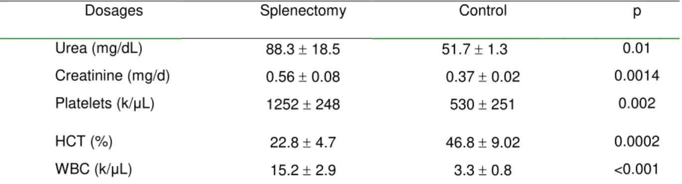
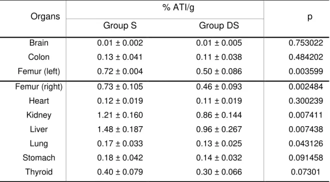
Table 1 - Results of 99mTc--DMSA biodistribution in each organ, and the p value to investigate the
existence of statistically significant differences between splenectomy and control groups.
Organs %ATI/g P
Splenectomy Control
Bladder 0.34 0.43 0.42 0.35 0.736
Blood 0.30 0.09 0.53 0.16 0.012
Femur 2.04 0.01 1.93 0.01 0.924
Heart 0.13 0.04 0.12 0.02 0.066
Kidney 2.10 0.30 3.27 0.81 0.041
Lung 0.23 0.05 0.12 0.06 0.008
Stomach 0.08 0.02 0.06 0.02 0.225
Thyroid 0.07 0.02 0.06 0.02 0.357
MeanStandard deviation; %ATI/g, percent of radioactivity per gram.
Table 2 – Values of urea, creatinine, platelets, hematocrit (HCT) and white blood cell (WBC) count at the 15th postoperative day following splenectomy, compared to controls.
Dosages Splenectomy Control p
Urea (mg/dL) 88.3 18.5 51.7 1.3 0.01
Creatinine (mg/d) 0.56 0.08 0.37 0.02 0.0014
Platelets (k/μL) 1252 248 530 251 0.002
HCT (%) 22.8 4.7 46.8 9.02 0.0002
WBC (k/μL) 15.2 2.9 3.3 0.8 <0.001
Meanstandard deviation; WBC, white blood cells; p-value from t test for independent samples; HCT, hematocrit.
27
day 15, the number of platelets and WBC was significantly higher in splenectomized rats than in controls (p<0.05). Hematocrit was significantly lower in splenectomized rats (22.84.7%) than in controls (46.89.02%; p=0.0002).
DISCUSSION
Splenectomy is recognized as having a significant immunomodulating effect, best described with regard to postsplenectomy sepsis and graft survival after renal transplant (Shaw & Print, 1989). During the last ten years, nonoperative management has became the primary method of preserving the spleen. Splenectomy is now required for around 50% of splenic trauma injuries (Velmahos et al., 2000). Sepsis following splenectomy, known as postsplenectomy infection syndrome and associated with a high mortality rate, is the most feared postoperative complication (Malangoni et al., 1984; Chhaikof & McCabe, 1985).
The white blood cell count (WBC) is an integral part of sepsis diagnosis. Early WBC trends alert the physician about the possibility of sepsis and allow prompt therapeutic response. However, postsplenectomy diagnosis of sepsis based on elevated WBC is confounded by the fact that leukocytosis is a physiologic response to splenectomy, similar to the phenomenon of elevated postsplenectomy platelet count (Bessler et al., 2004; Horowitz et al., 1992). Some reports suggest that postsplenectomy WBC in patients with sepsis is greater and more persistent than the WBC in patients without sepsis (Rutherford et al., 1994).
28
much feared complication of splenectomy. Therefore, confusion exists as to whether postsplenectomy leukocytosis should be considered a normal finding or a warning sign mandating treatment (Horowitz et al., 1992). Associated with hematuria, the animals in this study had significant kidney function impairment, due to the increase in urea and serum creatinine. These findings may explain the marked reduction in Tc-99m-DMSA uptake by renal parenchyma, reinforcing the characterization of postsplenectomy renal failure in rats.
Some physiological and pharmacological properties related to the radiobiocomplex sodium pertechnetate (Na99mTcO4) have been reported. After intravenous injection, it is weakly bound to serum proteins (70-80%). The pertechnetate ion (99mTcO4-) diffuses slowly through the capillary membranes to the interstitial fluids, from where it is cleared by various organs. In the kidneys, 99mTcO4 -is filtered in the glomeruli, but 86% -is reabsorbed in the proximal tubes. When associated with the radiotracer DMSA, the uptake will occur mainly in the kidneys. (Owunwanne et al., 1995; Zuckier et al., 2004).
Most investigators encourage using DMSA scintigraphy for the early diagnosis of many diseases, because of its cost-effectiveness and safety. A focal reduction or absence of uptake in one or more areas of the kidney is considered abnormal, indicating renal damage. In positive cases, DMSA scintigraphy is found to be highly sensitive in detecting multiple lesions (Piepsz et al., 1999; Stogianni et al., 2007). Besides leukocytosis and renal impairment evidenced by changes in urea and creatinine, we observed that the splenectomized animals had intense anemia associated with low hematocrit in all operated rats. In fact, they all showed clear signs of pale mucosa and hematuria at the 15th postoperative day. Certainly these hematologic findings contributed to the renal failure and to the low uptake of the radiopharmaceutical in the blood of splenectomized animals.
29 ACKNOWLEDEGMENT
The authors thank Michael Germain, from Canada, for his help in revision of English language.
RESUMO
Estudo com objetivo de avaliar se a esplenectomia altera a biodistribuição do DMSA-Tc-99m em ratos Wistar e a função renal. Usados 2 grupos: esplenectomia (n=6) e controle(n=6) animais não operados. Após 15 dias, administração de 0,1 ml de DMSA-Tc-99m via plexo orbital (0,66 MBq). Trinta minutos depois, retiradas biópsias rim, coração, pulmão, tireóide, estômago, bexiga, fêmur, sangue. Após pesadas as amostras, foi determinado o percentual de radioatividade/g (% ATI/g) em cada uma delas, com o Wizard Gama Counter Perkin-Elmer. Dosadas uréia e
creatinina sérica, hematócrito, plaquetas eleucócitos. Estatística pelo teste t, significância 0,05. Foi observada redução significante no %ATI/g no rim e sangue (p<0,05) dos animais esplenectomizados, aumento significante (p<0.05) da uréia (88,8±18,6mg/dL) e creatinina (0,56±0,08), comparado aos controles (51,5±1,6; 0,37±0,02mg/dL, respectivamente) assim como leucocitose, aumento de plaquetas e redução de hematócrito. Conclusão: em ratos a esplenectomia alterou a captação de DMSA-Tc-99m pelo rim, e a função renal.
REFERENCES
Araújo-Filho, I.; Rego A. C. M.; Brandão-Neto J.; Villarim-Neto A.; Egito E. S. T.; Azevedo I. M.; Medeiros A. C. (2007), Biodistribution of the Radiopharmaceutical Sodium Pertechnetate after Biliopancreatic Bypass with a Duodenal Switch. Braz. Arch. Biol. Technol., 50, 189-197.
30
Braga, A.C.S.; Oliveira, M.B.N.; Feliciano, G.D.; Reiniger, I.W.; Oliveria, J.F.; Silva, C.R.; Bernardo-Filho, M. (2000), The effect of drugs on the labeling of blood elements with Technetium-99m. Curr. Pharm. Des., 6, 1179-1191.
Brendolan, A.; Rosado, M.M.; Carsetti, R.; Selleri, L.; Dear, T.N. (2007), Development and function of the mammalian spleen. Bioessays., 29,166-177.
Chacon, D.A.; Araújo-Filho, I.; Villarim-Neto,A.; Rêgo,A.C.; Azevedo, I.M.; Bernardo-Filho, M.; Brandão-Neto, J.; Medeiros, A.C. (2007), Biodistribution of the radiophamarceutical sodium pertechnetate (Na99mTcO4) after massive small bowel resection in rats. Acta Cir. Bras., 22, 430-435.
Chaikof, E.L.; McCabe, C.J. (1985), Fatal overwhelming postsplenectomy infections. Am. J. Surg., 149,53-58.
Fonseca, A. S.; Frydman, J. N.; Rocha, V. C. and Bernardo-Filho, M. (2007), Acetylsalicylic acid decreases the labeling of blood constituents with technetium-99M. Acta. Biol. Hung., 2, 187-98.
Groshar, D.; Moskovitz, B.; Issaq, E.; Nativ, 0. (1997), Quantitative spect of DMSA uptake by the kidneys: assessment of reproducibility. Kidney Int., 52,817-820.
Horowitz, J.; Leonard, D.; Smith, J.; Brotman, S. (1992), Postsplenectomy leukocytosis: physiologic or an indicator of infection? Am. Surg., 58,387-390.
Majd, M.; Rushton, H.G. (1992), Renal cortical scintigraphy in the diagnosis of acute pyelonephritis. Semin. Nucl. Med., 22, 98–111.
Malangoni, M.A.; Dillon, L.D.; Klamer, T.W.; Condon, R.E. (1984), Factors influencing the risk of early and late serious infection in adults after splenectomy for trauma.
Surgery., 96,775-782.
Neves, RF., Moreno, SRF., Rebello, BM., Caldas LQA., Fonseca AS., Bernardo-Filho M., Medeiros AC. (2007), Effect of an Arctium lappa (burdock) extract on the labeling of blood constituents with technetium-99m and on the morphology of the red blood cells. Braz. Arch. Biol. Technol., 50, 167-174.
Owunwanne, A.; Patel, M.; Sadek, S. Preparation of radiopharmaceuticals. The handbook of radiopharmaceuticals. Chapman & Hall: London, 1995.
31
Petroianu, A.; Resende, V.; Da Silva, R.G. (2006), Late follow-up of patients submitted to subtotal splenectomy. Int. J. Surg., 4,172-178.
Piepsz, A.; Blaufox, M,D.; Gordon, I. (1999), Consensus on renal cortical scintigraphy in children with urinary tract infection. Semin. Nucl. Méd., 29,160–174.
Rebello, B.M.; Moreno, S.R.F.; Ribeiro, C.G., Neves, R.F., Fonseca, A.S., Caldas, L.Q.A., Bernardo-Filho, M., Medeiros, A.C. (2007), Effect of a peel passion fruit flour (Passiflora edulis f. flavicarpa) extract on the labeling of blood constituents with technetium-99m and on the morphology of red blood cells. Braz. Arch. Biol. Tchnol., 50, 153-159.
Rutherford, E.J.; Morris, J.A. Jr.; van Aalst, J.; Hall, K.S.; Reed, G.W.; Koestner, J.A. (1994), The white blood cell response to splenectomy and bacteraemia. Injury. 25,
289-292.
Shaw, J.H.; Print, C.G. (1989), Postsplenectomy sepsis. Br. J. Surg., 76:1074–1081.
Stogianni, A.; Nikolopoulos, P.; Oikonomou, I.; Gatzola, M.; Balaris, V.; Farmakiotis, D.; Dimitriadis, A. (2007), Childhood acute pyelonephritis: comparison of power Doppler sonography and Tc-DMSA scintigraphy. Pediatr. Radiol., 37, 685–690.
Stokland, E.; Jodal, U.; Sixt, R. (2007), Uncomplicated duplex kidney and DMSA scintigraphy in children with urinary tract infection. Pediatr. Radiol., 37, 826–828.
Tajiri, T.; Tate, G.; Enosawa, T.; Akita, H.; Ohike, N.; Masunaga, A.; Kunimura, T.; Mitsuya, T.; Morohoshi, T. (2007), Clinicopathological findings in fulminant-type pneumococcal infection: report of three autopsy cases. Pathol. Int., 57, 606-612.
Taylor, A. (1982), Quantitation of renal function with static imaging agents. Semin.
Nucl. Med., 12,330-344.
Velmahos, G.C.; Chan, L.S.; Kamel, E. (2000), Nonoperative management of splenic injuries. Arch. Surg.,135, 674-681.
Xavier-Holanda, C. M. C.; Holanda-Leite, R. C.; Nunes, R. A. S. N.; Oliveira, H. A.; Catanho, M. T. J. A.; Souza, G. M. L.; Bernardo-Filho, M. (2006), Effect of antimalarial drugas on the bioavailability of the methylenediphosphonic acid labeled with technetium99m (99mTc-MDP) in wistar rats. Braz. Arch. Biol. Technol., 49,
32
Zuckier, L.S.; Dohan, O.; Li, Y.; Chang, C.J.; Carrasco, N.; Dadachova, E. (2004), Kinetics of perrhenate uptake and comparative biodistribution of perrhenate, pertechnetate, and iodide by tissues in vivo. J. Nucl. Med., 45, 500-507.
4.2.2. Biodistribution of samarium-153-EDTMP in rats treated with docetaxel1
Biodistribuição de EDTMP-153-samário em ratos tratados com docetaxel
Arthur Villarim NetoI, Maria Kadja Meneses Torres AçucenaI, Kércia Regina Santos Gomes PereiraI, Amália Cínthia Meneses RêgoI, Ítalo Medeiros AzevedoII, Mário Bernardo-FilhoIII, Aldo Cunha MedeirosIV.
I Fellow PhD degree, Postgraduate Program in Health Sciences, Federal University of
Rio Grande do Norte (UFRN), Brazil.
II Statistician, Department of Surgery, Federal University of Rio Grande do Norte
(UFRN), Brazil.
III PhD, Full Professor, Department of Biophysics and Biometry, State University of
Rio de Janeiro, Brazil.
IV MD, PhD, Full Professor, Department of Surgery, UFRN, Brazil.
ABSTRACT
33
orbital plexus (25μCi). After 2 hours, the animals were killed and samples of the brain, thyroid, lung, heart, stomach, colon, liver, kidney and both femurs were removed. The percentage radioactivity of each sample (% ATI/g) was determined in an automatic gamma-counter (Wizard-1470, Perkin-Elmer, Finland). Results: On the 9th day after the administration of the 2nd chemotherapy cycle, the rats had a significant weight loss (314.50±22.09g) compared (p<0.5) to pre-treatment weight (353.66± 22.8). The % ATI/g in the samples of rats treated with samarium-153-EDTMP had a significant reduction in the right femur, left femur, kidney, liver and lungs of animals treated with docetaxel, compared to the control rats. Conclusion: The combination of docetaxel and samarium-153-EDTMP was associated with a lower response rate in the biodistribution of the radiopharmaceutical to targeted tissues. Further investigation into the impact of docetaxel on biodistribution of samarium-153-EDTMP would complement the findings of this study.
Key words: Docetaxel. Samarium. Bioavailability. Chemotherapy. Rat.
RESUMO
34
significante, passando de 353.66± 22.8g (controle/pré-tratamento) para 314,50±22,09g (p<0.5). Os % ATI/g nos órgãos dos ratos tratados com EDTMP-153–samário e docataxel tiveram redução significante nos fêmures direito e esquerdo, rim, fígado e pulmão, quando comparados com os não tratados com docetaxel. Conclusão: A combinação de docetaxel com EDTMP-153-samário foi associada com resposta mais baixa na biodistribuição do radiofármaco em órgãos alvo. Futuras investigações sobre o impacto do docetaxel na biodistribuição do EDTMP-153-samário poderão complementar os achados deste estudo.
Key words: Docetaxel. Samário. Biodistribuição. Quimioterapia. Rato.
________________________________________________________________ 1. Research performed at the Postgraduate Program in Health Sciences, UFRN, Brazil.
Introduction
Bone metastases are a frequent complication of cancer, occurring in up 70% of patients suffering from advanced breast or prostate cancer1. Bone metastases patients often present with severe bone pain, especially in the advanced stage of disease1,2. The major pain mechanism of small metastases appears to be the stimulation of nerve endings in the endosteum by a variety of chemical mediators. Larger bone metastases produce stretching of the periosteum, which leads to pain2. The resulting bone pain interferes with the patient’s quality of life and requires effective treatment. Unfortunately, various non-radiotherapeutic modalities such as analgesics, hormone therapy, orchidectomy, cytostatic and cytotoxic drugs, bisphosphonates, and surgery are not effective in all cases, especially in the late stage of the disease1,2. Systemic radionuclide therapy using boneseeking radiopharmaceuticals is considered a valuable and effective method for treating patients with widespread skeletal metastases and increased bone turnover, especially in patients with bone metastases from prostate and breast cancer 3,4,5.
35
Docetaxel is one of the taxane class chemotherapies that is being increasingly used to treat various solid tumors, including those of prostate and breast cancer. It acts by Bcl-2 protein inactivation in the metastatic cells, causing the death of tumor cells by apoptosis6. In patients with prostate cancer, docetaxel has been suggested in various studies as the first-line choice for treating metastatic hormone-refractory disease. This treatment increases survival, compared to previously accepted chemotherapy schemes 6.
A significant number of patients with metastatic bone disease are often subjected to a series of concomitant treatments, including the use of radioisotopes to relieve bone pain, associated with chemotherapy. Accordingly, the aim of this study was to assess the influence of docetaxel on the biodistribution of Sm-153-EDTMP, especially in the bones of rats, enabling a more thorough knowledge of its applicability.
Methods
We used male rats provided by the Health Science Center Vivarium, Federal University of Rio Grande do Norte, Brazil. The animals were randomly allocated into 2 groups of 6 rats each. They were weighed and placed in individual cages with water and food (Labina Purina ®) "ad libitum" and acclimatized in the lab for 7 days. The rats were kept under temperature (21ºC), humidity (60 - 70%) and lighting (12 / 12 hour cycle light / dark) control and they were handled in accordance with International Standards of Care and Use of Laboratory Animals, following the guidelines of the Brazilian College of Animal Experimentation.