Factors Controlling the Stable Nitrogen
Isotopic Composition (
δ
15
N) of Lipids in
Marine Animals
Elisabeth Svensson1¤*, Stefan Schouten1,2
*, Ellen C. Hopmans1, Jack J. Middelburg2, Jaap S. Sinninghe Damsté1,2
1Department of Marine Organic Biogeochemistry, NIOZ Royal Netherlands Institute for Sea Research, Den Burg (Texel), The Netherlands,2Department of Earth Sciences, Faculty of Geosciences, Utrecht University, Utrecht, The Netherlands
¤ Current address: Department of Geology & Planetary Science, University of Pittsburgh, Pittsburgh, Pennsylvania, United States of America
*esvensson@pitt.edu(ES);stefan.schouten@nioz.nl(SS)
Abstract
Lipid extraction of biomass prior to stable isotope analysis is known to cause variable changes in the stable nitrogen isotopic composition (δ15N) of residual biomass. However,
the underlying factors causing these changes are not yet clear. Here we address this issue by comparing theδ15N of bulk and residual biomass of several marine animal tissues (fish,
crab, cockle, oyster, and polychaete), as well as theδ15N of the extracted lipids. As
observed previously, lipid extraction led to a variable offset inδ15N of biomass (differences
ranging from -2.3 to +1.8‰). Importantly, the total lipid extract (TLE) was highly depleted in 15N compared to bulk biomass, and also highly variable (differences ranging from -14 to
+0.7‰). The TLE consisted mainly of phosphatidylcholines, a group of lipids with one nitro-gen atom in the headgroup. To elucidate the cause for the15N-depletion in the TLE, the δ15N of amino acids was determined, including serine because it is one of the main sources
of nitrogen to N-containing lipids. Serineδ15N values differed by -7 to +2‰from bulk
bio-massδ15N, and correlated well with the15N depletion in TLEs. On average, serine was less
depleted (-3‰) than the TLE (-7‰), possibly due to fractionation during biosynthesis of
N-containing headgroups, or that other nitrogen-N-containing compounds, such as urea and choline, or recycled nitrogen contribute to the nitrogen isotopic composition of the TLE. The depletion in15N of the TLE relative to biomass increased with the trophic level of the organisms.
Introduction
Stable isotopes of carbon and nitrogen (δ13C andδ15N) are routinely used in ecology to study a
wide range of subjects such as trophic interactions, energy flow, diet composition, feeding hab-its, and migration (see e.g. [1,2]; and references therein). Stable carbon isotopes are generally OPEN ACCESS
Citation:Svensson E, Schouten S, Hopmans EC, Middelburg JJ, Sinninghe Damsté JS (2016) Factors Controlling the Stable Nitrogen Isotopic Composition (δ15N) of Lipids in Marine Animals. PLoS ONE 11(1): e0146321. doi:10.1371/journal.pone.0146321
Editor:Candida Savage, University of Otago, NEW ZEALAND
Received:November 6, 2014
Accepted:December 16, 2015
Published:January 5, 2016
Copyright:© 2016 Svensson et al. This is an open access article distributed under the terms of the
Creative Commons Attribution License, which permits unrestricted use, distribution, and reproduction in any medium, provided the original author and source are credited.
Data Availability Statement:All relevant data are within the paper and its Supporting Information files. Data is also available atwww.pangaea.dedoi:10. 1594/PANGAEA.855456.
Funding:The research was funded by Waddenfonds for the project "Waddensleutels" (http://www. waddenfonds.nl/Home.2702.0.html(ES, SS, JSSD). The funder had no role in study design, data collection and analysis, decision to publish, or preparation of the manuscript.
used to distinguish between energy sources, such as terrestrial vs. aquatic, because differences inδ13C values are generated during primary production and largely conserved during
hetero-trophic processing (e.g. [3,4]). Nitrogen isotopes are mainly used to infer trophic transfers as each trophic step results in an increase in theδ15N signal of biomass (e.g. [5,6]). More recently,
theδ15N of specific amino acids has been used to estimate trophic levels [7–9].
For application of stable isotopes in trophic ecology, lipids are sometimes removed from bulk biomass prior to stable isotope analysis. This is done because lipids are depleted in13C compared to proteins and carbohydrates due to fractionation during lipid biosynthesis and because different tissues and organisms have variable lipid contents [10,11]. Interestingly, it has frequently been observed that lipid extraction also result in changes in theδ15N of the residual
biomass [12–15]. The change inδ15N bulk biomass with lipid extraction (Δδ15Nresidue-bulk) can
vary substantially (-2 to + 2.1‰[1]), but unlike the change inδ13C from lipid extraction (e.g.
[16–18]), no clear relationship betweenΔδ15Nresidue-bulkand parameters such as lipid content or
C:N ratio of the organisms has been found. The relatively large range inΔδ15Nresidue-bulkis
problematic as the change caused by lipid extraction is similar in magnitude to reported trophic fractionation (e.g. [6,19]) and diet-tissue discrimination factors (e.g. [6,15,20]). There is uncer-tainty about the cause for these changes. Some studies hypothesized that co-extraction of lipid-bound proteins leads to the removal of some amino acids (e.g. [13]), although this would imply that the extracted amino acids had a strongly different nitrogen isotopic composition compared to the remaining amino acids [14]. Another hypothesis is that lipid extraction leads to removal of cellular waste products (e.g. ammonia), which have quite different nitrogen isotopic compo-sitions than that of organic nitrogen [21]. However, no experimental evidence has been pro-vided to support these hypotheses.
In this study we investigate the cause of the changes seen in theδ15N following lipid
extrac-tion of tissues of several marine animals by determining theδ15N of bulk and residual,
lipid-free, biomass as well as of the total lipid extract (TLE). In addition, we identify the intact polar lipids present in the lipid extract to elucidate the sources of nitrogen to the lipid extracts. Finally, we determine theδ15N of amino acids to show thatδ15N of the total lipid extract relates
to theδ15N of serine and the source amino acid phenylalanine.
Materials and Methods
2.1 Samples
Four species of benthic invertebrates (Common cockle,Cerastoderma edule; Pacific oyster,
isotope analysis, samples were homogenized using a mortar and pestle or a ball mill grinder (Retsch, Düsseldorf, Germany).
2.2 Lipid extraction (TLE)
Total lipid extracts (TLE) were prepared as described in Svensson et al. [18]. In short, samples were extracted four times using dichloromethane (DCM) and methanol (MeOH) (2:1 v/v) and ultrasonication (1x10 min plus 3x5 min) and centrifuged at 1000xg, 2.5 min. Organic solvents were pipetted off after each extraction and combined as the total lipid extract (TLE). Residual biomass (lipid free) and TLEs were evaporated to dryness under a gentle stream of N2at room temperature.
2.3 Intact polar lipid analysis
Intact polar lipids (IPLs) were analyzed using HPLC/ESI/MS according to Sturt et al. [23] with some modifications as described in Schouten et al. [24] and Bale et al. [25]. In short, lipid extracts were re-dissolved in hexane:2-propanol:water (72:27:1, v/v/v) at a concentration of 2 mg mL-1and filtered through a 0.45μm regenerated cellulose (RC) filter (Alltech Associates Inc., Deerfield, IL) prior to injection. An Agilent 1200 series LC (Agilent, San Jose, CA),
equipped with thermostatted auto-injector and column oven, and coupled to a Thermo LTQ XL linear ion trap with Ion Max source with electrospray ionization (ESI) probe (Thermo Scientific, Waltham, MA), was used. Separation was achieved on a LiChrospher diol column (250 x 2.1 μm, 5μm particles; Alltech Associates Inc., Deerfield, IL) maintained at 30°C. The following elu-tion program was used with a flow rate of 0.2 mL min-1: 100% A for 1 min, followed by a linear gradient to 66% A: 34% B in 17 min, maintained for 12 min, followed by a linear gradient to 35% A: 65% B in 15 min, where A = hexane:2-propanol:formic acid:NH3aq(14.8M)
(79:20:0.12:0.04, v/v/v/v) and B = 2-propanol:water:formic acid:NH3aq(14.8M) (88:10:0.12:0.04, v/v/v/v). Total run time was 60 min with a re-equilibration period of 20 min in between runs. The lipid extracts were analyzed by an MS routine where a positive ion scan (m/z400–2000)
was followed by a data dependent MS2experiment where the base peak of the mass spectrum was fragmented (normalized collision energy 25, isolation width 5.0, activation Q 0.175). This was followed by a data dependent MS3experiment where the base peak of the MS2spectrum was fragmented under identical fragmentation conditions. This process was repeated on the 2nd to 4thmost abundant ions of the initial mass spectrum. Major IPL classes were identified as described in Brandsma et al. [26].
2.4 Stable isotope analysis
The stable nitrogen isotopic ratio was determined on bulk biomass (δ15Nbulk) and on residual
biomass after extraction (δ15Nresidue), as well as on the total lipid extracts (δ15NTLE). For δ15Nbulkandδ15Nresidue, ca. 0.4–0.8 mg of freeze dried, homogenized sample was weighed into
tin cups. These samples were analyzed forδ15N and percent total organic carbon (%TOC) and
percent total nitrogen (%TN) content in duplicate using isotope ratio monitoring mass spec-trometry (IRMS) with a Delta V Advantage-IRMS coupled to a Flash 2000 elemental analyzer (Thermo Scientific). Total lipid extracts were dissolved in ethyl acetate and pipetted into tin cups for a final weight of ca. 0.4 mg forδ15N determination. Ethyl acetate was allowed to
evapo-rate completely at room temperature (minimum 6 h) before folding the cups for analysis. Due to the low amount of nitrogen compared to carbon in the lipid extracts, the TLE fraction was analyzed forδ15N on a Delta XL isotope ratio MS (Thermo Finnigan) coupled to a Flash 1112
Stable isotope ratios are expressed using theδnotation in units per mil according to:
dð‰Þ¼ Rsample
Rstandard 1
where R =15N/14N, and expressed in per mil versus air. An acetanilide standard with aδ15N
value of 1.18‰(standard obtained from Arndt Schimmelmann, Indiana University; [27]), and
known %TOC and %TN content, was used for calibration. The average repeatability ofδ15N
determination was 0.2‰based on repeated analysis of the acetanilide standard. The pooled
standard deviation of replicate measurements (n = 2–5) were0.6‰for bulk and lipid-free
biomass, and0.9‰for total lipid extract.
2.5 Amino acid nitrogen isotope analysis
The method used for amino acid isotope analysis was a modified version of the method from Metges et al.[28] and Chikaraishi et al. [29]. In short, tissue samples were hydrolyzed in 6M HCl at 110°C for ca. 18 h. Hydrolyzates were then filtered (GHP Nanosep centrifugal filtration devices, Pall Co), and defatted using n-hexane:DCM (3:2, v/v). Samples were dried under a flow of N2with repeated additions of methanol. Carboxylic acid groups of amino acids were esterified by addition of 200μl 2-propanol:acetyl chloride (4:1, v/v) and heating at 110°C (2 h). Amine groups were subsequently acetylated using DCM:pivaloyl chloride (200μl; 4:1,v/v) at 110°C (2 h). After each derivatization step, any leftover reagents were removed by addition of DCM and drying under a gentle stream of N2(2x). Derivatized amino acids were dissolved in ca. 200μl bidistilled water and extracted using n-hexane:DCM (3:2, v/v), dried over MgSO4, diluted to a suitable concentration with dried (MgSO4) and de-gassed DCM, and stored at -20°C in amber vials until analysis.
The N-pivaloyl/2-propyl derivatives of amino acids were analyzed by gas chromatography/ combustion/isotope ratio mass spectrometry (GC/C/IRMS) using a Thermo Delta V Advan-tage connected to an Agilent 6890 GC. The GC and IRMS were interfaced via a combustion furnace (980°C), reduction furnace (650°C), and a liquid nitrogen cold trap to remove CO2. Separation of the derivatized amino acids was achieved on a DB-5ms column (Agilent J&W, 60 m x 0.32 mm i.d., 0.50μm film thickness; Agilent Technologies), using the following tempera-ture program: Initial temperatempera-ture 70°C for 1 min; ramp up to 140°C at 10°C min-1, dwell for 5 min; ramp up to 190°C at 2°C min-1; ramp up to 300°C at 10°C min-1, hold for 10 min. Carrier gas was helium at a continuous flow of 2 ml min-1(29 cm s-1). Injection volumes ranged from 0.4–2μl. An in-house standard mixture consisting of five amino acids (glycine, norleucine,
glu-tamic acid, phenylalanine, and tyrosine) with knownδ15N values (determined offline) was
used to evaluate daily system performance. Long-term reproducibility, based on the standard deviation of multiple injections (n = 52) of the in-house standard mixture was 0.7 (glycine), 0.9 (norleucine), and 1.2‰(phenylalanine, glutamic acid, and tyrosine).
2.6 Data analysis
Correlations were evaluated using Pearson correlation analysis. The non-parametric Kruskal-Wallis test was used to evaluate differences in isotopic compositions, because of the relative small dataset. For the few cases with larger sample size, a Student t-test was used and the results were similar. Data were non-transformed and evaluated at a 5% significance level. Statistical tests were done using XL-Stat version 2015.4.01.20780.
2.7 Ethical statement
This study was done with permission from the Dutch Fisheries Inspection of the Ministry of Agriculture, Nature and Food Quality (Visserijinspectie) and reviewed and approved by the Animal Experiments Committee (Dierenexperimentencommisie, DEC) under DEC protocol NIOZ 2010.03.
Results and Discussion
3.1
δ
15N contents of extracted biomass and lipid extracts
Lipids were extracted from biomass of several species of aquatic animals and theδ15N values
were determined both before (bulk biomass) and after lipid extraction (residual, lipid-free bio-mass), as well as that of the total lipid extract (TLE). Bulk biomassδ15N values for different
spe-cies and tissue types ranged, on average, from 6.7 to 17‰(Table 1). Theδ15N of residual biomass
after lipid extraction differed by -0.9 to + 1.8‰compared to bulk biomass (Δδ15Nresidue-bulk;
Fig 1andS1 Table) which is consistent with previous studies [30,31].
The TLEs were depleted in15N compared to bulk biomass in all samples except one (whole lugworm,Δδ15NTLE-bulk= +0.7‰), withΔδ15NTLE-bulkvalues ranging from -14 to +0.7‰(Fig 1,
Table 1, andS1 Table). The lipid content of the investigated tissues ranged from 1–40% with the
majority having lipid contents10% and lipids thus form a relative small portion of the total biomass. Furthermore, the relative amount of N in the lipid extract was small in most cases (average C:N ratio ranging from 14 to 63;Table 1) compared to bulk biomass (C:N ratio ranging from 3–8;Table 1) and highly variable. No correlation was observed betweenΔδ15Nresidue-bulk
and either the C:N ratio or %lipid of bulk biomass (Pearson correlation, r = -0.130 [P = 0.417], n = 40, and 0.239 [P = 0.132], n = 40, respectively).
Although there is a large variability in15N of the lipid extracts, on average they were signifi-cantly depleted in15N compared to biomass by approx. -7‰(Kruskal-Wallis test, p<0.001,
n = 26). TheΔδ15Nresidue-bulkof lipid-free biomass also varies, but the residue was on average
sig-nificantly enriched in15N by 0.4‰(Kruskal-Wallis test, p<0.001, n = 41) compared to the
Table 1. Ranges ofδ15N values and C:N ratios for bulk biomass, residual (lipid-free) biomass and lipids (total lipid extract) per species and tissue
type.n = number of individuals analyzed. Standard deviation ofδ15N values from replicate measurements were for bulk and lipid-free biomass0.6‰, for total lipid extract0.9.
Bulk biomass Residual biomass Total lipid extract
Species Tissue n %lipids δ15N C:N n δ15N C:N n δ15N C:N
Atlantic herring Gill 6 19–32 10.6–14.4a 4.8–8.0a 6 10.7–13.7 3.0–3.3 4 0.4–9.2 62.9
Muscle 6 7–40 11.4–16.2a 3.3–6.1a 6 11.4–16.3 2.4–3.1 5 -0.5–3.1 14.6–23.2 Brown trout Gill 6 4–11 13.9–16.3a 3.7–4.0a 6 14.2–16.7 3.2–3.5 3 7.2–11.0 19.5–42.2
Muscle 7 5–25 13.7–16.5a 3.2–5.1a 7 14.4–17.0 3.0–3.2 5 4.3–9.7 21.3
Twait shad Gill 2 6–9 14.7–16.7a 4.2–4.3a 2 14.8–17.0 3.3–3.6 n.d n.d
Muscle 2 6–7 15.8–17.1a 3.2–3.2a 2 16.7–17.9 3.1–3.1 1 10.3 14.1 Green shore crab Muscle 3 2–6 13.2–15.8 3.7–5.3 3 13.4–16.5 3.3–4.4 1 8.2 n.d Common cockle Muscle 5 4–5 11.3–12.6 4.1–4.9 5 11.7–12.7 3.7–4.3 2 6.9–7.2 n.d Pacific oyster Muscle 2 2–6 12.3–12.3 3.0–3.3 2 13.3–13.5 3.2–3.3 2 6.0–7.0 n.d
Lugworm Head 2 3–5 11.7–12.4 4.2–4.7 2 10.8–11.3 3.7–4.0 2 8.4–8.5 n.d
Whole 1 1 6.7 3.1 1 6.6 3.5 1 7.4 n.d
aData from Svensson et al. [18].
n.d. = not determined.
original biomass. This suggests that, in general, lipid extracts contain15N depleted nitrogen, which induces small but significant changes in15N in the residual biomass. Remarkably, however, there is a large variability in bothδ15N and C:N ratio of lipid extracts as well asΔδ15Nresidue-bulk
(Table 1). This may suggest that there is a mixture of different nitrogen sources in the lipid extract, such as lipids, lipid-bound protein and urea [12–14], all present in variable ratios and
with differentδ15N values. Below we investigate theδ15N of lipids as a potential source for the
variable depletion in15N in the lipid extract.
3.2 Sources of
15N depleted nitrogen in lipid extracts
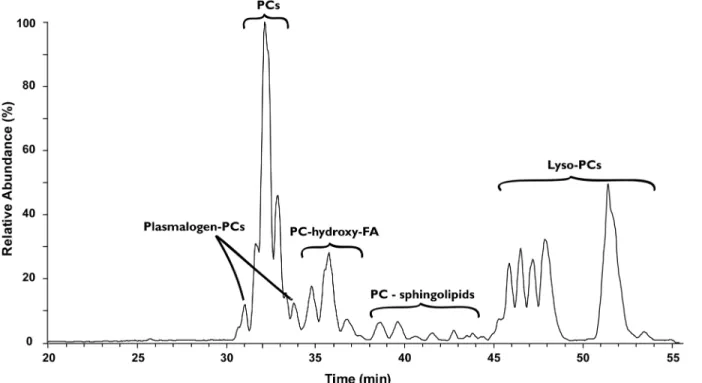
To investigate the origin of the15N depleted nitrogen in the TLE we investigated its lipid com-position by HPLC/ESI/MS. The majority of identified lipids in the TLEs comprised the nitro-gen-containing phosphatidylcholines (PC;Fig 2andS2 Table), which is a common lipid class in animal tissue (e.g. [32]). Taurine conjugated lipids as well as phosphatidylethanolamines (PE), betaines, and phosphatidylinositols (PI) were also detected in some species. With the exception of PI, all of these lipids contain one nitrogen atom in the lipid headgroup (Fig 3). The dominance of nitrogen-containing lipids suggests that they form an important source of nitrogen to the lipid extracts, although a contribution of nitrogen from other sources, such as extractable proteins and/or nitrogen containing waste products, cannot be excluded. Some evi-dence for the latter comes from the C:N ratio of the lipid extracts: the C:N ratio of some lipid extracts (Table 1) were lower than the theoretical C:N ratios of the identified lipids (25–50)
suggesting the presence of additional nitrogen.
To assess whether the N-containing headgroups, in particular PC, were15N-depleted com-pared to bulk biomass, as observed for the lipid extracts, the biosynthetic source of nitrogen for Fig 1.δ15N values of residual biomass, the total lipid extract and serine normalized to bulk biomass.Median values (symbols) and ranges of differences inδ15N of residual biomass, the total lipid extract (TLE) and serine compared to bulk biomass (Δδ15Nfraction-bulk) for the different animals.
these headgroups was considered. Headgroups like PC and PE derive their nitrogen either from amino acids, choline or from recycled nitrogen within the cell [33,34]. The amino acid serine in particular is interesting as it is one of two main sources of cellular nitrogen to many nitrogen-containing lipids (the other being choline), while for PE it is the only nitrogen source. If serine contributes a large fraction of nitrogen to the lipids during biosynthesis, theδ15N
val-ues of the precursor serine should be reflected in the lipid product. We, therefore, determined theδ15N of serine in a selection of tissues (Table 2). Serine was indeed depleted in15N
com-pared to bulk biomass (Fig 1) withΔδ15Nser-bulkranging from -1 to -8‰. This agrees with
pre-vious observations in animals that serine generally is15N depleted compared to total biomass (e.g. [35–37]). The range of depletion in15N of serine was highly variable and resembled the
variation inΔδ15NTLE-bulkof the total lipid extract. Indeed, theΔδ15Nser-bulkof serine is strongly
and significantly correlated withΔδ15NTLE-bulkof TLE (Pearson correlation, r = 0.94,
[P<0.001], n = 8). The strong correlation betweenΔδ15Nser-bulkandΔδ15NTLE-bulkis in line
with the biochemical evidence that serine is indeed an important source of nitrogen to the lipid pool and shows as well that theδ15N of lipid extracts were mainly determined by theδ15N of
N-containing lipids. It should be noted, however, that the extent of depletion of serine was less than for the lipids by 3–4‰in some tissues (Fig 1). These differences inδ15N between serine
and lipid extracts may be due to one or a combination of several factors: (i) a (large) contribu-tion of nitrogen recycled within the cell or originating from dietary choline, (ii) fraccontribu-tionacontribu-tion during biosynthesis of the headgroups from serine or choline. Further research using e.g. com-pound specific analysis of headgroups of lipids may shed further light on this.
The cause for the observed large variability in the15N depletion of N-containing lipids is not clear. However, it is noticeable that the organisms with higher trophic levels, such as fish, show a larger depletion in15N than those associated with lower trophic levels, such as Fig 2. LC–MS chromatogram showing identified intact polar lipids in brown trout (Salmo trutta) gill tissue.Base peak LC–MS chromatogram
(Gaussian smoothed) of MS1of intact polar lipids (IPLs) in brown trout (Salmo trutta) gill tissue showing the prevalence of the nitrogen-containing IPL phosphatidylcholine. PC = phosphatidylcholine.
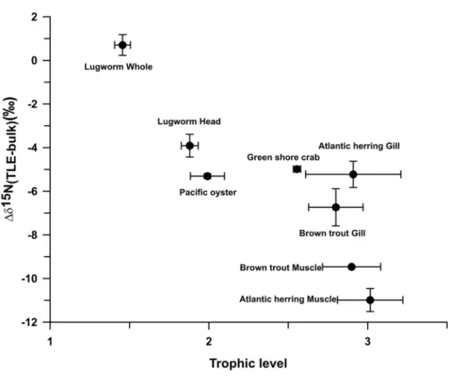
lugworms. Indeed, when we calculate trophic levels for the different organisms based on the
δ15N of glutamic acid and phenylalanine, using the equation of Chikaraishi et al. [29] for
marine food webs, we observe an increasing depletion in15N of the lipid extract relative to bio-mass with increasing trophic level (Fig 4). Further studies are needed to explore the cause for the large variability in the15N depletion of N-containing lipids and whether the observed cor-relation with trophic level is causal.
Conclusions
Our study shows that lipid extracts are generally highly depleted in15N compared to bulk bio-mass. The majority of lipids in the lipid extracts from biomass in this study consisted of glycer-ophospholipids with a phosphatidylcholine headgroup, a nitrogen-containing lipid which is Fig 3. Structures of identified intact polar lipids in lipid extracts of animal tissues.PC = phosphocholine.
Table 2. δ15N values of amino acids.Avg and s.d. = average and standard deviation of n injections. ALA = alanine; ASP = aspartic acid; GLU = glutamic acid; GLY = glycine; ILE = isoleucine; LEU = leucine; LYS = lysine; OH-PRO = hydroxy-proline; PHE = phenylalanine; PRO = proline; SER = serine; THR = threonine; TYR = tyrosine; VAL = valine.
Brown trout muscle
Brown trout gill
Atlantic herring muscle
Atlantic herring gill
Green shore crab
Pacific oyster Lugworm head
Lugworm whole
n Avg s.d. n Avg s.d. n Avg s.d. n Avg s.d. n Avg s.d. N Avg s.d. n Avg s.d. n Avg s.d.
ALA 6 29.3 0.9 8 27.5 0.6 4 26.3 0.9 3 22.5 0.5 5 23.3 2.2 4 23.0 0.7 3 20.9 0.9 4 18.1 1.4 ASX 5 22.6 1.1 8 21.4 0.7 3 21.7 0.7 3 18.0 0.9 5 20.2 1.3 4 19.9 0.3 3 20.0 1.7 4 18.2 1.2 GLU 6 26.5 0.7 8 27.2 0.5 4 26.6 1.0 3 23.9 0.5 5 24.1 0.9 4 22.7 0.2 3 19.4 1.1 4 17.4 0.9 GLY 6 7.6 0.8 8 8.8 0.3 4 5.0 0.8 3 5.2 0.9 5 10.0 1.4 4 9.8 0.6 3 9.9 0.9 4 9.1 1.0 ILE 5 26.0 0.8 5 25.7 0.6 3 25.7 0.1 n.d. 5 18.3 1.8 4 19.1 0.7 3 16.7 0.7 2 16.2 1.4 LEU 6 26.3 0.7 8 26.9 0.8 4 25.8 0.7 3 21.9 1.1 5 19.8 1.9 4 19.1 0.8 3 17.7 0.5 5 16.7 0.8 LYS 6 4.0 0.8 8 2.8 0.3 4 5.0 0.4 n.d. n.d. 4 8.0 0.8 3 3.6 0.5 2 2.1 0.2
OH-PRO n.d. 8 22.9 0.9 n.d. 3 20.5 1.8 n.d. n.d. n.d. n.d.
PHE 6 8.6 0.9 6 9.9 0.8 3 7.6 1.1 3 6.0 2.4 5 8.9 1.0 4 11.7 0.7 3 9.3 0.8 2 11.0 0.5
PRO n.d. 6 27.6 0.7 n.d. n.d. n.d. n.d. n.d. n.d.
SER 3 7.7 0.5 6 8.4 0.7 3 5.9 1.0 2 8.3 0.5 2 10.7 1.6 4 10.3 0.9 3 8.4 1.5 2 8.8 1.0
THR 5 -15.3 3.2 2 -22.2 0.0 3 -17.5 2.4 n.d. 1 -4.7 1 4.6 2 4.7 1.6 2 3.0 0.2
TYR 6 15.5 1.5 8 15.5 0.7 4 14.0 0.9 3 8.7 0.8 5 11.4 0.5 4 15.0 1.1 3 11.3 0.3 2 11.6 0.9 VAL 7 27.6 0.9 8 28.5 1.4 4 27.2 1.2 3 23.2 1.1 5 21.2 2.3 4 22.4 0.5 3 19.6 1.2 4 18.5 2.3 n.d. = not detected.
doi:10.1371/journal.pone.0146321.t002
Fig 4. Difference inδ15N of lipid extracts (TLE) and bulk biomass (Δδ15NTLE-bulk) plotted against trophic levels.Δδ15NTLE-bulkdata points represent averages of several individuals with the error bar
commonly found in animal tissues. The15N depletion in the lipids is likely a reflection of the
δ15N of the biosynthetic nitrogen sources such as the amino acid serine, which is also depleted
in15N compared to bulk biomass. The depletion ofδ15N of lipid extracts compared to bulk
bio-mass seems to be higher with organisms of a higher trophic level, the reasons for which need to be further explored.
Supporting Information
S1 Table. Stable nitrogen isotope values (δ15N), C:N ratios, and lipid content of bulk and residual biomass, and total lipid extracts of samples used in this study.Samples highlighted in yellow indicate those used for amino acid analysis.
(XLSX)
S2 Table. Fatty acid composition, indicated by number of carbons and degree of unsatura-tion, of most abundant intact polar lipids (IPLs) in different aquatic species in this study.
n.d. = not detected. PC: Phosphatidylcholine; PE: Phosphatidylethanolamine; MMPE: mono-methyl–PE; PI: Phosphatidylinositol; Plasmalogen: Fatty acid with vinyl linkage to glycerol
backbone; Lyso: IPL where one fatty acid has been removed (lysed); Sphingo: Sphingobase. See
Fig 3for structures. (DOCX)
Acknowledgments
We thank two anonymous reviewers for their comments, which substantially improved the manuscript. This study is part of the Waddensleutels project, funded by the Waddenfonds. We thank the NIOZ SIBES team for providing benthic samples, and Vânia Freitas, Hans Witte and Henk van der Veer for providing fish samples. Monique Verweij, Jort Ossebaar and Kevin Donkers are thanked for analytical assistance.
Author Contributions
Conceived and designed the experiments: ES SS. Performed the experiments: ES. Analyzed the data: ES SS ECH JJM. Contributed reagents/materials/analysis tools: SS ECH JSSD. Wrote the paper: ES SS ECH JJM JSSD.
References
1. Boecklen WJ, Yarnes CT, Cook BA, James AC. On the use of stable isotopes in trophic ecology. Annu Rev Ecol Evol Syst. 2011; 42: 411–440. doi:10.1146/annurev-ecolsys-102209-144726
2. Middelburg JJ. Stable isotopes dissect aquatic food webs from the top to the bottom. Biogeosciences. 2014; 11: 2357–2371. doi:10.5194/bg-11-2357-2014
3. Fry B, Scalan RS, Parker PL. Stable carbon isotope evidence for two sources of organic matter in coastal sediments: seagrasses and plankton. Geochim Cosmochim Acta. 1977; 41: 1875–1877. doi:
10.1016/0016-7037(77)90218-6
4. Hobson KA. Tracing origins and migration of wildlife using stable isotopes: a review. Oecologia. 1999; 120: 314–326.
5. DeNiro MJ, Epstein S. Influence of diet on the distribution of nitrogen isotopes in animals. Geochim Cosmochim Acta. 1981; 45: 341–351. doi:10.1016/0016-7037(81)90244-1
6. Minagawa M, Wada E. Stepwise enrichment of15N along food chains: Further evidence and the
rela-tion betweenδ15N and animal age. Geochim Cosmochim Acta. 1984; 48: 1135–1140. doi:10.1016/ 0016-7037(84)90204-7
8. Chikaraishi Y, Kashiyama Y, Ogawa NO, Kitazato H, Ohkouchi N. Metabolic control of nitrogen isotope composition of amino acids in macroalgae and gastropods: implications for aquatic food web studies. Mar Ecol Prog Ser. 2007; 342: 85–90. doi:10.3354/meps342085
9. McCarthy MD, Benner R, Lee C, Fogel ML. Amino acid nitrogen isotopic fractionation patterns as indi-cators of heterotrophy in plankton, particulate, and dissolved organic matter. Geochim Cosmochim Acta. 2007; 71: 4727–4744. doi:10.1016/j.gca.2007.06.061
10. DeNiro MJ, Epstein S. Mechanism of carbon isotope fractionation associated with lipid synthesis. Sci-ence. 1977; 197: 261–263. PMID:327543
11. Monson KD, Hayes JM. Carbon isotopic fractionation in the biosynthesis of bacterial fatty acids. Ozono-lysis of unsaturated fatty acids as a means of determining the intramolecular distribution of carbon iso-topes. Geochim Cosmochim Acta. 1982; 46: 139–149. doi:10.1016/0016-7037(82)90241-1
12. Pinnegar JK, Polunin NVC. Differential fractionation ofδ13C andδ15N among fish tissues: implications for the study of trophic interactions. Funct Ecol. 1999; 13: 225–231. doi:10.1046/j.1365-2435.1999.
00301.x
13. Sotiropoulos MA, Tonn WM, Wassenaar LI, Sotiropoulos MA, Tonn WM, Wassenaar LI. Effects of lipid extraction on stable carbon and nitrogen isotope analyses of fish tissues: potential consequences for food web studies, Effects of lipid extraction on stable carbon and nitrogen isotope analyses of fish tis-sues: potential consequences for food web studies. Ecol Freshw Fish. 2004; 13: 155–160. doi:10. 1111/j.1600-0633.2004.00056.x
14. Murry BA, Farrell JM, Teece MA, Smyntek PM. Effect of lipid extraction on the interpretation of fish com-munity trophic relationships determined by stable carbon and nitrogen isotopes. Can J Fish Aquat Sci. 2006; 63: 2167–2172. doi:10.1139/f06-116
15. Elsdon TS, Ayvazian S, McMahon KW, Thorrold SR. Experimental evaluation of stable isotope fraction-ation in fish muscle and otoliths. Mar Ecol Prog Ser. 2010; 408: 195–205. doi:10.3354/meps08518
16. McConnaughey T, McRoy CP. Food-web structure and the fractionation of carbon isotopes in the Bering Sea. Mar Biol. 1979; 53: 257–262. doi:10.1007/BF00952434
17. Logan JM, Jardine TD, Miller TJ, Bunn SE, Cunjak RA, Lutcavage ME. Lipid corrections in carbon and nitrogen stable isotope analyses: comparison of chemical extraction and modelling methods. J Anim Ecol. 2008; 77: 838–846. doi:10.1111/j.1365-2656.2008.01394.xPMID:18489570
18. Svensson E, Freitas V, Schouten S, Middelburg JJ, van der Veer HW, Sinninghe Damsté JS. Compari-son of the stable carbon and nitrogen isotopic values of gill and white muscle tissue of fish. J Exp Mar Biol Ecol. 2014; 457: 173–179. doi:10.1016/j.jembe.2014.04.014
19. Vander Zanden MJ, Rasmussen JB. Variation inδ15N andδ13C trophic fractionation: implications for aquatic food web studies. Limnol Oceanogr. 2001; 2061–2066.
20. Herzka SZ, Holt GJ. Changes in isotopic composition of red drum (Sciaenops ocellatus) larvae in response to dietary shifts: potential applications to settlement studies. Can J Fish Aquat Sci. 2000; 57: 137–147. doi:10.1139/f99-174
21. Logan JM, Lutcavage ME. A comparison of carbon and nitrogen stable isotope ratios of fish tissues fol-lowing lipid extractions with non-polar and traditional chloroform/methanol solvent systems. Rapid Commun Mass Spectrom. 2008; 22: 1081–1086. doi:10.1002/rcm.3471PMID:18327856
22. van der Veer H, Koot J, Aarts G, Dekker R, Diderich W, Freitas V, et al. Long-term trends in juvenile flat-fish indicate a dramatic reduction in nursery function of the Balgzand intertidal, Dutch Wadden Sea. Mar Ecol Prog Ser. 2011; 434: 143–154. doi:10.3354/meps09209
23. Sturt HF, Summons RE, Smith K, Elvert M, Hinrichs K-U. Intact polar membrane lipids in prokaryotes and sediments deciphered by high-performance liquid chromatography/electrospray ionization multi-stage mass spectrometry—new biomarkers for biogeochemistry and microbial ecology. Rapid Com-mun Mass Spectrom. 2004; 18: 617–628. doi:10.1002/rcm.1378PMID:15052572
24. Schouten S, Hopmans EC, Baas M, Boumann H, Standfest S, Konneke M, et al. Intact membrane lipids of“Candidatus Nitrosopumilus maritimus”, a cultivated representative of the cosmopolitan mesophilic Group I Crenarchaeota. Appl Environ Microbiol. 2008; 74: 2433–2440. doi:10.1128/AEM.01709-07
PMID:18296531
25. Bale NJ, Villanueva L, Hopmans EC, Schouten S, Sinninghe Damsté JS. Different seasonality of pelagic and benthic Thaumarchaeota in the North Sea. Biogeosciences. 2013; 10: 7195–7206. doi:10.
5194/bg-10-7195-2013
27. Schimmelmann A, Albertino A, Sauer PE, Qi H, Molinie R, Mesnard F. Nicotine, acetanilide and urea multi-level2H-,13C- and15N-abundance reference materials for continuous-flow isotope ratio mass
spectrometry. Rapid Commun Mass Spectrom. 2009; 23: 3513–3521. doi:10.1002/rcm.4277PMID:
19844968
28. Metges CC, Petzke K, Hennig U. Gas chromatography/combustion/isotope ratio mass spectrometric comparison of N -acetyl- and N-pivaloyl amino acid esters to measure15N isotopic abundances in
phys-iological samples: a pilot study on amino acid synthesis in the upper gastro-intestinal tract of minipigs. J Mass Spectrom. 1996; 31: 367–376. doi:10.1002/(SICI)1096-9888(199604)31:4<
367::AID-JMS310>3.0.CO;2-VPMID:8799283
29. Chikaraishi Y, Ogawa NO, Kashiyama Y, Takano Y, Suga H, Tomitani A, et al. Determination of aquatic food-web structure based on compound-specific nitrogen isotopic composition of amino acids. Limnol Oceanogr Methods. 2009; 7: 740–750. doi:10.4319/lom.2009.7.740
30. Bodin N, Le Loc’h F, Hily C. Effect of lipid removal on carbon and nitrogen stable isotope ratios in crus-tacean tissues. J Exp Mar Biol Ecol. 2007; 341: 168–175. doi:10.1016/j.jembe.2006.09.008
31. Ingram T, Matthews B, Harrod C, Stephens T, Grey J, Markel R, et al. Lipid extraction has little effect on theδ15N of aquatic consumers. Limnol Oceanogr Methods. 2007; 5: 338–343.
32. Cole LK, Vance JE, Vance DE. Phosphatidylcholine biosynthesis and lipoprotein metabolism. Biochim Biophys Acta BBA—Mol Cell Biol Lipids. 2012; 1821: 754–761. doi:10.1016/j.bbalip.2011.09.009
33. Elwyn D, Weissbach A, Henry SS, Sprinson DB. The biosynthesis of choline from serine and related compounds. J Biol Chem. 1955; 213: 281–295. PMID:14353926
34. Stekol JA. Biosynthesis of Choline and Betaine. Am J Clin Nutr. 1958; 6: 200–215. PMID:13533306
35. Germain LR, Koch PL, Harvey J, McCarthy MD. Nitrogen isotope fractionation in amino acids from har-bor seals: implications for compound-specific trophic position calculations. Mar Ecol Prog Ser. 2013; 482: 265–277. doi:10.3354/meps10257
36. Chikaraishi Y, Steffan SA, Ogawa NO, Ishikawa NF, Sasaki Y, Tsuchiya M, et al. High-resolution food webs based on nitrogen isotopic composition of amino acids. Ecol Evol. 2014; 4: 2423–2449. doi:10. 1002/ece3.1103PMID:25360278
37. Hoen DK, Kim SL, Hussey NE, Wallsgrove NJ, Drazen JC, Popp BN. Amino acid15N trophic


