212 Turk Neph Dial Transpl 2013; 22 (2): 212-214 Olgu Sunumu/Case Report
Can Vascular Access During Hemodialysis Cause Extremity Loss?
Hemodiyaliz S
ı
ras
ı
nda Damar Giri
ş
imi Ekstremite Kayb
ı
na Neden
Olabilir mi?
Nurkay KATRANCIOĞLU Oğuz KARAHAN
Umut Serhat SANRI
Cumhuriyet University School of Medicine, Department of Cardiovascular Surgery, Sivas, Turkey
doi: 10.5262/tndt.2013.1002.14
Correspondence Address:
Nurkay KATRANCIOĞLU
Cumhuriyet Üniversitesi Tıp Fakültesi, Kalp Damar Cerrahisi AD, Sivas, Turkey Phone : +90 505 242 42 05
E-mail : [email protected] Received : 01.10.2011
Accepted : 10.11.2011
vascular access during hemodialysis stresses vascular structures and can lead to pseudoaneurysm formations and circulation disorders in tissues (1,2). Native vascular grafts are more resistant than synthetic grafts for interventions such as recurrent cannulations. However, permanent applications can cause deterioration even in the ndothelial integrity of a native vascular graft (1-3). Pseudoaneurysm is one of the most important complication that can cause
INTRODUCTION
Hemodialysis is the most common method used in patients with chronic renal failure. Application of hemodialysis seems to be a simple intervention. Long-term vascular access, which can cause various complications in the vascular bed, is mandatory in patient with hemodialysis (1). However, the vascular access complications are a frequent cause of hospitalization in chronic hemodialysis patients (2). Repetitive
ABSTRACT
There is always the possibility of complications of vascular access in patients with hemodialysis. The pseudoaneurysm related with accidental arterial puncture is one of the most important complications in patients with hemodialysis and it can cause severe clinic manifestations that may go all the way to limb loss. We describe a 78-year-old woman undergoing regular dialysis treatment with renal failure. The patient had severe left arm pain starting suddenly after hemodialysis, with rapidly progressing cyanosis, and compartment syndrome caused by brachial artery pseudoaneurysm. Urgent surgical procedure was performed. The pseudoaneurysm was successfully repaired, left arm arterial circulation reestablished, and fasciotomy performed on the left arm for compartment syndrome. In the second session, debridement was performed and dermal graft applied to the necrotic lesions. The patient was discharged with full recovery within 2 months following the treatment. In this case, we emphasize that vascular access during hemodialysis has some complications that can even cause extremity loss..
KEY WORDS: Renal dialysis, Aneurysm, False, Complications, Blood circulation
ÖZ
Hemodiyaliz hastalarında vasküler girişim sırasında her zaman komplikasyon gelişme olasılığı
mevcuttur. Hatalı arteryal enjeksiyona bağlı psödoanevrizma gelişimi, ekstremite kaybına kadar ulaşan ciddi klinik sonuçlara yol açabilen, çok önemli komplikasyonlardan biridir. Biz 78 yaşında renal yetmezlik sebebiyle düzenli diyalize giren bir hastayı tanımladık. Hastada brakiyal arter anevrizmasının neden olduğu diyaliz sonrası ani başlayan ciddi sol kol ağrısı, hızlı ilerleyen, siyanoz ve kompartman sendromu mevcuttu. Acil cerrahi prosedür uygulandı. Psödoanevrizma başarı ile onarılarak, kolun dolaşımı tekrar sağlandı ve kompartman sendromu nedeniyle sol kola fasyotomi uygulandı. İkinci seansta nekrotik lezyonlara debritman ve dermal greft uygulandı. Tedaviyi takip eden 2 ay içerisinde hasta tam iyileşme ile taburcu edildi. Bu vakada hemodiyaliz sırasındaki vasküler erişimlerin ekstremite kaybına varabilen bazı komplikasyonları olduğunu vurgulamak istedik. .
213 Türk Nefroloji Diyaliz ve Transplantasyon Dergisi
Turkish Nephrology, Dialysis and Transplantation Journal
Katrancıoğlu N et al: Can Vascular Access During Hemodialysis Cause Extremity Loss?
Turk Neph Dial Transpl 2013; 22 (2): 212-214
limb (Figure 1 A,B). The color doppler ultrasound revealed 50x70x100 mm antecubital hematoma that surrounding the pulsatile pseudoaeurysm 27x41x56 mm in size in the left antecubital area, and an intact arteriovenous (AV) fi stula tract in the distal radiocephalic region. Urgent surgery was performed. The left radiocephalic AV fi stula tract was ligated and left brachial pseudoaneurysm repair and fasciotomy were performed by the cardiovascular and plastic surgery collaboration. After the surgical approach, the necrotic and bullous ulcers were debrided and peripheral vasodilator treatment was given during the clinical follow up period. The cyanotic appearance of the left upper limb recovered immediately after the surgery. The fasciotomy was repaired primarily and dermal graft implantation was performed on the antecubital necrotic area in the second session. The patient was discharged after the two months follow up period. There was no limb loss and no additional problem was detected eighth months after the operation (Figure 2 A,B). problems ranging from limb loss to life-threatening conditions
and should be controlled immediately for these kinds of risks (2,4). In this study, we aimed to present a case with extremity-threatening pseudoaneurysm that causing vascular access during hemodialysis.
CASE REPORT
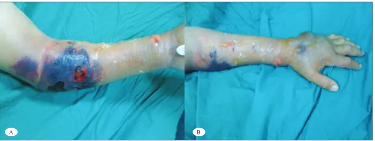
A 78-year old female patient with a side to-side radiocephalic AVF at the left antecubital region had been under hemodialysis for 2 years due to end stage chronic renal failure. The patient was admitted to another center for a complaint of painful mass in the left antecubital fossa that appeared after hemodialysis and the patient was referred to our clinic for cyanotic ulcer lesion and compartment syndrome in the left arm three days after the hemodialysis. On physical examination, bullous lesions, severe edema and necrotic ulcers were observed in the left upper
Figure 1 A-B: Severe necrosis and compartment syndrome due to brachial pseudoaneurysm after hemodialysis.
A B
Figure 2 A-B: Full recovery was achieved in the left arm eighth months after the successful repair of brachial pseudoaneurysm and appropriate dermal greft application.
214
Türk Nefroloji Diyaliz ve Transplantasyon Dergisi Turkish Nephrology, Dialysis and Transplantation Journal
Katrancıoğlu N et al: Can Vascular Access During Hemodialysis Cause Extremity Loss?
Turk Neph Dial Transpl 2013; 22 (2): 212-214
REFERENCES
1. Zibari GB, Rohr MS, Landreneau MD, Bridges RM, DeVault GA, Petty FH, Costley KJ, Brown ST, McDonald JC: Complications from permanent hemodialysis vascular access. Surgery 1988; 104(4): 681-686
2. Lapus TP, Trerotola SO, Savader SJ: Radial artery pseudoaneurysm complicating a brecia-cimino dialysis fi stula. Nephron 1996; 72: 673-675
3. Hein AN, Vesely TM: Use of the percutaneous thrombolytic device for the treatment of thrombosed pseudoaneurysms during mechanical thrombectomy of hemodialysis grafts. J Vasc Interv Radiol 2002; 13: 201-204
4. Moszkowicz A, Behrens G, Gueyikian S, Patel NH, Ferral H: Occlusion of a rapidly expanding hemodialysis graft pseudoaneurysm with placement of a stent graft. Semin Intervent Radiol 2007; 24(1): 34-37
5. Rooijens PP, Burgmans JP, Yo TI, Hop WC, de Smet AA, van den Dorpel MA, Fritschy WM, de Groot HG, Burger H, Tordoir JH: Autogenous radial-cephalic or prosthetic brachial-antecubital forearm loop AVF in patients with compromised vessels? A randomized, multicenter study of the patency of primary hemodialysis access. J Vasc Surg 2005; 42(3): 481-486
6. Kutay V, Ekim H, Karadağ M, Öztürk V, Kırali K, Yakut C: Kronik Böbrek yetmezlikli hastalarda görülen arteriyovenöz fi stül komplikasyonları ve cerrahi tedavisi. Turkish J Thorac Cardiovasc Surg 2004; 12: 115-118
7. Schanzer H, Skladany M: Vascular Access For Dialysis. Haımovıcı
H (Ed), Haımovıcı’s Vascular Surgery Principles and Techniques. (4th ed). 1996; 1028-1041
8. Topal M, Özdemir N: Acute complications of hemodialysis. Turkiye Klinikleri J Int Med Sci 2006; 2(4): 24-29
9. Padberg FT Jr, Calligaro KD, Sidawy AN: Complications of arteriovenous hemodialysis access: Recognition and management. J Vasc Surg 2008; 48(5): 55-80
10. Yasim A, Kabalci M, Eroglu E, Zencirci B: Complication of hemodialysis graft: Anastomotic pseudoaneurysm: A case report. Transplant Proc 2006; 38(9): 2816-2818
11. Yildirim S, Nursal TZ, Yildirim T, Tarim A, Caliskan K: Brachial artery pseudoaneurysm: A rare complication after haemodialysis therapy. Acta Chir Belg 2005; 105(2): 190-193
12. Paulson EK, Sheafor DH, Kliewer MA, Nelson RC, Eisenberg LB, Sebastian MW, Sketch MH Jr: Treatment of iatrogenic femoral arterial pseudoaneurysms: Comparison of US guided thrombin injection with compression repair. Radiology 2000; 215: 403-408
DISCUSSION
The number of patients with end-stage renal failure has been increasing each year (5). The best treatment for renal failure is kidney transplant but patients are obliged to undergo hemodialysis due to lack of transplant donors; (6). Currently, AV
fi stulas are the gold standard vascular access for hemodialysis. These patients are dependent on regular and continuous vascular intervention to continue their treatment. The professionalism and experience of dialysis staff are also as important as surgical techniques in this regard (5-7).
Most of the complications related to vascular access after hemodialysis are related with a failure of hemostasis at the puncture sites in a reasonable. This situation may manifest as external bleeding, hematoma, or pseudoaneurysm (8,9). Pseudoaneurysm is an important complication of AV fi stulas that can cause problems ranging from bleeding to life-threatening conditions. The incidence of pseudoaneurysm is estimated at 2% to 10% of venous access grafts (9,10). It is more common in prosthetic grafts than in autogenous access. This complication may also occur in every needle puncture during routine hemodialysis. Since, pseudoaneurysm is located often at the puncture site, determination of the vascular puncture site is crucial to prevent vascular access complications. (9). In our case, there was direct puncture to brachial artery, which rapidly enlarged and lead to the compartment syndrome, instead of an arteriovenous fi stula.
There are various treatment strategies in pseudoaneurysm management. A small puncture site pseudoaneurysm may usually resolve conservatively. Surgical intervention may be necessary if it is enlarging or acutely expanding. Anastomotic pseudoaneurysms almost always require intervention (9). Ultrasound-guided compression repair, percutaneous injection of thrombin, endovascular covered stent exclusion, aneurysmectomy and surgical repair are various treatment options (11,12). Moszkowicz and his collegues (4) reported their brachial artery pseudoaneurysm cases. They used various treatment methods and according to their report only one patient had limb loss ( 4th & 5th digits). However, surgical interventions
are preferable in case of compartment syndrome causing extremity ischemia, as in our case.