of Missing Anterior Teeth with a Removable Partial Denture
Adriana da Fonte Porto Carreiro, DDS, MSc, PhD,1 Ana Lucia Machado, DDS, MSc, PhD,2 Eunice Teresinha Giampaolo, DDS, PhD,2 Ivone Lima Santana, DDS, MSc, PhD,3& Carlos Eduardo Vergani, DDS, MSc, PhD2
1Assistant Professor, Department of Dentistry, Rio Grande do Norte Federal University (UFRN), Natal, Rio Grande do Norte, Brazil 2Associate Professor, Department of Dental Materials and Prosthodontics, Araraquara Dental School, S ˜ao Paulo State
University (UNESP), Araraquara, Sao Paulo, Brazil
3Assistant Professor, Department of Dentistry, Maranh ˜ao Federal University (UFMA), S ˜ao Luis, Maranh ˜ao, Brazil
Keywords
Removable partial denture; esthetics; dual path; path of insertion; surveyor.
Correspondence
Ana Lucia Machado, Department of Dental Materials and Prosthodontics, Araraquara Dental School, S ˜ao Paulo State University (UNESP), Rua Humaita, 1680 Araraquara, Sao Paulo 14801-903, Brazil. E-mail: [email protected]
Accepted 11 July 2007
doi: 10.1111/j.1532-849X.2008.00332.x
Abstract
The dual path of insertion concept for removable partial denture (RPD) design may be used in esthetically demanding situations. When compared to conventional RPDs, the main advantage of this design is the minimal use of clasps. This clinical report describes the treatment of a patient with an anterior maxillary edentulous area using a dual path RPD. The diagnostic cast was surveyed to ensure the adequacy of the undercuts on the mesial surfaces of the anterior abutments, where rigid minor connectors were placed. Inverted V-shaped canine cingulum rest seats were prepared to provide resistance to tooth movement during function. The dual path RPD concept allows excellent esthetic results, minimizes tooth preparation, and reduces the tendency toward plaque accumulation in a Kennedy class IV partially edentulous arch.
The decision to replace anterior missing teeth presents a den-tist with numerous alternatives including fixed, removable, and implant-based approaches. When a conventional clasp-type re-movable partial denture (RPD) is the selected treatment, placing retaining elements on the abutment teeth results in an undesir-able display of metal. An RPD that incorporates a dual path of insertion may offer some advantages in these esthetically demanding situations,1-9including the absence of buccal clasp arms placed on the anterior abutment teeth. In addition, the re-duced coverage of the tooth surface results in advantages, such as reduced plaque formation and caries incidence.10
Although the design principles and theoretical considera-tions of dual path RPDs have been widely reported, and clinical cases have been described,1-4,6-8,10-15
there is still reluctance by the majority of dentists to use this concept more often, when indicated.13,14
The lack of sufficient understanding of the concept for both the dentist and laboratory technician and the difficulty in transferring the clinical/survey information to the dental laboratory are reasons for the limited application of the rotational path concept among dentists.13Another plau-sible reason could be that a more detailed description of clin-ical and laboratory steps involved in the fabrication of dual
path RPDs is not provided in most of the above-cited clinical reports.
This clinical report describes in detail the treatment of a patient with an anterior maxillary edentulous area treated by a dual path RPD. The presence of combined anterior and posterior modification areas and the need to fabricate a full crown for one of the abutment teeth make this report unique.
Clinical report
Intraoral examination
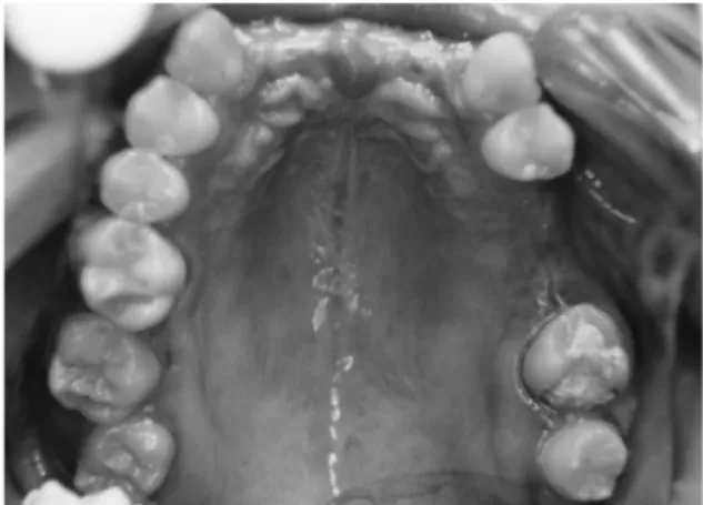
Figure 1 Preoperative occlusal view of missing teeth: maxillary incisors, left second premolar, and left first molar.
The planned abutment teeth (teeth #2, 6, 11, 12, and 15) ex-hibited healthy gingiva and no mobility (Fig 1). The intraoral examination also demonstrated the presence of stable contacts in intercuspal position. The patient had good oral hygiene, and periodontal probing revealed a normal and intact periodontium. The panoramic radiograph and bitewing radiographs revealed no visible pathologies.
Surveying
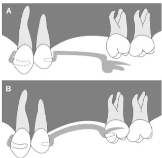
An impression was made with irreversible hydrocolloid ma-terial, and a diagnostic cast was fabricated. To determine the dual path accurately, the diagnostic cast was first surveyed at 0◦ tilt. This initial survey was made to ensure the adequacy of the undercuts on the mesial surfaces of the anterior abutments, as well as the distobuccal surface of tooth #2. The surveyor blade was used to locate proximal undercuts on the anterior abut-ments (Fig 2A). The retentive area on the distobuccal surface of tooth #2 was determined using the conventional 0.25-mm undercut gauge.16 The path of insertion of the conventional retainer (circumferential clasp) was indicated by the cemented-pin method.17The cast was then tilted down posteriorly, using the surveyor blade to determine the degree of tilt necessary to eliminate the proximal undercuts on the anterior abutment teeth (Fig 2B). This tilt was used to determine the straight path of in-sertion the denture would follow, along with the initial seating, thus allowing the framework access to the anterior undercuts.3
Figure 2 Diagnostic cast on a surveyor. (A) At 0◦tilt, surveyor blade
demonstrates an undercut on the mesial surface of the abutment; (B) cast is tilted down posteriorly until undercut is eliminated.
Figure 3Analysis of the distal proximal surface of the left first premolar to accommodate the rotational path. The first tip of a caliper is placed at the rotational center of the anterior abutment, and the second tip is placed in the proximal undercut and rotated occlusally.
The path of insertion for the rigid anterior retainer was indicated by vertical lines on the three sides of the diagnostic cast.
As in this case a modified edentulous area was present, the distal proximal surface of tooth #12 was analyzed to accom-modate the rotational path. For this analysis, the first tip of a divider was placed at the rotational center of the anterior abut-ment. The second tip was then extended to the marginal ridge of tooth #12 and rotated in an occlusogingival direction. As the caliper rotated, the space that appeared between the distal surface of tooth #12 and the second tip indicated the excessive blockout required to allow seating of the framework (Fig 3). Although guiding planes are not usually recommended for dual path RPDs, the recontouring of the distal surface was indicated to minimize the amount of relief required in this area.
Mouth preparation
After surveying was completed, the mouth preparation was made following the treatment plan outlined in the diagnostic cast. A metal ceramic crown restoration was made for tooth #15. The wax pattern was surveyed to provide adequate contour. To avoid any interference to the denture rotation, the mesial and
lingual surfaces were made flat (Fig 4), and the mesial surface was analyzed by means of a divider as described for tooth #12. A mesial rest seat was also prepared in the wax pattern. The distobuccal retentive area for the circumferential clasp arm was determined in the surveyor before glazing, by using the 0.25-mm undercut gauge.
Cingulum rests were prepared on the canines. The depth of the preparation was sufficient to allow a rest thickness of 1.5 to 2 mm.3,6,8The rest seat outline form was made asymmetri-cal to provide resistance to tooth movement in all directions.2 These preparations were greater than one-half the mesial-distal width of the tooth and had relatively parallel axial walls.6,8 Conventional rests were prepared on teeth #2 and 12.
Prosthesis fabrication
After the metal ceramic crown of tooth #15 was cemented, and the other abutment teeth were prepared, an impression was made with irreversible hydrocolloid, and a master cast was obtained. The dual path of insertion was then determined by surveying the master cast in the same manner as described for the diagnostic cast. Also, the dual path was recorded on the master cast, and the framework outline was drawn for the laboratory’s convenience. To obtain a better esthetic result, no clasp arm was placed on tooth #12.
All hard and soft tissue undercuts were blocked out, except the proximal undercuts bounding the anterior edentulous space. Using a set of dividers, the proximal areas were blocked out to the required degree. When the framework returned from the laboratory, it was tried in the mouth (Figs 5 and 6) by seating the anterior rigid metal portion until intimate contact with the proximal undercuts (Fig 7A) and then rotating the framework toward the tissue until it was fully seated on the posterior abutments (Fig 7B). The rotational axis is defined by the rotational centers located at the gingival extension of the minor connectors placed on both sides of the arch.2Try-in of the metal framework revealed a highly retentive prosthesis that exhibited sufficient resistance to displacement.
The denture teeth were arranged and waxed into place, and the necessary occlusal adjustments were carried out. The fin-ished prosthesis was inserted, and minor occlusal adjustments were made. The patient was instructed about oral hygiene and
Figure 5Occlusal view of the framework in place.
Figure 6Minor connectors in intimate contact with undercut areas.
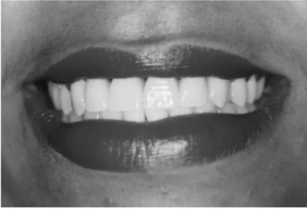
how to properly insert and remove the prosthesis. She was re-called 3 and 15 days after insertion. The evaluation of oral health revealed no problems. The patient had speech accom-modation and was pleased with the prosthesis (Fig 8). Recalls up to 3 years revealed the same favorable conditions.
Discussion
The treatment for an anterior partially edentulous arch by means of a fixed partial denture generally is the method of choice18 because of better esthetic results and satisfactory patient ac-ceptance9; however, when the edentulous span is of greater magnitude, the loss of anterior residual bone is excessive, or financial considerations are adverse, a fixed restoration is contraindicated.19Although implant-supported prostheses have demonstrated good results20for the treatment of partially eden-tulous patients, surgical procedures are necessary. Also, local and systemic factors such as quantity and quality of bone and
Figure 8 Frontal view of the patient showing the favorable esthetic result.
hormonal influence on calcium metabolism must be considered as they can influence the osseointegration process.21Therefore, in many instances, an RPD can be a valid alternative.
Retention needed for a conventional clasp-type partial den-ture is accomplished by placing retaining elements on the buc-cal surfaces of the teeth. When anterior abutment teeth are involved, the esthetic appearance may be compromised. The rotational path RPD design permits the elimination of the buc-cal clasp arms. This enhances esthetics without compromising the biomechanical principles of the RPD. Precision or semi-precision attachments can sometimes be used as another al-ternative approach. Unfortunately, the use of attachments has some disadvantages, such as increased cost and time, exten-sive preparation of the abutment tooth, and the need for precise clinical techniques and technical skill.22
Acetal resin clasps also offer an alternative for patients con-cerned about the retainer’s metallic color23; however, defor-mation of acetal resin direct retainers has been found to be significantly greater than their metal alloy counterparts.24 In addition, it has been reported that the retentive force for an acetal resin clasp may not be sufficient for RPDs due to the sig-nificantly low retention force required for removal.25Another mean of limiting clasp display in anterior edentulous regions is the “spring clasp,” also known as the Twin-Flex technique, which consists of a wire clasp soldered into a channel that is cast in the major connector.18,26
In addition, Valplast, a ther-moplastic nylon material,27can be used in combination with a metal frame to allow the esthetic benefit of replacing the buccal arm; however, the biomechanical properties of these types of clasps and the possible long-term effects on the abutments still remain to be investigated.
Although the dual path RPD presents excellent esthetic and functional results, there are some requirements necessary for a successful treatment. These requirements include (i) using spe-cially designed rests seats; (ii) critical finishing and polishing on the rigid retainer because it must be in intimate contact with the proximal surface of the abutment; (iii) the need for proximal undercuts on anterior abutments; and (iv) anterior single-tooth replacements usually are not practical with a rotational path RPD because of lack of access of the metal framework to cin-gulum rest areas.28In addition, the dual path design should not
be indicated for Kennedy class I and class II RPDs with an-terior modification spaces. In this situation, the rigid retainers will usually torque the abutments during rotational movements in function.5Therefore, the dual path of insertion requires care-ful patient selection, treatment planning, and mouth prepara-tion.8When these principles are followed, clinical experience has shown long-term success. Patients followed for 10 years or more revealed that the rigid retainers demonstrated sup-port, stability, retention, adequate encirclement, and passivity at rest.13
Conclusion
The clinical results demonstrated that the dual path RPD concept could be successfully applied to the treatment of a Kennedy class IV partially edentulous arch. This design sat-isfies functional and esthetic requirements because the use of a conventional clasp is minimized without compromising the retention of the prosthesis. In addition, the tooth coverage is decreased, thus improving esthetics and resulting in less plaque formation.
References
1. King GE, Barco MT, Olson RJ: Inconspicuous retention for removable partial dentures. J Prosthet Dent 1978;39:505-507
2. Jacobson TE: Satisfying esthetic demands with rotational path partial dentures. J Am Dent Assoc 1982;105:460-465 3. Daniel RE, Granata JS: The rotational-path removable partial
denture. Compend Contin Educ Dent 1985;6:719-723 4. Chow TW, Clark RKF, Clark DA, et al: A rotational path of
insertion for Kennedy class IV removable partial dentures. Br Dent J 1988;164:180-183
5. Krol AJ, Finzen FC: Rotational path removable partial dentures: part 2. Replacement of anterior teeth. Int J Prosthodont 1988;1:135-142
6. Levitt J, Harle TJ: Rotational path removable partial denture: a case report. Univ Tor Dent J 1991;5:11-13
7. Halberstam SC, Renner RP: The rotational path removable partial denture: the overlooked alternative. Compend Contin Educ Dent 1993;14:544-552
8. Jacobson TE: Rotational path partial denture design: a 10-year clinical follow-up-part I. J Prosthet Dent 1994;71:271-277 9. Baharav H, Ben-Ur Z, Laufer BZ, et al: Removable partial
denture with a lateral rotational path of insertion. Quintessence Int 1995;26:531-533
10. Firtell DN, Jacobson TE: Removable partial dentures with rotational paths of insertion: problem analysis. J Prosthet Dent 1983;50:8-15
11. Jacobson TE, Krol AJ: Rotational path removable partial denture design. J Prosthet Dent 1982;48:370-376
12. Schwartz RS, Murchison DG: Design variations of the rotational path removable partial denture. J Prosthet Dent 1987;58:336-338
13. Jacobson TE: Rotational path partial denture design: a 10-year clinical follow-up-part II. J Prosthet Dent 1994;71:278-282 14. Reagan SE, Dao TM: Oral rehabilitation of a patient with
congenital partial anodontia using a rotational path partial denture: report of a case. Quintessence Int 1995;26:181-185 15. Ivanhoe JR: Laboratory considerations in rotational path
16. Stern WS: Guiding planes in clasp reciprocation and retention. J Prosthet Dent 1975;34:408-414
17. Wagner AG, Forgue EG: A study of four methods of recording the path of insertion of removable partial dentures. J Prosthet Dent 1976;35:267-272
18. Belles DM: The twin-flex clasp: an esthetic alternative. J Prosthet Dent 1997;77:450-452
19. Wostmann B, Budtz-Jorgensen E, Jepson N, et al: Indications for removable partial dentures: a literature review. Int J Prosthodont 2005;18:139-145
20. Telleman G, Meijer HJ, Raghoebar GM: Long-term evaluation of hollow screw and hollow cylinder dental implants: clinical and radiographic results after 10 years. J Periodontol
2006;77:203-310
21. Wood MR, Vermilyea SG: Committee on Research in Fixed Prosthodontics of the Academy of Fixed Prosthodontics. A review of selected dental literature on evidence-based treatment planning for dental implants: report of the Committee on Research in Fixed Prosthodontics of the Academy of Fixed Prosthodontics. J Prosthet Dent 2004;92:447-462
22. McGivney GP, Carr AB (eds): McCracken’s Removable Partial Prosthodontics (ed 10). St. Louis, MO, Elsevier, 2004, pp. 97-142 23. Chu CH, Chow TW: Esthetic designs of removable partial
dentures. Gen Dent 2003;51:322-324
24. Wu JC, Latta GH Jr, Wicks RA, et al: In vitro deformation of acetyl resin and metal alloy removable partial denture direct retainers. J Prosthet Dent 2003;90:586-590
25. Arda T, Arikan A: An in vitro comparison of retentive force and deformation of acetal resin and cobalt-chromium clasps. J Prosthet Dent 2005;94:267-274
26. Santana-Pen´ın U, Mora J: An esthetically attractive twin-flex clasp for removable partial dentures. J Prosthet Dent 1998;80:367-370
27. Parvizi A, Lindquist T, Schneider R, et al: Comparison of the dimensional accuracy of injection-molded denture base materials to that of conventional pressure-pack acrylic resin. J Prosthodont 2004;13:83-89