INTRODUCTION
Microbial biofilm is the main etiologic factor of chronic atrophic candidiasis, also known as denture stomatitis (1). The fitting surface of the denture is the main reservoir of Candida albicans (2). Therefore, biofilm control with an adequate hygiene of the oral mucosa and the prosthesis would avoid microbial adhesion. Irregularities and porosities present on denture surfaces offer a favorable niche to retain stain and microbial plaque (3).
The use of chemical disinfectants is usually
Effect of Denture Cleansers on Metal Ion Release
and Surface Roughness of Denture Base Materials
Letícia Resende DAVI Daniela Nair Borges FELIPUCCI
Raphael Freitas de SOUZA Osvaldo Luiz BEZZON Cláudia Helena LOVATO-SILVA
Valéria Oliveira PAGNANO Helena de Freitas Oliveira PARANHOS
Department of Dental Materials and Prosthodontics, Ribeirão Preto Dental School, USP - University of São Paulo, Ribeirão Preto, SP, Brazil
Chemical disinfectants are usually associated with mechanical methods to remove stains and reduce biofilm formation. This study evaluated the effect of disinfectants on release of metal ions and surface roughness of commercially pure titanium, metal alloys, and heat-polymerized acrylic resin, simulating 180 immersion trials. Disk-shaped specimens were fabricated with commercially pure titanium (Tritan), molybdenum-titanium (Vi-Star), nickel-chromium (Fit Cast-SB Plus), and nickel-chromium-beryllium (Fit Cast-V) alloys. Each cast disk was invested in the flasks, incorporating the metal disk to the heat-polymerized acrylic resin. The specimens (n=5) were immersed in these solutions: sodium hypochlorite 0.05%, Periogard, Cepacol, Corega Tabs, Medical Interporous, and Polident. Deionized water was used as a control. The quantitative analysis of metal ion release was performed using inductively coupled plasma mass spectrometry (ELAN DRC II). A surface analyzer (Surftest SJ-201P) was used to measure the surface roughness (µm). Data were recorded before and after the immersions and evaluated by two-way ANOVA and Tukey’s test (α=0.05). The nickel release proved most significant with the Vi-Star and Fit Cast-V alloys after immersion in Medical Interporous. There was a significant difference in surface roughness of the resin (p=0.011) after immersion. Cepacol caused significantly higher resin roughness. The immersion products had no influence on metal roughness (p=0.388). It could be concluded that the tested alloys can be considered safe for removable denture fabrication, but disinfectant solutions as Cepacol and Medical Interporous tablet for daily denture immersion should be used with caution because it caused greater resin surface roughness and greater ion release, respectively.
Key Words: denture hygiene, commercially pure titanium, nickel-chromium alloys, heat-polymerized acrylic resin.
associated with mechanical methods, and their efficacy in removing stains and reducing biofilm formation on the surface irregularities of dentures has been reported (4). Effervescent tablets are classified as chemical soak-type products, and when dissolved in water the sodium perborate readily decomposes to form an alkaline peroxide solution. This peroxide solution subsequently releases oxygen, thereby enabling a mechanical cleaning by the oxygen bubbles in addition to the chemical cleaning (1). Nevertheless, the factors contributing to the infrequent use of alkaline peroxide include insufficient information being provided to the patient, high cost, and
restricted market access.
Sodium hypochlorite (NaOCl) diluted in water has been indicated for complete denture disinfection. This method is effective in reducing Candida albicans in patients with denture stomatitis and prevention (5), depending on the concentration and the immersion time. Household bleaches are recommended for occasional overnight soaking, so their use as everyday chemical denture disinfection products would demand a study of lower concentrations during longer periods.
Antimicrobial mouthwashes are also indicated as denture cleansers (6). Chlorhexidine digluconate, cetylpyridinium chloride, or triclosan/copolymer solutions significantly inhibit microbial colonization, but they lack effectiveness against mature biofilm with Candida albicans (7).
Possible deleterious effects on the denture materials after immersion in cleansing solutions can occur (8). Metal alloys can corrode or stain as a result of surface contact with the chlorine or oxygen present in some commercial cleansers (9). Nickel-chromium alloys appear to be an appropriate substitute for gold alloys, and they offer low cost and lower specific weight. Better physical properties, such as mechanical resistance, hardness, and corrosion resistance, motivated the development of those alloys (10). In spite of the advantages of titanium alloys, like biocompatibility, corrosion resistance, low specific weight, low modulus of elasticity, low thermal conductivity, high mechanical resistance (11), as well as great acceptance by patients, there have been reports of tarnish (surface discoloration) (12) and corrosion (surface pitting) (13).
The purpose of this study was to evaluate the effect of disinfectants on the release of metal ions and surface roughness of commercially pure titanium, metal alloys, and heat-polymerized acrylic resin, by simulating 180 consecutive hygiene immersion trials. The null hypothesis tested was that immersion in disinfectant solutions would not influence metal ion release or the surface roughness of denture materials.
MATERIAL AND METHODS
Specimen Fabrication
Disk-shaped wax patterns (GEO; Renfert GmbH, Hilzingen, Germany) (12 x 3 mm) were sprued, invested, and casted in each metal (Table 1) according to the manufacturer’s instructions. After casting, the metal disks were finished with 180-grit sandpaper (Norton Abrasives, Saint-Gobain, France) in a water-cooled polishing machine (AROTEC, Cotia, SP, Brazil) and then washed to remove any metal particles or sand granules.
Metallic flasks were previously prepared by Teflon rectangular matrices (38 x 18 x 4 mm) and then invested with type IV dental stone (Durone; Dentsply Dentsply Ind. e Com. Ltda., Petrópolis, RJ, Brazil). The Teflon matrices were removed and the cast disks were inserted in the flasks in the left side of each rectangular mold. Before packing the heat-polymerized acrylic resin Lucitone 550 (Dentsply Ind. Com. Ltda.), the mold was isolated with two coats of a liquid separating medium (Cel-Lac; SS White, Rio de Janeiro, RJ, Brazil).
Thirty-five specimens of each metal were prepared. The resin was handled, packed, and pressed into the mold according to the manufacturer’s instructions. The polymerization cycle was undertaken in by immersion in water at 73°C for 90 min and at 94°C for 30 min. All flasks were allowed to cool at room temperature before opening. After polymerization, the specimens were immersed in distilled water at 37 ± 1°C for 50 ± 2 h for residual monomer reduction (14).
Excess resin was trimmed, and one of the surfaces was finished using 180-, 220-, 400-, 600- and 1200-grit sandpapers (Norton Abrasives) in the polishing machine, followed by polishing cloths soaked with 1-µm diamond suspension (Fortel Ind. Com., São Paulo, SP, Brazil).
Immersion Procedures
The specimens were distributed into groups
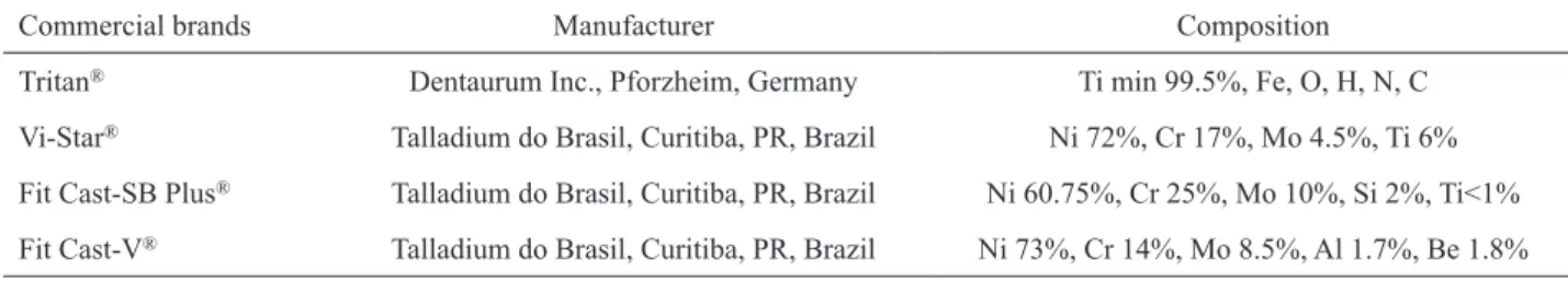
Table 1. Manufacturer and composition of the metals.
Commercial brands Manufacturer Composition
Tritan® Dentaurum Inc., Pforzheim, Germany Ti min 99.5%, Fe, O, H, N, C
Vi-Star® Talladium do Brasil, Curitiba, PR, Brazil Ni 72%, Cr 17%, Mo 4.5%, Ti 6%
Fit Cast-SB Plus® Talladium do Brasil, Curitiba, PR, Brazil Ni 60.75%, Cr 25%, Mo 10%, Si 2%, Ti<1%
(n=5) and immersed in one of the following cleanser solutions: 0.05% NaOCl (Q’Boa; Anhembi S/A, Osasco, SP, Brazil) for 10 min; 0.12% chlorhexidine digluconate (Periogard; Colgate-Palmolive Ind., São Bernardo do Campo, SP, Brazil) for 10 min; cetylpyridinium chloride 0.500 mg (Cepacol; Sanofi-Aventis Farmacêutica Ltda., São Paulo, SP, Brazil) for 10 min; Corega Tabs (sodium perborate and enzyme; Stafford-Miller Ind., Rio de Janeiro, RJ, Brazil) for 5 min; Medical Interporous (citric acid; MST-Laboratories AG, Liechtenstein) for 15 min; and Polident 3 Minute (sodium perborate and enzyme; GlaxoSmithKline, Clifton, NJ, USA) for 3 min. The control group was immersed in 200 mL of deionized water for 15 min. The 0.05% NaOCl solution was prepared by mixing 200 mL of deionized water and 6 mL of Q’Boa (2% NaOCl). The Periogard and Cepacol groups were immersed in 50 mL of the solution. The effervescent cleansers were prepared according to the manufacturer’s directions, by adding one tablet to 200 mL of warm deionized water (40ºC) (15). The five specimens of each group were immersed at one time in the same container with the surface to be measured facing upward, and the solution covered all specimens.
After immersion, the resin specimens were removed from the chemical solutions, thoroughly washed in deionized water, dried with absorbent paper, and then this procedure of immersion was repeated. All experiments simulated 180 consecutive hygiene immersion trials.
Metal Ion Release Test
The quantitative analysis of metal ion release was analyzed using inductively coupled plasma mass spectrometry (ICP-MS - ELAN DRC II; Perkin Elmer-Sciex, Norwalk, CT, USA). Each cleanser solution was collected before the specimens’ immersion, as was one sample from each group at the end of the immersions. The solutions were submitted to analyses, and the spectrometer was calibrated to recognize the following chemical elements: aluminum (Al), chromium (Cr), nickel (Ni), beryllium (Be), molybdenum (Mo), and titanium (Ti).
Surface Roughness Test
A surface analyzer (Surftest SJ-201P; Mitutoyo Corporation, Japan) was calibrated at a sample length of 0.8 mm, 4.0 mm percussion of measure, and 0.5 mm/s,
was used to measure the roughness of the resin and the metal of each specimen before (baseline) and after
immersion to obtain the ΔRa (roughness differences). The stylus was moved across the specimen surface, and three lines were recorded with a distance of 1 mm between each scanning line. The mean Ra was calculated from three lines as the mean roughness of the specimen. The resolution of the record data was 0.01 µm.
Statistical Analysis
The ΔRa values were subjected to statistical analysis by two-way analysis of variance (ANOVA) and Tukey’s post-hoc test for pairwise comparisons
(α=0.05), using the statistical program SPSS 12.0 (SPSS
Inc., Chicago, IL, USA).
RESULTS
Ion Release
Table 2 presents the concentration of the chemical elements Al, Cr, Ni, Be, Mo, and Ti for each experimental group. The negative values indicate that the results were below the detection limit of the element.
A comparison of all solutions used for immersion showed that the most significant ion release was obtained for the elements Ni and Be, verified on specimens with Vi-Star and Fit Cast-V alloys after immersion in Medical Interporous.
Surface Roughness
Two-way ANOVA showed a statistically significant difference of surface roughness of the acrylic resin of the specimens treated with several denture cleansers and the acrylic resin of the specimens made with different metals. Further analysis by the Tukey’s test indicated significantly higher surface roughness of the acrylic resin for the specimens treated with Cepacol.
Table 3 presents the mean resin ΔRa and standard deviations of each cleanser solution.
Two-way ANOVA data showed a statistically significant difference of surface roughness of the metal, but no significant difference was found for the solutions.
DISCUSSION
Table 2. Concentration of the chemical elements (µg/L).
Solution Element Pure solution Tritan® Vi-Star® Fit Cast-SB Plus® Fit Cast-V®
Deionized water
Al 2.120 3.365 5.719 6.528 8.616
Cr - 1.663 16.426 8.933 29.849 9.027
Ni 0.309 1.184 123.971 4.746 103.917
Be - 0.033 - 0.033 13.760 0.098 13.170
Mo - 0.035 0.041 0.170 2.081 0.126
Ti 7.905 14.700 5.235 25.477 12.235
0.05% NaOCl
Al 35.693 13.867 16.787 16.565 10.754
Cr 47.431 120.913 71.254 117.650 73.196
Ni 1.359 1.326 136.465 3.533 8.359
Be - 0.033 - 0.033 10.140 - 0.016 5.569
Mo 0.810 0.806 5.421 20.658 7.915
Ti 25.671 13.384 17.332 15.172 15.272
Periogard
Al 24.901 26.670 22.996 23.487 20.409
Cr 204.858 218.452 214.227 211.387 202.206
Ni 1.525 1.960 72.056 5.855 71.358
Be - 0.033 - 0.033 11.385 0.016 10.533
Mo 0.460 0.660 0.830 1.150 0.739
Ti 155.666 167.167 166.439 119.296 119.296
Cepacol
Al 47.986 47.324 26.897 22.428 25.740
Cr 204.407 207.836 218.265 248.897 228.738
Ni 3.661 4.035 68.637 6.032 69.397
Be - 0.016 0.000 10.189 0.016 9.009
Mo 1.675 10.089 9.507 11.070 11.314
Ti 4208.823 4057.171 4186.263 4507.776 4339.946
Corega
Al 1618.054 1359.746 1325.059 1477.116 1562.753
Cr 4.810 10.214 11.241 6.114 9.339
Ni 3.810 3.606 27.708 6.533 20.540
Be 0.213 0.115 3.276 0.164 2.113
Mo 1.816 1.637 1.799 3.378 2.112
Ti 1113.910 966.972 984.425 1078.533 1247.182
Medical interporous
Al 43.639 52.706 51.346 58.803 35.198
Cr 58.554 79.951 55.093 78.095 64.133
Ni 2.068 3.178 896.233 12.489 1005.456
Be - 0.033 0.016 122.282 0.066 135.144
Mo 0.413 0.473 1.822 4.627 2.554
Ti 27.247 25.338 24.788 25.460 20.530
Polident
Al 1535.044 1394.532 1084.073 1102.185 1140.217
Cr 22.275 31.831 19.737 34.799 25.075
Ni 3.945 4.946 32.340 5.730 38.521
Be 0.000 0.229 3.505 0.262 4.259
Mo 1.185 1.965 1.950 3.270 2.173
the null hypothesis of the study because immersion in disinfectant solutions did influence metal ion release, and it exhibited significantly higher resin surface roughness, but it did not affect the roughness of the metal surface. Daily hygiene for a removable denture should include brushing, but also immersion in disinfectant solutions for biofilm removal and decontamination because this process reduces the pathogenesis of the microorganisms of the prosthesis surface (16). Peracini et al. (17) observed that the patients do not have correct professional instructions about how to clean their dentures. Denture cleaning by immersion in chemical solutions should not involve any physical, mechanical, or chemical change in the denture materials (8).
The immersion of a removable denture in commercial bleaches is indicated for patients’ home care, particularly because they are inexpensive and easy to use (18). The chemical solutions reduce the pathogenicity of the microorganisms present on the surface of the prosthesis (19) and the clinical signs of denture stomatitis, and they also control biofilm formation. The deleterious effects of NaOCl can seriously change the shape and the mechanical resistance of the metallic components of the prosthesis (18) caused by the chloride ions’ attack on metal surfaces. At a concentration of 0.05% for NaOCl, an antimicrobial effect was detected (19), so that same concentration was used in this study.
The effervescent tablets are efficient in removing biofilm and stains (5), but the alkaline peroxide solution can alter the resin properties if not correctly used. The water temperature used to prepare the solutions is a critical factor, resulting in whitening of the acrylic resin when patients use hot water (20). Other research found that water absorption on acrylic surfaces was caused
by hot alkaline peroxide solution, which resulted in irreversible surface whitening when the specimens were left to dry (15). In the present study, the solutions were prepared with warm water (40ºC), as recommended by the manufacturer.
Another factor involved in the mechanical properties is the residual methyl methacrylate monomer in the polymerized acrylic resin, which has a plasticizing effect. Acrylic resin plasticizers leach out when they are in contact with chlorine-containing solutions (21). The specimens in this study were immersed in distilled water to eliminate residual monomers (14).
The mouthwashes Cepacol and Periogard were also evaluated in this study because of their antimicrobial effects have been scientifically proven (7,22). The results showed that the surface roughness of the resin increased when the specimens were soaked in Cepacol solutions.
The ion release test revealed that, when comparing all solutions used for immersion, Ni and Be were the most significantly released elements, which was verified on specimens of Vi-Star and Fit Cast-V alloys after immersion in Medical Interporous. Although the manufacturer of the Medical Interporous tablets has indicated its safe use for metallic structures, this tablet can lead to surface alteration of those alloys.
Complete immersion of dentures in denture cleansers may adversely affect the surface roughness of denture base resins and metals. The retention of Candida albicans on smooth and rough acrylic resins was compared, and a higher numbers of cells were observed on roughened surfaces (23). To produce a flat, smooth surface in the specimens of the present study, a sequence of sandpapers was used for finishing and polishing cloths embedded in 1-µm diamond suspension to polish the specimens. Thus, the findings of this study show that the alterations could be related to the denture cleansers. In addition to that, some studies have shown that immersion in a low concentration of NaOCl did not change the surface roughness of acrylic resin. According to Azevedo et al. (24), no alterations in the surface roughness of acrylic resin were found after 7 days of immersion in 1% NaOCl. Also, the results of Lima et al. (21) showed no alterations after immersion in 0.5% NaOCl. In the present study, significantly higher surface roughness of the resin was obtained in the specimens treated with Cepacol.
Considering the methodological limitations of this study, no simulation of the denture inside the patients’ mouth was conducted, i.e., no immersion in
Table 3. Mean resin ΔRa (µm) and standard deviations for each denture cleanser. Different uppercase letters represent significant differences (p<0.05).
Solution Mean Standard deviation
Corega tabs® -0.005 A 0.04
Periogard® -0.003 A 0.03
Deionized water 0.004 AB 0.04
0.05% NaOCl 0.011 AB 0.04
Polident® 0.016 AB 0.03
Medical interporous® 0.020 AB 0.04
saline solution. Such simulation would remove chemical substances from the specimens, and as a result, any possible chemical degradation would be the same or even less. This way, a different result would not be expected by means of a period of simulated denture use. Possibly the damage on acrylic resin and metallic components is slighter in vivo due to the amount of time inside the oral cavity and combination with other forms of hygiene, such as brushing or tap water. Future studies should include long-term clinical trials in order to evaluate whether denture bases are really damaged by immersion in denture cleansers (25).
Further research with biofilm is needed to determine whether it has any influence on the disinfectant solutions used in this study. In addition, other conditions of the oral environment should be simulated in vitro, such as continuous cyclic loading. Also, the testing period should be longer for the simulation of long-term use, and the association with mechanical cleaning methods could show potential interactions, i.e., significant changes in roughness when brushing is associated with one of the tested cleansers. Also, in vivo studies could determine whether daily use of a cleanser may cause mucosal irritation and allergies.
Under the conditions of the present study, it could be concluded that the tested alloys can be considered safe for removable denture finishing, but disinfectant solutions such as Cepacol and Medical Interporous tablet for daily denture immersion should be used with caution because it caused greater resin surface roughness and greater ion release, respectively.
RESUMO
Desinfetantes químicos são normalmente associados a métodos mecânicos para remover manchas e reduzir a formação do biofilme. Este estudo avaliou o efeito de desinfetantes na liberação de íons metálicos e na rugosidade superficial do titânio comercialmente puro, ligas metálicas e resina acrílica termopolimerizável, simulando 180 ensaios de imersões. Espécimes em formato de discos foram confeccionados com titânio comercialmente puro (Tritan), liga de níquel-cromo-molibdênio-titânio (Vi-Star), liga de níquel-cromo (Fit Cast-SB Plus) e liga de níquel-cromo-berílio (Fit Cast-V). Os espécimes (n=5) foram imersos nestas soluções: hipoclorito de sódio a 0,05%, Periogard, Cepacol, Corega Tabs, Medical Interporous e Polident. Como controle, foi utilizada a água deionizada. A análise quantitativa de liberação de íons metálicos foi realizada por meio de espectrometria de massa com plasma indutivamente acoplado (ELAN DRC II). O rugosímetro (Surftest SJ-201P) foi utilizado para medir a rugosidade superficial (μm). Os dados foram registrados antes e depois das imersões e avaliados por ANOVA com dois fatores e teste de Tukey (α=0,05). A liberã̧o de ńquel provou ser mais
expressiva nas ligas Vi-Star e Fit Cast-V após a imersão em Medical Interporous. Houve diferença significante na rugosidade superficial da resina (p=0,011) após a imersão. O Cepacol causou maior rugosidade superficial de forma significativa. Os produtos de imersão não influenciaram nos resultados da rugosidade do metal (p=0,388). Pode-se concluir que as ligas metálicas testadas podem ser consideradas seguras para a fabricação de próteses removíveis, mas as soluções desinfetantes como o Cepacol e a pastilha Medical Interporous para a imersão diária da prótese devem ser utilizados com cautela, pois causaram maior rugosidade superficial da resina e maior liberação de íons, respectivamente.
REFERENCES
1. Budtz-Jorgensen E. Materials and methods for cleaning dentures. J Prosthet Dent 1979;42:619-623.
2. Nikawa H, Jin C, Makihira S, Egusa H, Hamada T, Kumagai H. Biofilm formation of Candida albicans on the surfaces of deteriorated soft denture lining materials caused by denture cleansers in vitro. J Oral Rehabil 2003;30:243-250.
3. Jagger DC, Al-Akhazam L, Harrison A, Rees JS. The effectiveness of seven denture cleansers on tea stain removal from PMMA acrylic resin. Int J Prosthodont 2002;15:549-552.
4. Sarac D, Sarac YS, Kurt M, Yuzbasioglu E. The effectiveness of denture cleansers on soft denture liners colored by food colorant solutions. J Prosthodont 2007;16:185-191.
5. Moore TC, Smith DE, Kenny GE. Sanitization of dentures by several denture hygiene methods. J Prosthet Dent 1984;52:158-163.
6. Nikawa H, Hamada T, Yamashiro H, Kumagai H. A review of in vitro and in vivo methods to evaluate the efficacy of denture cleansers. Int J Prosthodont 1999;12:153-159.
7. Sreenivasan PK, Mattai J, Nabi N, Xu T, Gaffar A. A simple approach to examine early oral microbial biofilm formation and the effects of treatments. Oral Microbiol Immunol 2004;19:297-302.
8. Pisani MX, Macedo AP, Paranhos Hde F, Silva CH. Effect of experimental Ricinus communis solution for denture cleaning on the properties of acrylic resin teeth. Braz Dent J 2012;23:15-21. 9. Rodrigues Garcia RC, Joane Augusto de S Jr., Rached RN, Del Bel
Cury AA. Effect of denture cleansers on the surface roughness and hardness of a microwave-cured acrylic resin and dental alloys. J Prosthodont 2004;13:173-178.
10. Morris HF, Manz M, Stoffer W, Weir D. Casting alloys: the materials and “the clinical effects”. Adv Dent Res 1992;6:28-31. 11. Mori T, Togaya T, Jean-Louis M, Yabugami M. Titanium for
removable dentures. I. Laboratory procedures. J Oral Rehabil 1997;24:338-341.
12. Sutton AJ, Rogers PM. Discoloration of a titanium alloy removable partial denture: a clinical report. J Prosthodont 2001;10:102-104. 13. Zavanelli RA, Pessanha Henriques GE, Ferreira I, De Almeida
Rollo JM. Corrosion-fatigue life of commercially pure titanium and Ti-6Al-4V alloys in different storage environments. J Prosthet Dent 2000;84:274-279.
14. American Dental Association. Revised American Dental Association Specification nº 12 for denture base polymers. JADA 1975;90:451-458.
15. Devlin H, Kaushik P. The effect of water absorption on acrylic surface properties. J Prosthodont 2005;14:233-238.
Prosthodontists. J Prosthodont 2011;20:S1-S12.
17. Peracini A, Andrade IM, Paranhos H de F, Silva CH, de Souza RF. Behaviors and hygiene habits of complete denture wearers. Braz Dent J 2010;21:247-252.
18. Keyf F, Gungor T. Comparison of effects of bleach and cleansing tablet on reflectance and surface changes of a dental alloy used for removable partial dentures. J Biomater Appl 2003;18:5-14. 19. Barnabe W, de Mendonca Neto T, Pimenta FC, Pegoraro LF,
Scolaro JM. Efficacy of sodium hypochlorite and coconut soap used as disinfecting agents in the reduction of denture stomatitis, Streptococcus mutans and Candida albicans. J Oral Rehabil 2004;31:453-459.
20. Robinson JG, McCabe JF, Storer R. Denture bases: the effects of various treatments on clarity, strength and structure. J Dent 1987;15:159-165.
21. Lima EM, Moura JS, Del Bel Cury AA, Garcia RC, Cury JA. Effect of enzymatic and NaOCl treatments on acrylic roughness and on biofilm accumulation. J Oral Rehabil 2006;33:356-362.
22. Andre RF, Andrade IM, Silva-Lovato CH, Paranhos H de F, Pimenta FC, Ito IY. Prevalence of mutans streptococci isolated from complete dentures and their susceptibility to mouthrinses. Braz Dent J 2011;22:62-67.
23. Verran J, Maryan CJ. Retention of Candida albicans on acrylic resin and silicone of different surface topography. J Prosthet Dent 1997;77:535-539.
24. Azevedo A, Machado AL, Vergani CE, Giampaolo ET, Pavarina AC, Magnani R. Effect of disinfectants on the hardness and roughness of reline acrylic resins. J Prosthodont 2006;15:235-242. 25. de Souza RF, de Freitas Oliveira Paranhos H, Lovato da Silva CH, Abu-Naba’a L, Fedorowicz Z, Gurgan CA. Interventions for cleaning dentures in adults. Cochrane Database Syst Rev 2009;CD007395.