Submitted30 July 2015 Accepted 7 April 2016 Published11 May 2016 Corresponding author
Shaobin Gu, [email protected]
Academic editor Chandan Goswami
Additional Information and Declarations can be found on page 14
DOI10.7717/peerj.1983
Copyright 2016 Gu et al.
Distributed under
Creative Commons CC-BY 4.0 OPEN ACCESS
Screening of cytoprotectors against
methotrexate-induced cytogenotoxicity
from bioactive phytochemicals
Shaobin Gu1,2, Ying Wu1,3and Jianbo Yang4
1College of Food and Bioengineering, Henan University of Science and Technology, Luoyang, China 2Luoyang Engineering and Technology Research Center of Microbial Fermentationon, Luoyang, China 3Henan Engineering Research Center of Food Material, Luoyang, China
4Rice Research Institute, Anhui Academy of Agricultural Science, Hefei, China
ABSTRACT
As a well known anti-neoplastic drug, the cytogenotoxicity of methotrexate (MTX) has received more attention in recent years. To develop a new cytoprotector to reduce the risk of second cancers caused by methotrexate, an umu test combined with a micronucleus assay was employed to estimate the cytoprotective effects of ten kinds of bioactive phytochemicals and their combinations. The results showed that allicin, proanthocyanidins, polyphenols, eleutherosides and isoflavones had higher antimutagenic activities than other phytochemicals. At the highest dose tested, the MTX
genetoxicity was suppressed by 34.03%∼67.12%. Of all the bioactive phytochemical
combinations, the combination of grape seed proanthocyanidins and eleutherosides from Siberian ginseng as well as green tea polyphenols and eleutherosides exhibited stronger antimutagenic effects; the inhibition rate of methotrexate-induced
genotoxi-city separately reached 74.7±6.5% and 71.8±4.7%. Pretreatment of Kunming mice
with phytochemical combinations revealed an obvious reduction in micronucleus and
sperm abnormality rates following exposure to MTX (p<0.01). Moreover, significant
increases in thymus and spleen indices were observed in cytoprotector candidates in treated groups. The results indicated that bioactive phytochemicals combinations had the potential to be used as new cytoprotectors.
SubjectsBiochemistry, Plant Science, Oncology
Keywords Umu test, Cytoprotector, Methotrexate, Phytochemical
INTRODUCTION
risk of second tumors (Schmiegelow et al., 2009). Being a structural analogue of folic acid (FA), MTX competes with the normal substrate FA for the binding site on dihydrofolate reductase (DHFR), which is the critical enzyme involved in the synthesis of essential DNA precursors such as thymidylates and purines. Suppression of DHFR leads to depletion of tetrahydrofolates which are required for the synthesis of purines and thymidilate and thereby perturb the DNA synthesis (Aggarwal et al., 2006). On the other hand, the deficiency of folates induced by MTX interfering in the folate metabolism is often accompanied by genotoxic damage including strand breaks, chromosomal abnormalities, extensive incorporation of uracil in place of thymine into the DNA, defective DNA repair, anomalous DNA methylation patterns and increased somatic mutation rates (Vinson & Hales, 2002;Branda et al., 2001;Knock et al., 2008;Kapiszewska et al., 2005;Branda et al., 2007). In addition, Coleshowers’s work indicated that MTX causes oxidative stress by reducing the activities of superoxide dismutase, catalase and glutathione reductase
(Coleshowers et al., 2010).Gressier et al. (1994) demonstrated that MTX increases the
amount of hydrogen peroxide and other free radicals which may lead to toxicity thus accelerating the rate of cellular damage. Several studies suggested that oxidative stress in the pathogenesis of MTX-induced damage play an important role in the various organs (Vardi, Parlakpinar & Ates, 2012). Thus, the development of efficient protective agents against methotrexate-induced cytogenotoxicity has attracted more and more attention.
In recent years, a number of natural plant products have been investigated as
cytoprotectors to defend normal cells from the damage induced by MTX.Horie et al.
(2006)reported that aged garlic extract could protect IEC-6 cells from the MTX-induced
intestinal damage. Verschaeve & Van Staden (2008)found that apricot andβ-carotene
treatment could alleviate the impairment of oxidative stress and ameliorate MTX-induced
intestine damage.Chang et al. (2013)showed that MTX-induced apoptosis of IEC-6 cells
could be repressed by the pre-treatment of lutein. Daggulli et al. (2014)observed that
carvacrol significantly reduced deleterious effects of MTX on testicular tissue. Vardi,
Parlakpinar & Ates (2012) demonstrated that chlorogenic acid treatment may protect against the impairment of oxidative stress and ameliorate MTX-induced cerebellar damage in rats. It appears that proanthocyanidin from Vitis vinifera can protect the small intestine of rats and inhibits methotrexate-induced oxidative stress (Gulguna et al., 2010). There is evidence indicating that grape seed extract could ameliorate the MTX-induced oxidative
injury in the rat liver (Cetin et al., 2008). Contrasting with garlic, apricot, Origanum
onites L. panax ginseng, chlorogenic acid, proanthocyanidin and American ginseng, the cytoprotective effect of those bioactive phytochemicals against methotrexate-induced cytogenotoxicity has been rarely reported, such as eleutherosides (Siberian ginseng root), gingerols (ginger root), ginkgo flavone (ginkgo leaf), ginsenosides (ginseng root), polyphenols (green tea), polysaccharides (reishi mushroom), and isoflavones (soybean). Actually, ginger phenolic compounds exhibited potential antioxidant properties, moderate
activity against xanthine oxidase, monoamine oxidase-A andα-glucosidase, and protection
of PC-12 and primary rat liver cells against H2O2-induced damage (Peng et al., 2012).
Ginkgo flavonoids might protect against apoptosis of hippocampal neurons through
(Guo etal., 2015). Ginsenoside can increase the activities of antioxidant enzymes such as T-SOD and GSH-Px and decrease the level of lipid peroxidation such as TBARS and protein carbonyl to block oxidative pathways (Zhao, Li & Li, 2011). Green tea polyphenols can protect against oxidative stress/damage and bladder cell death (Coyle et al., 2008). The antimutagenic and antioxidant properties of mushrooms polysaccharides have
been also reported in literature (Delmanto et al., 2001;Kozarski et al., 2011;Wang et al.,
2015). The antioxidant ability of soybean isoflavone is well known. However, most of the previous studies are focused on the cytoprotection of individual plant extracts or combined with other agents, such as Κ-carotene and quercetin. Studies on the protective potentials of various bioactive phytochemicals against MTX-induced damage, such as Siberian ginseng eleutherosides, chrysanthemum chlorogenic acid, ginger gingerols, grape seed proanthocyanidins, green tea polyphenols and so on, especially plant extract combinations have received less attention. To develop a more efficient protective agent against MTX-induced cytogenotoxicity, cytoprotective activity of ten different extracts and
some extracts combinations were evaluatedin vivoandin vitrotests. The combination of
grape seed proanthocyanidins and eleutherosides from Siberian ginseng as well as green tea polyphenols and eleutherosides owned potentials to be used as new cytoprotectors.
MATERIALS AND METHODS
Materials
S. typhimuriumTA1535/pSK1002 was kindly provided by Dr. Yoshimitsu Oda (Osaka Prefectural Institute of Public Health, Osaka, Japan). 4-NQO was used as a positive control. DMSO served as control and solvent. All bioactive phytochemicals (Chrysanthemum chlorogenic acid, garlic allicin, ginger root gingerols, ginkgo leaf flavone, ginseng root ginsenosides, grape seed proanthocyanidins, reishi mushroom polysaccharides, Siberian ginseng root eleutherosides, soybean isoflavones) were purchased from Changsha Active Ingredients Group Inc (Changsha, China). The purity of tested compounds was more than
95%. Kunming specific pathogen-free mice (4–6 weeks old, average body weight 19 ±
2 g) were provided by the Henan Laboratory Animal Center. License number: SCXK (Yu) 2005-0001. The present study was conducted in accordance with the principles outlined in
the National Institutes of HealthGuide for the Care and Use of Laboratory Animals.
Umu test
To investigate protective effects of ten phytochemical compounds and their combinations against MTX induced Genotoxicity, an umu test was applied in the study. Doses of phytochemical compounds refer to the recommended dosage of Pharmacopoeia of the People’s Republic of China (2010 English Edition). According to the compatibility theory
of traditional Chinese medicine (Li & Xu, 2011), pairwise combinations of phytochemical
compounds with cytoprotective effects were prepared by dissolving two plant extracts in DMSO solvent. The active ingredient content of each phytochemical compound is 1 mg/ml in the plant extract combinations. The umu test was performed according to the method of
according to the following equation:
IU=1,000(A420−1.75×A550)/25×0.1×A600.
Induction ratio (R) was calculated according to the following equation:
R=sample IU/control IU.
Assay of bone marrow micronucleus and indices of thymus, spleens in mice
The total of 32 mice were randomly divided into four groups with eight each group (four males and four females). The combination of bioactive phytochemicals was prepared by dissolving the two bioactive phytochemicals in DMSO, later diluted it with distilled
water to an active ingredient content of 50 mg L−1for each bioactive phytochemical. The
combination of bioactive phytochemicals was administered one week prior to the MTX exposure. Treatment group I: mice were given a combination of green tea polyphenols and eleutherosides from Siberian ginseng (0.2 ml/10 g W, i.g. once daily) for 15 days, and a
single dose of MTX (2 mg kg−1, i.p. once daily) was added on the 8th day. Treatment group
II: mice were given a combination of grape seed proanthocyanidins and eleutherosides from Siberian ginseng for 15 days, and MTX was administered on the 8th day in a similar manner. Model group: animals received distilled water instead of bioactive phytochemicals combinations for 15 days and the same MTX protocol applied to this group on the 8th day. Control group: mice were given distilled water through 15 days and physiological saline instead of MTX was administered on the 8th day in a similar manner. Twelve hours after the final doses, the animals were euthanized by cervical dislocation.
The micronucleus assay was performed according to the method of Schmid (1975).
Femurs were removed from the animals, bone marrow extracted with fetal calf serum
and maintained at 37 ◦C. The material was homogenized, transferred to a centrifuge tube
and centrifuged at 1,000×g for 5 min. The supernatant was discarded and samples were
prepared with the remaining cells. Then, samples were allowed to air dry and 24 h later they were fixed in absolute methanol for 5 min. They were then stained with 2% Giemsa stain diluted with distilled water. Slides were coded and from dry slides about 1,500 tinge blue coloured polychromatic erythrocytes were scanned from each animal, and the incidence of micronuclei in polychromatic (PCEs) and normochromatic (NCEs) erythrocytes was counted and the PCEs/NCEs ratios were scored.
The thymus and spleen indices were assayed according to the method (Zhang et al., 2003) and calculated as follows:
thymus or spleen index=thymus or spleen weight/body weight×1,000.
Sperm deformity test in mice
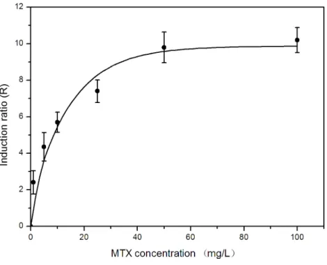
Figure 1 Effects of methotrexate dose on umu gene expression inS. typhimuriumTA1535/pSK1002.
sacrificed by cervical dislocation to get the bilateral epididymis. Sperm deformity test was carried out according to the following method of Wyrobek et al. Two sperm suspensions were prepared from the caudal of each testis by mincing the caudal in physiological saline (Wyrobek et al., 1983) . The sperm was spread on a slide glass and stained with 1% Eosine Y for 45 min after which the slides were air dried. A total of 1,000 sperm cells of mice were
assessed for morphological abnormalities under oil immersion at×1,000 magnification.
Sperm head morphology was scored under the category of normal, sperm without hook, amorphous head, banana head and triangular head.
Statistical analysis
Values are presented as means ±standard deviation (SD).The data were analyzed for
statistical significance usingt-test (SPSS 13.0 for Windows). Apvalue of less than 0.05 was
deemed as significant.
RESULTS
Screening of cytoprotector derived bioactive phytochemicals based on SOS/umu test
To ensure the reliability of screening results of cell protective agent from bioactive phytochemicals plant extracts and avoid the interference on genetic toxicity test from the growth inhibition effect, it is necessary to select the appropriate concentration of MTX
in SOS/umu test. Our previous study suggested that growth inhibition ofS. typhimurium
TA1535/pSK1002 was clearly observed while the concentration of MTX above 100 mg
L−1. Herein, we investigated the genotoxicity of MTX within the concentration range
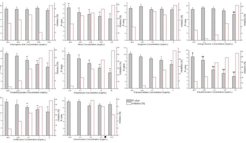
Figure 2 Effects of bioactive phytochemicals on umu gene expression inSalmonella typhimuriumTA1535/pSK1002 exposed to 50 mg L−1
methotrexate.Inhibition (%)=100×(Rcontrol−Rsample)/Rcontrol;∗p<0.05,∗∗p<0.01.
gene expression level and MTX genetoxicity wasn’t linear relationship. Before the MTX
concentration approached 50 mg L−1,Rvalue has already reached the platform. This
result is similar to the kinetics of induction of the umu operon by mitomycin C in S. typhimuriumNM2009 (Oda et al., 1995). In order to ensure the reliability of screening
results of cell protective agent from plant extracts, 50 mg L−1MTX was chosen in subsequent
SOS/umu test.
Effects of ten different plant extracts on umu gene expression inSalmonella typhimurium
TA1535/pSK1002 exposed to MTX were shown in Fig. 2. It was demonstrated that
allicin, proanthocyanidins, polyphenols, eleutherosides and isoflavones showed stronger antimutagenic activities than other five kinds of plant extracts. At the highest dose tested,
the MTX genetoxicity was inhibited by 34.03%∼67.12%. Subsequently, the pairwise
combinations of the five bioactive phytochemicals with antimutagenic activities were prepared by dissolving two plant extracts in DMSO solvent. The concentration of
effective components of each extracts reached 1 g L−1. According to the above methods,
antimutagenic activity of various combinations of plant extracts was determined by umu
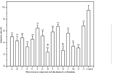
test. Antimutagenic potential of plant extract combinations were illustrated inFig. 3. Of
Figure 3 Effects of phytochemicals and phytochemical combinations on umu gene expression inS. typhimuriumTA1535/pSK1002 exposed to 50 mg L−1methotrexate. A, Allicin (1 g L−1); B, Allicin+ Grape seed proanthocyanidins; C, Allicin+Green tea polyphenols; D, Allicin+Eleutherosides; E, Allicin +Soybean isoflavones; F, Grape seed proanthocyanidins (1 g L−1); G, Grape seed proanthocyanidins+
Green tea polyphenols; H, Grape seed proanthocyanidins+Eleutherosides; I, Grape seed proanthocyani-dins+Soybean isoflavones; J, Green tea polyphenols (1 g L−1); K, Green tea polyphenols+
Eleuthero-sides; L, Green tea polyphenols+Soybean isoflavones; M, Eleutherosides (1 g L−1); N, Eleutherosides+
Soybean isoflavones; O, Soybean isoflavones (1 g L−1). a,p<0.01 as compared to Eleutherosides
treat-ment group; b,p<0.05 as compared to Eleutherosides treatment group;∗
p<0.05,∗∗
p<0.01 as com-pared to control.
extract exhibited higher cytoprotective activity. The inhibition rate of
methotrexate-induced genotoxicity separately reached 74.7 ± 6.5% and 71.8 ± 4.7%. The both
combinations of plant extracts were selected as cell protective agent candidates, and the cytoprotective effects of candidates would be subsequently assessed by micronucleus
test and sperm malformation testin vitro.
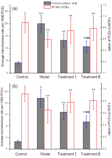
Evaluation of the antimutagenic potentials of cytoprotector candidates by micronucleus test
The micronucleus assay is internationally recognized as the standard method to detect the mutagenicity of chemicals. In order to assess the protective effects of the candidate cytoprotectors against the genotoxicity caused by MTX, micronucleus assay was employed in the next trial. With administration of the candidate plant extract combinations, the data of the bone marrow polychromatic erythrocyte micronucleus test in mice exposed to MTX
were presented inFig. 4. Whether male or female, there were statistically significant increases
(p<0.01) in the frequency of micronucleated polychromatic erythrocytes and ratio of
polychromatic erythrocytes (PCE) to normochromatic erythrocytes (NCE) in model group and control group. However, the treatment of the candidate cytoprotectors markedly restrained the incidence of mice bone marrow micronucleus, and improved the ratio of PEC and NEC. Furthermore, there were significant differences between treatment groups
Figure 4 Effect of cytoprotector candidates on incidence of micronucleated polychromatic erythro-cytes in bone marrow cells of mice treated with methotrexate.(A) male mice; (B) female mice; (1) Con-trol Group=saline; Model Group=methotrexate+normal saline; Treatment Group I=methotrexate +combination of polyphenols and eleutherosides; Treatment Group II=methotrexate+combination of proanthocyanidins and eleutherosides. (2)1P<0.05,11P<0.01 vs control. (3)∗P<0.05,∗∗P<0.01
vs Model Group. (4) #P<0.05, ##P<0.01 vs Treatment I.
Figure 5 Effect of cytoprotector candidates on abnormalities of male Kunmin mice sperm head after consecutive 7 days of methotrexate exposure.(1) Control Group=saline; Model Group=methotrexate +normal saline; Treatment Group I=methotrexate+combination of polyphenols and eleutherosides; Treatment Group II=methotrexate+combination of proanthocyanidins and eleutherosides. (2)1P< 0.05,11P<0.01 vs control. (3)∗P<0.05,∗∗P<0.01 vs Model Group.
Influence of cytoprotector candidates on reproductive toxicity induced by MTX
To investigate whether the combinations of bioactive phytochemicals treatment could strengthen or weaken the reproductive toxicity induced by MTX, sperm tests had been adopted. The method is one reliable and easy way to detect reproductive toxicity. In this study, the kinds of abnormal sperm in any group were mainly no hooks, amorphous,
bananas and triangular head heads. As could be seen fromFig. 5, the incidence of mouse
sperm head deformity of treatment groups had no significant difference from that of
the control (p>0.05). Significant differences could be observed between the treatment
groups and model groups (p<0.01). The summary of no hooks and amorphism heads
Figure 6 Effect of cytoprotector candidates on thymus and spleen indices of mice exposed to
Methotrexate.(A) male mice; (B) female mice; (1) Control Group=saline; Model Group=methotrexate +normal saline; Treatment Group I=methotrexate+combination of polyphenols and eleutherosides; Treatment Group II=methotrexate+combination of proanthocyanidins and eleutherosides. (2) 1P<0.05,11P<0.01 vs control. (3)∗P
<0.05,∗∗P
<0.01 vs Model Group.
Influence of cytoprotector candidates on immune organ indices
As cytoprotector candidates, the effect of two combinations of bioactive phytochemicals
on thymus and spleen indices of mice exposed to MTX was shown inFig. 6. The thymus
and spleen indices were similar between control and the treatment groups in both male
and female mice. MTX exposure reduced thymus and spleen indices of mice. Kawai
lymphocytes at dose≥5 mg/kg. However, there was a significant difference between the
treatment plus control group and the model group (p<0.01). The combination of grape
seed proanthocyanidins and Siberian ginseng eleutherosides obviously diminished the effects of MTX exposure on indices of thymus and spleens in mice. And, the increase of spleens index in the treatment of grape seed proanthocyanidins and Siberian ginseng eleutherosides combination was higher than that of green tea polyphenols and Siberian ginseng eleutherosides combination administration. Our findings implied that the two combinations of bioactive phytochemicals not only could affect the immune organ indices of experimental animal Kunming mice, but also could effectively relieve the immune toxicity caused by MTX.
DISCUSSION
The effective estimating for antimutagenic properties of bioactive phytochemicals includes
the bacterial gene mutation assay Ames test (Verschaeve & Van Staden, 2008; Horn &
Vargas, 2003), mammalian cell culture benzo(a)pyrene metabolism assay (Cassady et al., 1988), the mammalian cell gene mutation assay (Mersch-Sundermann et al., 2004), and thein vitromicronucleus assay (Serpeloni et al., 2008). Compared with animal-cell-based systems, microbe-based assays for screening cytoprotectors present several advantages, such as the simplicity of the procedures, the relatively short time needed to obtain results, and cost-effective. Nevertheless, except for the Ames test, there are few reports in the literature describing the studies of evaluation for antimutagenic properties of bioactive phytochemicals by umu test so far. In this study, we selected a short-term bacterial test system, the umu test, to evaluate the antimutagenic potential of plant extracts. Moreover, based on comparison of the umu test results (486 chemicals) with the Ames test (274
compounds) as well as rodent carcinogenicity data (179 compounds), Reifferscheid &
Heil (1996)found good agreement between the umu test and the Ames test results. Thus umu test could be developed an effective high-throughput screening assay to evaluate antimutagenic potential of phytochemicals same as Ames test and comet assay.
Natural plant medicine is an important resource to develop new cytoprotectors. Cytoprotective effects of many bioactive phytochemicals have been proved in the past several decades. Isoflavones from soybean seeds have showed antimutagenic activity in S. typhimuriumTA1535/pSK1002 and TA100 (Miyazawa et al., 1999). Garlic extract has been proved that it could reduce apoptotic cell injury induced by MTX. It should be noted that garlic was most popular supplement in US households (Amagase et al., 2001). Meanwhile, polyphenols from green tea (Chinese Gunpowder and Japanese
Sencha) exhibited high antimutagenic activity in the Ames test as well as inS. cerevisiae
bioactive phytochemicals against methotrexate-induced cytogenotoxicity has been reported rarely, such as Siberian ginseng eleutherosides, ginger gingerols, ginkgo flavone, ginseng ginsenosides, mushroom polysaccharides, and soybeam isoflavones. Our results suggested that allicin, proanthocyanidins, polyphenols, eleutherosides and isoflavones showed stronger antimutagenic activities than other five kinds of plant extracts. Moreover, it was demonstrated results single eleutherosides and its combinations with proanthocyanidins, polyphenols had strong antimutagenic effect. It is well known that the detrimental effects of MTX was partly due to its direct toxic action by increasing reactive oxygen species production, although the exact mechanisms of methotrexate-induced toxicity had not yet been elucidated to date (Oktem, 2006). Coleshowers’s work indicated that MTX causes oxidative stress by reducing the activities of superoxide dismutase, catalase and glutathione
reductase (Coleshowers et al., 2010).Gressier et al. (1994)demonstrated that MTX increases
the amount of hydrogen peroxide and other free radicals which may lead to toxicity thus accelerating the rate of cellular damage. Several studies have well confirmed the contribution of oxidative stress in the pathogenesis of MTX-induced damage in the various organs (Vardi, Parlakpinar & Ates, 2012). It is supposed that the oxidative stress induced by MTX was reduced and the cell damage caused by free radical accumulation was alleviated owing to potential antioxidant activity of the grape seed proanthocyanidins, green tea polyphenols,
soybean isoflavone and Siberian ginseng eleutherosides.Gulguna et al. (2010)demonstrated
that proanthocyanidin fromVitis viniferacan inhibits methotrexate-induced oxidative
stress. Yu et al. (2016)found that a polyphenol-rich Herb (Scutellariae radix) ingestion
increased the systemic exposure and mean residence time of MTX via modulation on efflux transporters multidrug resistance–associated protein 2 and breast cancer resistance
protein.Chiang et al. (2005) observed that the coadministration of Pueraria lobata root
decoction (an isoflavone-rich herb) significantly decreased the elimination and resulted in markedly increased exposure of MTX in rats. Recently, it was proved that eleutherosides (as one kind of polyphenols) were promising chemical substances with antioxidant properties. Substantially, grape seed proanthocyanidins, green tea polyphenols, soybean isoflavone and Siberian ginseng eleutherosides belong to the members of polyphenols which have strong antioxidant capacity. In this radical scavenging mechanism, polyphenols sacrificially reduce
ROS/RNS, such as•OH, O•−
2 , NO•, or OONO−after generation, preventing damage to
biomolecules or formation of more reactive ROS (Perron & Brumaghim, 2009). Previous research found that water extract of Siberian ginseng showed significant antioxidant activity
and protective effect against oxidative DNA damage induced by H2O2(Park et al., 2006).
Moreover, the methanol extract of root, stem, and leaf ofEleutherococcous senticosusshowed
inhibitory effects on the mutagenicity induced by 2-AF or Trp-P-1 inS. typhimuriumTA98
(Park et al., 2002). Therefore, we inferred that the cytoprotector candidates may play a key role in attenuating the methotrexate-induced cytogenotoxicity due to their antimutagenic and antioxidant activity.
decreases in sperm abnormality rate in the case of MTX exposure.Akram et al. (2012) ever reported that American ginseng extract treatment exhibited therapeutic effects on sperm parameters in rats treated with Cyclophosphamide (CP), which is an antineoplastic agent and immunosuppressive medicine in the treatment of various types of tumors, and autoimmune diseases such as systemic lupus erythematosus, rheumatoid arthritis and multiple sclerosis (Tripathi & Jena, 2009). Moreover, ginseng has been demonstrated
to have a cytoprotective effects against these toxins, in which administration ofPanax
ginseng extract was reported to markedly decrease the 2,3,7,8-tetrachlorodibenzo-p-dioxin-induced pathological and genotoxical damages in rat testes (Lee et al., 2007). Recent studies had confirmed that green tea and soybean extracts showed protective
effects against reproductive toxicity induced by CP.Fahmy et al. (2014)found a significant
decrease in the percentage of sperm abnormalities in orally administrated soybean extracts. Zanchi et al. (2015)demonstrated that polyphenols improved CP-induced damage on reproductive system, and its effect is probably due to high concentrations of catechins and antioxidant activity.
Although some evidence suggested that Siberian ginseng may not stimulate immune function (Wang et al., 2003), it has been indicated that polyphenols from green tea and grape seed proanthocyanidin have apparent immunomodulatory effects. Sheikhzadeh found that decaffeinated green tea in lower doses of administration could enhance the immunity of rainbow trout (Sheikhzadeh et al., 2011). The dietary administration of green tea supplementation positively raises the cellular immune responses and disease resistance of kelp grouper Epinephelus bruneus to Vibrio carchariae (Harikrishnan, Balasundaram & Heo, 2011).Tong et al. (2011) demonstrated that grape seed proanthocyanidins (GSPs) could improve functional activation of the immune system, and the antitumor effects of GSPs were achieved by immunostimulating properties. Our findings implied that the two combinations of phytochemical compounds not only could affect the immune organ indices of experimental animal Kunming mice, but also could effectively relieve the immune toxicity caused by MTX. According to the above results, it can be deduced that the combination of grape seed proanthocyanidins and Siberian ginseng eleutherosides as well as green tea polyphenols and eleutherosides was able to weaken the reproductive toxicity induced by MTX.
CONCLUSIONS
in the cytoprotector candidates treated groups. The results implied that phytochemicals combination of proanthocyanidins, eleutherosides and polyphenols could be used as new cytoprotectors.
ACKNOWLEDGEMENTS
We are grateful to Dr. Yoshimitsu Oda for the generous gift of S. typhimurium
TA1535/pSK1002.
ADDITIONAL INFORMATION AND DECLARATIONS
Funding
This work was supported by the Foundation for University Key Teacher by Henan Province (Grant No. 2014GGJS-056) and the National Natural Science Foundation of China (Grant No. U1304307). The funders had no role in study design, data collection and analysis, decision to publish, or preparation of the manuscript.
Grant Disclosures
The following grant information was disclosed by the authors: Henan Province: 2014GGJS-056.
National Natural Science Foundation of China: U1304307.
Competing Interests
The authors declare there are no competing interests.
Author Contributions
• Shaobin Gu conceived and designed the experiments, performed the experiments,
analyzed the data, contributed reagents/materials/analysis tools, wrote the paper, prepared figures and/or tables, reviewed drafts of the paper.
• Ying Wu performed the experiments, contributed reagents/materials/analysis tools,
reviewed drafts of the paper.
• Jianbo Yang conceived and designed the experiments.
Animal Ethics
The following information was supplied relating to ethical approvals (i.e., approving body and any reference numbers):
Kunming specific pathogen-free mice were provided by the Henan Laboratory Animal Center. License number: SCXK (Yu) 2005-0001.The present study was conducted in accordance with the principles outlined in the National Institutes of Health Guide for the Care and Use of Laboratory Animals.
Data Availability
The following information was supplied regarding data availability:
Supplemental Information
Supplemental information for this article can be found online athttp://dx.doi.org/10.7717/
peerj.1983#supplemental-information.
REFERENCES
Aggarwal P, Naik S, Mishra KP, Aggarwal A, Misra R. 2006.Correlation between
methotrexate efficacy&toxicity with C677T polymorphism of
themethylenetetrahy-drofolate gene in rheumatoid arthritis patients on folate supplementation.Indian
Journal of Medical Research124:521–526.
Akram H, Ghaderi Pakde F, Ahmadi A, Zare S. 2012.Beneficial effects of american
ginseng on epididymal sperm analyses in cyclophosphamide treated rats.Cell Journal
14:116–121.
Amagase H, Petesch BL, Matsuura H, Kasuga S, Itakura Y. 2001.Intake of garlic and its
bioactive components.The Journal of Nutrition131:955S–962S.
Attia SM, Helal GK, Abd-Ellah MF, Mansour AM, El-sayed E-SM. 2008.The effects
of oral grape seed extract on Cisplatin-induced cytogenotoxicity in mice.Saudi
Pharmaceutical Journal16:161–167.
Branda RF, O’Neill JP, Brooks EM, Powden C, Naud SJ, Nicklas JA. 2007.The effect
of dietary folic acid deficiency on the cytotoxic and mutagenic responses to methyl methanesulfonate in wild-type and in 3-methyladenine DNA glycosylase-deficient
Aag null mice.Mutation Research615:12–17DOI 10.1016/j.mrfmmm.2006.09.007.
Branda RF, O’Neill JP, Brooks EM, Trombley LM, Nicklas JA. 2001.The effect of folate
deficiency on the cytotoxic and mutagenic responses to ethyl methanesulfonate
in human lymphoblastoid cell lines that differ in p53 status.Mutation Research
473:51–71DOI 10.1016/S0027-5107(00)00138-X.
Buchbinder RI, Barber M, Heuzenroeder L, Wluka AE, Giles G, Hall S, Harkness A,
Lewis D, Littlejohn G, Miller MH, Ryan PF, Jolley D. 2008.Incidence of melanoma
and other malignancies among rheumatoid arthritis patients treated with methotrex-ate.Arthtitis and Rheumatism59:794–799DOI 10.1002/art.23716.
Bunkova R, Marova I, Nemec M. 2005.Antimutagenic properties of green tea.Plant
Foods for Human Nutrition60:25–29DOI 10.1007/s11130-005-2539-7.
Cassady JM, Zennie TM, Chae YH, Ferin MA, Portuondo NE, Baird WM. 1988.Use of a
mammalian cell culture benzo(a)pyrene metabolism assay for the detection of poten-tial anticarcinogens from natural products: inhibition of metabolism by biochanin A,
an isoflavone from Trifolium pratense L.Cancer Research48:6257–6261.
Cetin A, Kaynar L, Koçyiğit I, Hacioğlu SK, Saraymen R, Oztürk A, Orhan O, Sağdiç O.
2008.The effect of grape seed extract on radiation-induced oxidative stress in the rat
liver.The Turkish Journal of Gastroenterology19(2):92–98.
Chang CJ, Lin JF, Chang HH, Lee GA, Hung CF. 2013.Lutein protects against
methotrexate-induced and reactive oxygen species-mediated apoptotic cell injury
Chiang HM, Fang SH, Wen KC, Hsiu SL, Tsai SY, Hou YC, Chi YC, Chao PD.
2005.Life-threatening interaction between the root extract of Pueraria lobata
and methotrexate in rats.Toxicology and Applied Pharmacology 209:263–268
DOI 10.1016/j.taap.2005.04.015.
Choudhury RC, Palo AK. 2004.Modulatory effects of caffeine on
methotrexate-induced cytogenotoxicity in mouse bone marrow.Environmental Toxicology and
Pharmacology15:79–85DOI 10.1016/j.etap.2003.10.001.
Coleshowers CL, Oguntibeju OO, Ukpong M, Truter JE. 2010.Effects of methotrexate
on antioxidant enzyme status in a rodent model.Medical Technology SA24:5–9.
Coyle CH, Philips BJ, Morrisroe SN, Chancellor MB, Yoshimura N. 2008.Antioxidant
effects of green tea and its polyphenols on bladder cells.Life Sciences83:12–18
DOI 10.1016/j.lfs.2008.04.010.
Daggulli M, Dede O, Utangac MM, Bodakci MN, Hatipoglu NK, Penbegul N,
Sancaktu-tar AA, Bozkurt Y, Türkçü G, Yüksel H. 2014.Protective effects of carvacrol against
methotrexate-induced testicular toxicity in rats.International Journal of Clinical and
Experimental Medicine7:5511–5516.
Delmanto RD, Lima PLA, Sugui MM, Da Eira AF, Salvadori DMF, Speit G, Ribeiro
LR. 2001.Antimutagenic effect ofAgaricus blazeiMurrill mushroom on the
genotoxicity induced by cyclophosphamide.Mutation Research496:15–21
DOI 10.1016/S1383-5718(01)00228-5.
Fahmy MA, Hassan NHA, Melek FR, Hassan ZM, Al-Ashaa HA. 2014.Studies
on the genotoxic effect of nickel chloride in mice and the possible protective
role of soybean seeds extracts.Global Journal of Pharmacology 8:625–634
DOI 10.5829/idosi.gjp.2014.8.4.85195.
Gressier B, Lebegue S, Brunet C, Luyckx M, Dine T, Cazin M, Cazin JC. 1994.
Pro-oxidant properties of methotrexate: evaluation and prevention by an antioxidant
drug.Pharmazie49:679–681.
Gulguna M, Erdemb O, Oztasc E, Kesikd V, Balamtekina N, Vurucua S, Kula M,
Kismetd E, Koseoglu V. 2010.Proanthocyanidin prevents methotrexate-induced
intestinal damage andoxidative stress.Experimental and Toxicologic Pathology
62:109–115DOI 10.1016/j.etp.2009.02.120.
Guo MJ, Suo YR, Gao Q, Du H, Zeng WY, Wang YJ, Hu XT, Jiang XJ. 2015.The
protective mechanism of Ginkgolides and Ginkgo flavonoids on the TNF-α
induced apoptosis of rat hippocampal neurons and its mechanismsin vitro.Heliyon
1(1):e00020DOI 10.1016/j.heliyon.2015.e00020.
Harikrishnan R, Balasundaram C, Heo MS. 2011.Influence of diet enriched with green
tea on innate humoral and cellular immune response of kelp grouper (Epinephelus
bruneus) to Vibrio carchariae infection.Fish and Shellfish Immunology 30:972–979
DOI 10.1016/j.fsi.2011.01.029.
Horie T, Li T, Ito K, Sumi S, Fuwa T. 2006.Aged garlic extract protects against
methotrexate-induced apoptotic cell injury of IEC-6 cells.The Journal of Nutrition
Horn RC, Vargas VM. 2003.Antimutagenic activity of extracts of natural substances in
the Salmonella/microsome assay.Mutagenesis18:113–118
DOI 10.1093/mutage/18.2.113.
Kapiszewska M, Kalemba M, Wojciech U, Milewicz T. 2005.Uracil misincorporation
into DNA of leukocytes of youngwomenwith positive folate balance depends on plasma vitamin B12 concentrations and methylenetetrahydrofolate reductase
polymorphisms. A pilot study.Journal of Nutritional Biochemistry16:467–478
DOI 10.1016/j.jnutbio.2005.01.018.
Kawai R. 2014.Studies on primary and secondary responses to a T-cell-dependent
antigen, keyhole limpet hemocyanin (KLH), in immunotoxicology evaluation. PhD dissertation, Kyoto University, Kyoto, 63–65.
Knock E, Deng L, Wu Q, Lawrance AK, Wang XL, Rozen R. 2008.Strain differences in
mice highlight the role of DNA damage in neoplasia induced by low dietary folate. Journal of Nutrition138:653–658.
Kozarski M, Klaus A, Niksic M, Jakovljevic D, Helsper JPFG, Van Griensven LJLD.
2011.Antioxidative and immunomodulating activities of polysaccharide extracts
of the medicinal mushrooms Agaricus bisporus, Agaricus brasiliensis, Ganoderma
lucidum and Phellinus linteus.Food Chemistry129:1667–1675
DOI 10.1016/j.foodchem.2011.06.029.
Lee JH, Sul D, Oh E, Jung WW, Hwang KW, Hwang TS, Lee KC, Won NH. 2007.Panax
ginseng effects on DNA damage, CYP1A1 expression and histopathological changes
in testes of rats exposed to 2,3,7,8-tetrachlorodibenzo-p-dioxin.Food and Chemical
Toxicology45:2237–2244DOI 10.1016/j.fct.2007.05.019.
Li Z, Xu C. 2011.The fundamental theory of traditional Chinese medicine and
the consideration in its research strategy.Frontiers of Medicine5:208–211
DOI 10.1007/s11684-011-0126-x.
Mersch-Sundermann V, Knasmüller S, Wu XJ, Darroudi F, Kassie F. 2004.Use of a
human-derived liver cell line for the detection of cytoprotective, antigenotoxic and
cogenotoxic agents.Toxicology198:329–340DOI 10.1016/j.tox.2004.02.009.
Miyazawa M, Sakano K, Nakamura SI, Kosaka H. 1999.Antimutagenic activity of
isoflavones from soybean seeds (Glycine max Merrill).Journal of Agricultural and
Food Chemistry 47:1346–1349DOI 10.1021/jf9803583.
Oda Y, Nakamura S, Oki I, Kato T, Shinagawa H. 1985.Evaluation of the new system
(umu-test) for the detection of environmental mutagens and carcinogens.Mutation
Research147:219–229 DOI 10.1016/0165-1161(85)90062-7.
Oda Y, Yamazaki H, Watanabe M, Nohmi T, Shimada T. 1995.Development of
high sensitive umu test system: rapid detection of genotoxicity of promutagenic
aromatic amines bySalmonella typhimuriumstrain NM2009 possessing high
O-acetyltransferase activity.Mutation Research334:145–156
DOI 10.1016/0165-1161(95)90005-5.
Oktem F. 2006.Methotrexate-induced renal oxidative stress in rats: the role of a novel
antioxidant caffeic acid phenethyl ester.Toxicology and Industrial Health22:241–247
Padmanabhan S, Tripathi DN, Vikram A, Ramarao P, Jena GB. 2008.Cytotoxic and
genotoxic effects of methotrexate in germ cells of male Swiss mice.Mutation Research
655:59–67DOI 10.1016/j.mrgentox.2008.07.003.
Park JS, Oh CH, Koh HY, Choi DS. 2002.Antimutagenic effect of extract of
Eleuthe-rococcus senticosus Maxim.Korean Journal of Food Science and Technology
34:1110–1114.
Park HR, Park E, Rim AR, Jeon KI, Hwang JH, Lee SC. 2006.Antioxidant activity of
extracts fromAcanthopanax senticosus.African Journal of Biotechnology5:2388–2396.
Peng F, Tao Q, Wu X, Dou H, Spencer S, Mang C, Xu L, Sun L, Zhao Y, Li H,
Zeng S, Liu G, Hao X. 2012.Cytotoxic, cytoprotective and antioxidant effects
of isolated phenolic compounds from fresh ginger.Fitoterapia83:568–585
DOI 10.1016/j.fitote.2011.12.028.
Perron NR, Brumaghim JL. 2009.A review of the antioxidant mechanisms of polyphenol
compounds related to iron binding.Cell Biochemistry and Biophysics53:75–100
DOI 10.1007/s12013-009-9043-x.
Reifferscheid G, Heil J. 1996.Validation of the SOS/umu test using test results of 486
chemicals and comparison with the Ames test and carcinogenicity data.Mutation
Research369:129–145 DOI 10.1016/S0165-1218(96)90021-X.
Schmid W. 1975.The micronucleus test.Mutation Research31:9–15
DOI 10.1016/0165-1161(75)90058-8.
Schmiegelow K, AI-Modhwahi I, Andersen MK, Behrendtz M, Forestier E, Hasle H, Heyman M, Kristinsson J, Nersting J, Nygaard R, Svendsen AL, Vettenranta K, Weinshilboum R, Nordic Society for Paediatric Haematology and Oncology. 2009.
Methotrexate/6-mercaptopurine maintenance therapy influences the risk of a second malignant neoplasm after childhood acute lymphoblastic leukemia: results from the
NOPHO ALL-92 study.Blood113:6077–6084DOI 10.1182/blood-2008-11-187880.
Serpeloni JM, Bisarro dos Reis M, Rodrigues J, Campaner dos Santos L, Vilegas W,
Varanda EA, Dokkedal AL, Cólus IM. 2008.In vivoassessment of DNA damage
and protective effects of extracts from Miconia species using the comet assay and
micronucleus test.Mutagenesis23:501–507DOI 10.1093/mutage/gen043.
Sheikhzadeh N, Nofouzi K, Delazar A, Oushani AK. 2011.Immunomodulatory
effects of decaffeinated green tea (Camellia sinensis) on the immune system of
rainbow trout (Oncorhynchus mykiss).Fish Shellfish Immunol 31:1268–1269
DOI 10.1016/j.fsi.2011.09.010.
Tong H, Song X, Sun X, Sun G, Du F. 2011.Immunomodulatory and antitumor
activities of grape seed proanthocyanidins.Journal of Agricultural & Food Chemistry
59(21):11543–11547.
Tripathi DN, Jena GB. 2009.Intervention of astaxanthin against
cyclophosphamide-induced oxidative stress and DNA damage: a study in mice.Chemico-Biological
Interactions180:398–406DOI 10.1016/j.cbi.2009.03.017.
Vardi N, Parlakpinar H, Ates B. 2012.Beneficial effects of chlorogenic acid on
methotrexate-induced cerebellar Purkinje cell damage in rats.Journal of Chemical
Verschaeve L, Van Staden J. 2008.Mutagenic and antimutagenic properties of extracts
from South African traditional medicinal plants.Journal of Ethnopharmacology
119:575–587DOI 10.1016/j.jep.2008.06.007.
Vinson RK, Hales BF. 2002.Expression and activity of the DNA repair enzyme
uracil DNA glycoslyase during organogenesis in the rat conceptus and
follow-ing methotrexate exposurein vitro.Biochemical Pharmacology64:711–721
DOI 10.1016/S0006-2952(02)01252-2.
Wang H, Actor JK, Indrigo J, Olsen M, Dasgupta A. 2003.Asian and Siberian ginseng as
a potential modulator of immune function: anin vitrocytokine study using mouse
macrophages.Clinica Chimica ACTA327:123–128
DOI 10.1016/S0009-8981(02)00343-1.
Wang JH, Xu JL, Zhang JC, Liu Y, Sun HJ, Zha X. 2015.Physicochemical properties
and antioxidant activities of polysaccharide from floral mushroom cultivated in
Huangshan Mountain.Carbohydrate Polymers131:240–247
DOI 10.1016/j.carbpol.2015.05.052.
Wyrobek AJ, Gordon LA, Burkhart JG, Francis MW, Kapp Jr RW, Letz G, Malling HG,
Topham JC, Whorton MD. 1983.An evaluation of the mouse sperm morphology
test and other sperm tests in non-human mammals. A report of the United States
Environmental Protection Agency Gene-Tox Programme.Mutation Research
115:1–72DOI 10.1016/0165-1110(83)90014-3.
Yu CP, Hsieh YC, Shia CS, Hsu PW, Chen JY, Hou YC, Hsieh YW. 2016.Increased
systemic exposure of methotrexate by a polyphenol-rich herb via modulation on efflux transporters multidrug resistancee associated protein 2 and breast
cancer resistance protein.Journal of Pharmaceutical Sciences105:343e–349e
DOI 10.1016/j.xphs.2015.11.031.
Zanchi MM, Manfredini V, D dos Santos Brum, Vargas LM, Spiazzi CC, Soares MB,
Izaguirry AP, Santos FW. 2015.Green tea infusion improves
cyclophosphamide-induced damage on male mice reproductive system.Toxicology Reports2:252–260
DOI 10.1016/j.toxrep.2014.12.016.
Zhang QB, Li N, Zhou GF, Lu XL, Xu ZH, Li ZE. 2003.In vivoantioxidant activity
of polysaccharide fraction from Porphyrahaitanesis (Rhodephyta) in aging mice. Pharmacological Research48:151–155DOI 10.1016/S1043-6618(03)00103-8.
Zhao H, Li Q, Li Y. 2011.Long-term ginsenoside administration prevents memory
loss in aged female C57BL/6J mice by modulating the redox status and up-regulating
the plasticity-related proteins in hippocampus.Neuroscience183:189–202