DOI: 10.1590/0004-282X20150121
ARTICLE
Lipopolysaccharide-induced memory impairment
in rats is preventable using 7-nitroindazole
O déficit de memória induzido por lipossacarídeos em ratos é prevenido por nitroindazol
Akbar Anaeigoudari1, Mohammad Naser Shafei1, Mohammad Soukhtanloo2, Hamid Reza Sadeghnia3, Parham Reisi4, Farimah Beheshti1,Reza Mohebbati5, Seyed Mojtaba Mousavi5, Mahmoud Hosseini5
A large number of individuals in the world particularly
the elderly people sufer from diferent degrees of learning
and memory impairments. Although precise causes have
remained unknown until now, inlammation and oxidative
stress may at least in part be responsible for learning and
memory deicits1. Systemic inlammation promotes the pro
-duction of various cytokines such as tumor necrosis factors-α
(TNF-α), interleukin-1β (IL-1β) and IL-6 in the brain. It is
doc-umented that TNF-α impairs spatial learning and memory,
when it is administrated intracerebroventricularly before
wa-ter maze training. In addition, injection of TNF-α into the
hippocampus disrupted hippocampal-dependent working memory which was indicated by an enhanced number of er-rors and longer latencies to perform the three-panel runway task2. In human also inlammation is considered to contribute
in pathogenesis disorders including Alzheimer’s disease (AD),
Parkinson disease, multiple sclerosis3 and epilepsy4. In
mul-tiple animal models, exposure to immune system stimulating
1Neurocognitive Research Center, School of Medicine, Mashhad University of Medical Sciences, Mashhad, Iran;
2Department of Biochemistry, School of Medicine, Mashhad University of Medical Sciences, Mashhad, Iran;
3Pharmacological Research Center of Medicinal Plants, School of Medicine, Mashhad University of Medical Sciences, Mashhad, Iran;
4Department of Physiology, School of Medicine, Isfahan University of Medical Sciences, Isfahan, Iran;
5Neurogenic Inlammation Research Center, School of Medicine, Mashhad University of Medical Sciences, Mashhad, Iran.
Correspondence: Mahmoud Hosseini; Neurocognitive Research Center and Department of Physiology, School of Medicine, Mashhad University of Medical Sciences, Mashhad, Iran Mashhad 9177948564, Islamic Republic of Iran; E-mail: hosseinim@mums.ac.ir
Conflict of interest: There is no conlict of interest to declare.
Received 30 October 2014; Received in inal form 30 April 2015; Accepted 20 May 2015.
ABSTRACT
Inlammation and oxidative stress have important roles in memory impairment. The effect of 7-nitroindazole (7NI) on lipopolysaccharide (LPS)-induced memory impairment was investigated. Rats were used, divided into four groups that were treated as follows: (1) control (saline); (2) LPS; (3) 7NI-LPS; and (4) 7NI before passive avoidance (PA). In the LPS group, the latency for entering the dark compartment was shorter than in the controls (p < 0.01 and p < 0.001); while in the 7NI-LPS group, it was longer than in the LPS group (p < 0.01 and p < 0.001). Malondialdehyde (MDA) and nitric oxide (NO) metabolite concentrations in the brain tissues of the LPS group were higher than in the controls (p < 0.001 and p < 0.05); while in the 7NI-LPS group, they were lower than in the LPS group (p < 0.001 and p < 0.05, respectively). The thiol content in the brain of the LPS group was lower than in the controls (p < 0.001); while in the 7NI-LPS group, it was higher than in the LPS group (p < 0.001). It is suggested that brain tissue oxidative damage and NO elevation have a role in the deleterious effects of LPS on memory retention that are preventable using 7NI.
Keywords: learning, memory, lipopolysaccharide, 7NI.
RESUMO
Inlamação e estresse oxidativo tem importante papel no déicit de memória. O efeito do 7-nitroindazol (7NI) no déicit de memória induzido por lipossacarídeos (LPS) foi investigado. Foram utilizados ratos que foram divididos em quatro grupos e tratados da seguinte maneira: (1) controles (solução salina); (2) LPS; (3) 7NI-LPS; e (4) 7NI antes da esquiva passiva (PA). No grupo LPS, a latência para entrar no compartimento escuro foi mais curta que nos controles (p < 0,01 e p < 0,001); enquanto no grupo 7NI-LPS, a latência foi maior que aquela do grupo LPS (p < 0,01 e p < 0,001). Concentrações de malondialdeído (MDA) e metabólitos do ácido nítrico (NO) no tecido cerebral do grupo LPS foram maiores que aquelas dos controles (p < 0,001 e p < 0,05); enquanto no grupo 7NI-LPS, as concentrações foram menores do que no grupo LPS (p < 0,001 e p < 0,05, respectivamente). O conteúdo cerebral de tiol no grupo LPS foi menos do que nos controles (p < 0,001); enquanto no grupo 7NI-LPS, este conteúdo foi maior que no grupo LPS (p < 0,001). Sugere-se que o dano oxidativo cerebral e o aumento de NO tenham um papel nos efeitos deteriorativos dos LPS na memória de retenção, e que isto possa ser prevenido com o uso de 7NI.
pathogens such as viral and bacterial or viral coat proteins
and bacterial endotoxins results in learning and memory def
-icits2. Lipopolysaccharide (LPS) obtained from the cell wall
of gram-negative bacteria has been shown that promotes
the production of inlammatory cytokines that in turn lead to excessive production of free radicals and oxidative stress. Intraperitoneal injection of LPS also leads to neuroinlamma
-tion, hippocampus apoptosis, cognitive deicits and learning
and memory impairments5.
Nitric oxide (NO) is a difusible gaseous messenger which synthesized from amino acid L-arginine by the three difer
-ent isoforms of nitric oxide synthase (NOS) including; neuro
-nal NOS (nNOS), inducible NOS (iNOS) and endothelial NOS (eNOS). he nNOS is expressed in the neurons of the cerebel
-lum, hypothalamus, striatum, cerebral cortex and hippocam -pus as well as in the astrocytes. In the brain of individuals
with AD, nNOS is aberrantly expressed in vulnerable pyrami
-dal cells, astrocytes and nerve cells6,7. In physiological
con-centrations, NO plays a neuroprotective role in the nervous
system whereas, it promotes apoptosis and cell death in high
concentrations through stimulating of the superoxide anion
formation in the mitochondria8. It has been suggested that
overproductions of NO take place due to activation of nNOS by the stimuli such as endotoxins and cytokines9. It has also
been reported that both nNOS and eNOS isoforms afect
learning and memory10.
7-nitroindazol (7NI) has been considered as a
selec-tive nNOS inhibitor as well as a nonselecselec-tive NOS inhibitors (nNOS/eNOS)10,11. It has been shown that 7NI induces
learn-ing and memory deicits in diferent behavioral experiments
such as, Morris water maze(MWM), radial maze, passive
avoidance (PA) and elevated plus maze tests12.
Researchers have reported that LPS increases the
pro-duction of mediators such as NO, prostaglandin E2 (PGE2) and reactive oxygen species (ROS) through stimulating the
macrophages13. LPS also increases the level of NO through
promoting the release of pro-inlammatory cytokines such
as TNF-α and IL-1β from macrophages and leucocytes14. It
has been reported that the direct intracerebral injection of
LPS increases the levels of NO metabolites in the brain which is prevented by NOS inhibitors particularly iNOS inhibitors
such as aminoguanidine15. Despite these reports the aim
of this study was further evaluation of the efect of 7NI in
LPS-induced memory impairment.
METHOD
Animals and drugs
hirty-six male Wistar rats (8 weeks oldand 200-250g
weight) were keptin standard conditions (22 ± 2ºC
temper-ature and 12 h light/dark cycle). Working with the animals
was conducted in accordance with procedures approved by Mashhad Medical University Committee on Animal
Research (NO: A.H/1393/380). he animals were divided into four groups: (1) control (2) LPS, (3) 7NI-LPS and (4) 7NI (n = 9-10 in each group). LPS was dissolved in saline and injected (1 mg/kg; i.p.) 2 h before retention trail. 7NI was dissolved in saline supplemented with 3% dimethyl sulfox
-ide (DMSO) and injected (30 mg/kg; i.p.) 30 minutes before
LPS or saline in 7NI-LPS and 7NI groups respectively. In the LPS group, the animals were injected by saline
supplement-ed with 3% DMSO (2 mL/kg) instead of 7NI. he control and 7NI groups received 2 mL/kg of saline instead of LPS.
LPS and 7NI were purchased from Sigma (Sigma Aldrich
Chemical Co.). Other chemicals such as DMSO and those
which were used for biochemical assessments were pur-chased from Merck Company.
Passive avoidance (PA) test
PA apparatus is included of light and dark compart-ments separated by a guillotine door. In pre-acquisition trial, the animals were placed individually into the light compartment, the door was opened and the animals were
allowed to explore the compartments for 300 s during two
consecutive days. In the training trail, the rats were placed in the light compartment, the door was opened after 15 s and the latencies to enter the dark compartment were re-corded. When the animals were entered completely into the dark compartment, the door was closed and an electrical
shock (2 mA, 2 s) was delivered to the loor of the compart -ment. At 3, 24, 48 and 72 h later, the animals were again lo-cated in the light compartment for 15 s, after opening the guillotine door they gave access to the dark compartment
within a period of 300 s. hen the time latency to enter into
the dark room as well as the time spent by the animals in the light and dark compartments were recorded and
de-ined as retention trail16,17.
Biochemical assessment
he animals were sacriiced after a deep anesthesia, the
brains were removed, weighed, and submitted to determine
of total thiol (SH) content, malondialdehyde (MDA), and NO metabolites (NO2 or NO3) concentrations and the activity of
superoxidedismutase (SOD).
MDA assessment
MDA level is as an index of lipid peroxidation. MDA reacts
with thiobarbituric acid (TBA) as a TBA reactive substance
(TBARS) and produces a red complex. Briely, 1 mL of the brain homogenates was added to 2 mL of a complex solution containing TBA/trichloroacetic acid (TCA)/hydrochloric acid (HCL) and it was then boiled in a water bath for 40 min -utes. After reaching to the room temperature, the solution
was centrifuged at 1000 g for 10 minutes. he absorbance was read at 535 nm. he MDA concentration was calculated ac
-cording to follow equation18.
Determination of total thiol contents
DTNB (2, 2’-dinitro-5, 5’-dithiodibenzoic acid) reagent
which reacts with the SH group, was used to determine the total thiol contents. he produced yellow complex has a peak
absorbance at 412 nm. In brief, 50 μL of tissue homogenates
were added to 1 mL Tris-EDTA) ethylenediaminetetraace
-tic acid) bufer (pH = 8.6) and the absorbance was read at 412 nm against Tris-EDTA bufer alone (A1). hen, 20 μL of
10 mM solution of DTNB were mixed with the solution and
it was stored in room temperature for 15 minutes and the
absorbance was read again (A2). he absorbance of DTNB re -agent was also read as a blank (B). Total thiol concentration
(mM) was calculated as follow equation18.
Total thiol concentration
(mM) = (A2 – A1 – B) × 1.07 / 0.05 × 13.6
Determination of NO metabolites (NO2/NO3)
he Griess reaction was used to assayNOmetabolites.
Briely, standard curves for nitrates (Sigma St. Louis, Missouri,
USA) were prepared, and the samples (100 µL the tissue
sus-pension) were added to the Griess reagent. he proteins were
subsequently precipitated by addition of 50 µL of 10% TCA.
he contents were then vortex-mixed and centrifuged, and the supernatants were transferred to a 96-well lat-bottomed
microplate. Absorbance was read at 520 nm using a
micro-plate reader, and the inal values were calculated from stan
-dard calibration plots19,20.
Determination of SOD activity
SOD activity was measured by the procedure described
by Madesh and Balasubramanian. A colorimetric assay
in-volving generation of superoxide by pyrogallol auto-oxidation and the inhibition of superoxide-dependent reduction of the
tetrazolium dye, MTT (3-(4, 5-dimethylthiazol-2-yl)
2,5-di-phenyltetrazolium bromide) to its formazan by SOD was measured at 570 nm. One unit of SOD activity was deined
as the amount of enzyme causing 50% inhibition in the MTT reduction rate.
Statistical analysis
All data were expressed as means ± SEM. he data were evaluated by two-way ANOVA. here were two groups (control × LPS) and two treatments (Vehicle × 7NI). Diferences were considered statistically signiicant when p < 0.05.
RESULTS
PA results
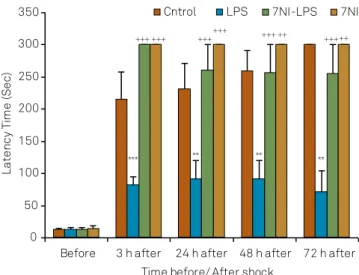
he results showed that there were no signiicant dif -ferences before receiving the shock in the latency for en-tering the dark compartment between the groups. Using
two way ANOVA, the results showed that there was also a signiicant main efects for LPS on latency to enter the
dark compartment at all 3( f(1,35) = 336.166; p < 0.001),
24( f(1,30) = 43.51; p < 0.001), 48( f(1,35) = 37.58; p < 0.001)
and 72( f(1,35) = 19.66; p < 0.001) hours after receiving
shock. 7NI also signiicantly afected latency to enter
the dark compartment at all 3( f(1,35) = 336.166; p < 0.001),
24( f(1,35) = 22.66; p < 0.001), 48 ( f(1,35) = 18.207; p < 0.001)
and 72 ( f(1,35) = 18.76;p < 0.001) hours after receiving
shock. here was a signiicant interaction between 7NI
and LPS on latency to enter the dark compartment at
all 3( f(1,35) = 336.166;p < 0.001), 24( f(1,35) = 20.18; p < 0.001),
48( f(1,35) = 16.16; p < 0.001) and 72 ( f(1,35) = 8.13; p < 0.01)
hours after receiving shock. he results also showed that
the latency to enter the dark compartment in LPS group
was signiicantly lower control group at 3, 24, 48 and 72 hours after receiving shock (p < 0.001 and p < 0.01). he la -tencyin 7NI-LPS and 7NI groups was longer than LPS group
(p < 0.001 and p < 0.01). here was no signiicant diference
between control, 7NI and 7NI-LPS groups (Figure 1).
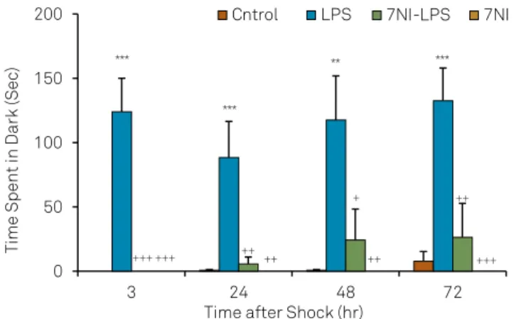
Using two way ANOVA, the results showed that there was also a signiicant main efects for LPS on the total time
spent in dark compartment at all 3( f(1,35) = 19.13; p < 0.001),
24( f(1,35) = 7.71; p < 0.01), 48( f(1,35) = 10.47; p < 0.01) and
72( f(1,35) = 18.80; p < 0.001) hours after receiving the shock.
7NI also signiicantly afected the total time spent in dark
compartment at all 3( f(1,35) = 19.13; p < 0.001), 24( f(1,35) = 6.18;
p < 0.01), 48 ( f(1,35) = 5.56; p < 0.05)and 72 ( f(1,35) = 10.68; p < 0.01)
hours after receiving the shock. here was a signiicant inter -action between 7NI and LPS on the total time spent in dark
LPS: lipopolysaccharide; 7N1: 7-nitroindazole.
Figure 1. Comparison of latency for entering to the dark compartment before and at the 3, 24, 48 and 72 hours after receiving the shock in the experimental groups. Data are presented as Mean ± SEM (n = 9-10 in each group). The animals of LPS and 7NI-LPS groups were treated by 1 mg/kg LPS, 2 h before the retention phase in passive avoidance test. The animals of 7NI and 7NI-LPS groups received 30 mg/kg 7NI, 30 minutes before saline and LPS respectively. The control group received saline. **p < 0.01, *** p < 0.001 compared to control group. ++ p < 0.01, +++ p < 0.001 compared to LPS group.
0 50 100 150 200 250 300 350
Before 3 h after 24 h after 48 h after 72 h after Time before/ After shock
La
te
nc
y
Ti
me
(S
ec
)
Cntrol LPS 7NI-LPS 7NI
*** ** ** **
compartment at all 3( f(1,35) = 19.13; p < 0.001), 24( f(1,35) = 5.99;
p < 0.05), 48( f(1,35) = 4.43; p < 0.05) and 72 ( f(1,35) = 7.98; p < 0.01)
hours after receiving shock. he total time spent in dark com
-partment by the animals LPS group was signiicantly longer
than control group at 3, 24, 48 and 72 hours after receiving
shock (p < 0.01, p < 0.001). he total time spent in dark com
-partment in 7NI and 7NI-LPS groups was signiicantly lower than LPS group (p < 0.05, p < 0.01, p < 0.001) but there was no signiicant diference between control, 7NI and 7NI-LPS
groups (Figure 2).
Using two way ANOVA, the results showed that there was also a signiicant main efects for LPS on the total time
spent in light compartment at all 3( f(1,30) = 71.09; p < 0.001),
24( f(1,30) = 10.45; p < 0.01), 48( f(1,30) = 13.06; p < 0.001) and
72( f(1,30) = 19.27; p < 0.001) hours after receiving the shock.
7NI also signiicantly afected the total time spent in light
compartment at all 3( f(1,30) = 71.09; p < 0.001), 24( f(1,30) = 8.56;
p < 0.01), 48 ( f(1,30) = 6.03; p < 0.05)and 72 ( f(1,30) = 12.13;
p < 0.01)hours after receiving the shock. here was a sig
-niicant interaction between 7NI and LPS on the total time
spent in light compartment at all 3( f(1,30) = 71.09; p < 0.001),
24( f(1,30) = 8.32; p < 0.01), 48( f(1,30) = 5.88; p < 0.05) and
72( f(1,30) = 9.14; p < 0.01)hours after receiving the shock. he
total time spent in light compartment by the animals which received LPS was lower than the rats of the control group
at 3, 24, 48 and 72 hour after receiving the shock (p < 0.001)
whereas, in the 7NI and 7NI-LPS groups was higher than
LPS group (p < 0.05, p < 0.01, p < 0.001). No signiicant dif -ference was observed between control, 7NI and 7NI-LPS groups (Figure 3).
Biochemical results
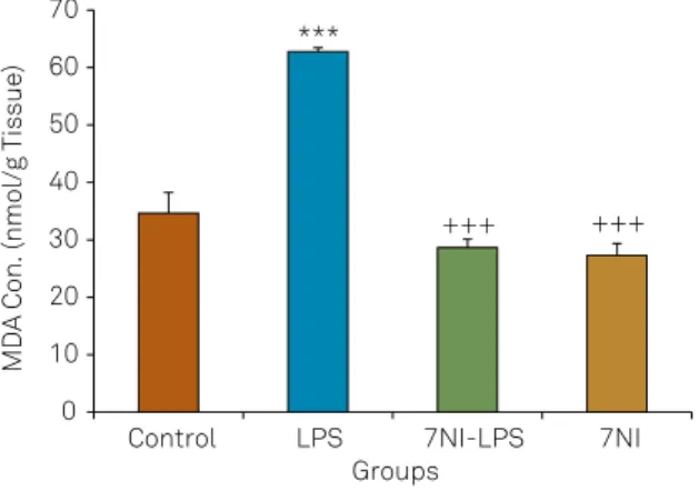
Using two way ANOVA, the results showed that there was a signiicant main efects for LPS on the MDA concen
-tration in the brain tissues ( f(1,35) = 28.57; p < 0.001). 7NI also
signiicantly afected the MDA concentration in the brain
tissues ( f(1,35) = 56.64; p < 0.001). here was a signiicant in
-teraction between 7NI and LPS on the MDA concentration ( f(1,35) = 23.62; p < 0.001). he results also showed that the
MDA concentration in the brain tissues of LPS group was
higher than the control group (p < 0.001). Injection of 7NI be -fore LPS decreased MDA concentration in the brain tissues
in comparison with LPS group (p < 0.001) however, there was no signiicant diference between the control, 7NI and
7NI-LPS groups (Figure 4).
Using two way ANOVA, the results showed that there was a signiicant main efects for LPS on the total thiol con
-tents in the brain tissues ( f(1,35) = 4.08; p < 0.05). 7NI also sig
-niicantly afected the total thiol contents in the brain tissues
( f(1,35) = 19.57;p < 0.01). here was a signiicant interaction be
-tween 7NI and LPS on the total thiol contents ( f(1,35) = 15.25;
p < 0.001). he total thiol contents in the brain tissues of LPS group was lower than the control group (p < 0.001). Injection
of 7NI increased the total thiol contents in 7NI-LPS group
compared with LPS group (p < 0.001). here was no signii
-cant diference between the control, 7NI and 7NI-LPS groups (Figure 5). here were no signiicant diferences in SOD activ -ity in the brain tissues between four groups (Figures 6).
Using two way ANOVA, the results showed that there was a signiicant main efects for LPS on NO metabolites concen
-trations in the brain tissues ( f(1,18) = 7.68; p < 0.05). 7NI also
LPS: lipopolysaccharide; 7N1: 7-nitroindazole.
Figure 2. Comparison of the total time spent in dark component at 3, 24, 48 and 72 hours after receiving the shock in the experimental groups. Data are presented as Mean ± SEM (n = 9-10 in each group). The animals of LPS and 7NI-LPS groups were treated by 1 mg/kg LPS, 2 h before the retention phase in passive avoidance test. The animals of 7NI and 7NI-LPS groups received 30 mg/kg 7NI,30 minutes before saline and LPS. The control group received saline. ** p < 0.01, *** p < 0.001 compared to control group. + p < 0.05, ++ p < 0.01, +++ p < 0.001 compared to LPS group.
0 50 100 150 200
3 24 48 72
Time Spent
in
Dark
(Sec)
Time after Shock (hr)
***
+++ +++ ++ ++
+
++ ***
++
+++ ***
**
Cntrol LPS 7NI-LPS 7NI
LPS: lipopolysaccharide; 7N1: 7-nitroindazole.
Figure 3. Comparison of the total time spent in light component at 3, 24, 48 and 72 hours after receiving the shock in the experimental groups. Data are presented as Mean ± SEM (n = 9-10 in each group). The animals of LPS and 7NI-LPS groups were treated by 1 mg/kg LPS, 2 h before the retention phase in passive avoidance test. The animals of 7NI and 7NI-LPS groups received 30 mg/kg 7NI, 30 minutes before saline and LPS respectively. The control group received saline. *** p < 0.001 compared to control group. + p < 0.05, ++ p < 0.01, +++ p <0.001 compared to LPS group.
0 50 100 150 200 250 300 350
3 24 48 72
Time Sepent
in Light (Sec)
Time after Shock (hr)
Cntrol LPS 7NI-LPS 7NI
***
*** ***
++++++ ++ +++ + ++ ++ +++
signiicantly afected NO metabolites concentrations in the
brain tissues ( f(1,18) = 4.02; p < 0.05). here was no a signiicant
interaction between 7NI and LPS on the NO metabolites con
-centrations ( f(1,18) = 3.25; p > 0.05). NO metabolites concen
-trations, in the brain tissues of LPS group were higher than
control group (p < 0.05).7NI decreased the concentrations of
these metabolites in 7NI-LPS group in comparison with LPS
group (p < 0.05) however, no signiicant diferences were seen in NO metabolites concentrations between the control, 7NI
and 7NI-LPS groups (Figure 7).
DISCUSSION
In the present study, LPS impaired the passive avoidance
memory retention. his inding was supported by decreas -ing the time latency to enter the dark compartment and
enhancing the total time spent in dark room and reducing the total time spent in light room in LPS group in comparison with control group. It has been indicated that injection of LPS
into the lateral ventricle of the rat,s brain attenuates
memo-ry retention trail21. Researcher has also reported that i.p.
ad-ministration of LPS-induced the passive avoidance memory
retention impairment22. In an another study, intraperitoneal
acute injection of LPS resulted in retention deicit after 24 h
and seventh day of its injection in both of passive avoidance
and elevated plus maze tasks23. All these indings conirm the
results of our study. During inlammation responses, microg
-lia as the principle efectors of immune system in the brain, release pro-inlammatory cytokines which directly afects
neuronal function including long-term potentiation,
glu-tamate release and cell signaling pathways24. LPS has been
shown that triggers the overproduction of pro-inlammatory
cytokines such as TNFα, IL-1β, IL-613,25. It has also been
report-ed that IL-1 receptors antagonist ameliorates learning and
MDA: malondialdehyde; LPS: lipopolysaccharide; 7N1: 7-nitroindazole.
Figure 4. Comparison of the MDA concentrations in cortical tissues of four groups. Data are presented as Mean ± SEM (n = 9-10 in each group). *** p < 0.01 compared to control group. +++ p < 0.001 compared to LPS group.
0 10 20 30 40 50 60 70
Control LPS 7NI-LPS 7NI
MDA Con. (nmol/g Tissue)
Groups ***
+++ +++
LPS: lipopolysaccharide; 7N1: 7-nitroindazole.
Figure 5. Comparison of the total thiol concentrations in cortical tissues of four groups. Data are presented as Mean ± SEM (n= 9-10 in each group). *** p < 0.01 compared to control group. +++ p < 0.001 compared to LPS group.
0 5 10 15 20 25 30 35
Control LPS 7NI-LPS 7NI
Total Thiol Con. (nM)
Groups ***
+++
+++
SOD: superoxide dismutase; LPS: lipopolysaccharide; 7N1: 7-nitroindazole.
Figure 6. Comparison of the SOD level in cortical tissues between four groups. Data are shown as Mean ± SEM (n = 9-10 in each group). There is no signiicant difference between groups.
0 0,2 0,4 0,6 0,8 1
control LPS 7NI-LPS 7NI
SOD A
ctivity (U/ml)
Groups
LPS: lipopolysaccharide; 7N1: 7-nitroindazole.
Figure 7. Comparison of the nitric oxide metabolites level in cortical tissues between four groups. Data are shown as Mean ± SEM (n= 5-6 in each group) * p < 0.05 compared to control group. + p < 0.05 compared to LPS group.
0 10 20 30 40
Control LPS 7NI-LPS 7NI
NO
2
/NO
3
Conc. (µmol/ml)
Groups *
memory deicits when it is administrated before LPS26. Due
to a high density cytokines receptors, hippocampus has been
considered to be very vulnerable to inlammation27. Using
animal models, it has been indicated that administration of cytokines or other immune system stimuli including LPS deteriorate hippocampus-dependent learning and memory
process24. Recently, the brain tissues oxidative damage was
considered to have an important role in deleterious efects
of LPS on learning and memory16. he cerebral cortex and
hippocampus which play crucial roles in learning and
cog-nition have been shown to be very sensitive to the oxidative
stress28. In our study, the level of MDA increased in LPS group
in comparison with the control while, the total thiol contents
diminished which conirms the contribution of brain tissues oxidative damage in impairing efects of LPS on memory re -tention which was seen in the present study. We observed
no signiicant diference in the activity SOD in the brain tis -sues between groups. It has been previously shown that LPS
disturbs the oxidative status and energy metabolism in the
brain29. In has also been reported that LPS increases the level
of MDA and reduces glutathione (GH) while, does not change the activity of SOD30.
NO has been shown to act as a cytotoxic agent if its pro
-duction gets out of control15. NO reacts with oxygen species
including superoxide (O -2
.) to produce peroxynitrite31. NO
and its derived-oxygen species starts damaging biochemical events including lipid peroxidation, protein oxidation, and ox
-idation of thiols, which inally lead to activation/deactivation
of various enzymatic systems32. It has also been reported that
LPS triggers the production of hydroxyl radical (OH•), nitric oxide metabolites (NOx-), superoxide (O2•) and other reac
-tive oxygen/nitrogen species as well as inlammatory cyto
-kines14. In contrast, NO as a retrograde messenger plays an
essential role in learning and memory processes, although its
efects are incompatible6. However, other reports conirmed
that a several fold concentrations of NO in the brain disturbs
the retention of acquired task in rats33. In this study, the level
of NO metabolites (NO2/NO3) in LPS-treated group was
high-er than control group. hus, it is possible that excessive pro
-duction of NO by probably inducing an oxidative procedure
has an important role in LPS-induced memory retention im-pairment which was seen in the present study. In the
pres-ent research, besides of prevpres-ention of impairing efects of LPS on memory retention, 7 NI prevented the enhancing efect of LPS on the brain levels of NO metabolites (NO2/NO3) in the
animals of 7NI-LPS group compared to LPS group. In agree with our results, it has been shown that i.p. injection of 7-NI reduced zinc-induced cell death in cerebellar Purkinje cells
of the rats34. In the current study, injection of 7-NI before LPS
resulted in a reduction of MDA concentration and enhance-ment of total thiol concentrations. According to the results of
present study, 7NI prevented oxidative damage thus it seems that 7-NI improves memory retention deicits through atten
-uating of the efect of LPS on brain tissues oxidative dam -age. Regarding the results of current study, it seems that the
impairing efects of LPS on memory retention impairment is partly through inducing the activity of nNOS which is pre -ventable by 7NI however, all of these mechanisms should be investigated in the future studies.
Finally, it is concluded that overproduction of NO and brain tissues oxidative damage which probably take place following LPS-induced inlammation, are important con
-tributing factors in the efects ofLPS on memory retention impairment. Regarding the protective efect of 7NI against
LPS-induced memory retention impairment in the present
study, which was accompanied with improving of oxidative stress criteria and lowering of NO metabolites, the mediatory efects of nNOS could be postulated.
Acknowledgments
he results described in this paper were from a PhD student’s thesis. he authors would like to thank the Vice
Presidency of Research of Mashhad University of Medical
Sciences for their inancial support.
References
1. Yan WW, Chen GH, Wang F, Tong JJ, Tao F. Long-term acarbose administration alleviating the impairment of spatial learning and memory in the SAMP8 mice was associated with alleviated reduction of insulin system and acetylated H4K8. Brain Res. 2015;1603:22-31. doi:10.1016/j.brainres.2015.01.042
2. Yirmiya R, Goshen I. Immune modulation of learning, memory, neural plasticity and neurogenesis. Brain Behav Immun. 2011;25(2):181-213. doi:10.1016/j.bbi.2010.10.015
3. Kipnis J, Derecki NC, Yang C, Scrable H. Immunity and cognition: what do age-related dementia, HIV-dementia and ‘chemo-brain’ have in common? Trends Immunol. 2008;29(10):455-63. doi:10.1016/j.it.2008.07.007
4. Walker L, Sills GJ. Inlammation and epilepsy: the foundations for a new therapeutic approach in epilepsy? Epilepsy Curr. 2012;12(1):8-12. doi:10.5698/1535-7511-12.1.8
5. Zarifkar A, Choopani S, Ghasemi R, Naghdi N, Maghsoudi AH, Maghsoudi N et al. Agmatine prevents LPS-induced spatial memory impairment and hippocampal apoptosis. Eur J Pharmacol. 2010;634(1-3):84-8. doi:10.1016/j.ejphar.2010.02.029
6. Guix FX, Uribesalgo I, Coma M, MuÑoz FJ. The physiology and pathophysiology of nitric oxide in the brain. Prog Neurobiol. 2005;76(2):126-52. doi:10.1016/j.pneurobio.2005.06.001
7. Lüth HJ, Münch G, Arendt T. Aberrant expression of NOS isoforms in Alzheimer’s disease is structurally related to nitrotyrosine formation. Brain Res. 2002 Oct 25;953(1-2):135-43. doi:10.1016/S0006-8993(02)03280-8
8. Brown GC, Borutaite V. Nitric oxide, cytochrome c and mitochondria. Biochem Soc Symp. 1999;66:17-25.
10. Akar F, Mutlu O, Komsuoglu Celikyurt I, Bektas E, Tanyeri P, Ulak G et al. Effects of 7-NI and ODQ on memory in the passive avoidance, novel object recognition, and social transmission of food preference tests in mice. Med Sci Monit Basic Res. 2014;20:27-35. doi:10.12659/MSMBR.890438
11. Yu SY, Zhang M, Luo J, Zhang L, Shao Y, Li G. Curcumin ameliorates memory deicits via neuronal nitric oxide synthase in aged mice. Prog Neuropsychopharmacol Biol Psychiatry. 2013;45:47-53. doi:10.1016/j.pnpbp.2013.05.001
12. Yildiz Akar F, Ulak G, Tanyeri P, Erden F, Utkan T, Gacar N. 7-Nitroindazole, a neuronal nitric oxide synthase inhibitor, impairs passive-avoidance and elevated plus-maze memory performance in rats. Pharmacol Biochem Behav. 2007;87(4):434-43. doi:10.1016/j.pbb.2007.05.019
13. Bak MJ, Truong VL, Kang HS, Jun M, Jeong WS. Anti-inlammatory effect of procyanidins from wild grape (Vitis amurensis) seeds in LPS-induced RAW 264.7 cells. Oxid Med Cell Longev. 2013;2013:409321. doi:10.1155/2013/409321
14. Hou CC, Lin H, Chang CP, Huang WT, Lin MT. Oxidative stress and pyrogenic fever pathogenesis. Eur J Pharmacol. 2011;667(1-3):6-12. doi:10.1016/j.ejphar.2011.05.075
15. Yamada K, Komori Y, Tanaka T, Senzaki K, Nikai T, Sugihara H et al. Brain dysfunction associated with an induction of nitric oxide synthase following an intracerebral injection of lipopolysaccharide in rats. Neuroscience. 1999;88(1):281-94. doi:10.1016/S0306-4522(98)00237-1
16. Pourganji M, Hosseini M, Soukhtanloo M, Zabihi H, Hadjzadeh MA. Protective role of endogenous ovarian hormones against learning and memory impairments and brain tissues oxidative damage induced by lipopolysaccharide. Iran Red Crescent Med J. 2014;16(3):e13954. doi:10.5812/ircmj.13954
17. Naghibi SM, Hosseini M, Khani F, Rahimi M, Vafaee F, Rakhshandeh H et al. Effect of aqueous extract of Crocus sativus L. on
morphine-induced memory impairment. Adv Pharmacol. Sci. 2012;2012:494367. doi:10.1155/2012/494367
18. Khodabandehloo F, Hosseini M, Rajaei Z, Soukhtanloo M, Farrokhi E, Rezaeipour M. Brain tissue oxidative damage as a possible mechanism for the deleterious effect of a chronic high dose of estradiol on learning and memory in ovariectomized rats. Arq Neuropsiquiatr. 2013;71(5):313-9. doi:10.1590/0004-282X20130027
19. Azizi-Malekabadi H, Hosseini M, Soukhtanlo M, Sadeghian R, Fereidoni M, Khodabandehloo F. Different effects of scopolamine on learning, memory, and nitric oxide metabolite levels in hippocampal tissues of ovariectomized and Sham-operated rats. Arq Neuropsiquiatr. 2012;70(6):447 doi:10.1590/S0004-282X2012000600012
20. Sadeghian R, Fereidoni M, Soukhtanlo M, Azizi-Malekabadi H, Hosseini M. Decreased nitric oxide levels in the hippocampus may play a role in learning and memory deicits in ovariectomized rats treated by a high dose of estradiol. Arq Neuropsiquiatr. 2012;70(11):874-9. doi:10.1590/S0004-282X2012001100010
21. Lee B, Sur B, Park J, Kim SH, Kwon S, Yeom M, et al. Ginsenoside rg3 alleviates lipopolysaccharide-induced learning and memory impairments by anti-inlammatory activity in rats. Biomol Ther (Seoul). 2013;21(5):381-90. doi:10.4062/biomolther.2013.053
22. Rostami F, Oryan S, Ahmadiani A, Dargahi L. Morphine
preconditioning protects against LPS-induced neuroinlammation
and memory deicit. J Mol Neurosci. 2012;48(1):22-34. doi:10.1007/s12031-012-9726-4
23. Jain NK, Patil CS, Kulkarni SK, Singh A. Modulatory role of cyclooxygenase inhibitors in aging- and scopolamine or lipopolysaccharide-induced cognitive dysfunction in mice. Behav Brain Res. 200218;133(2):369-76. doi:10.1016/S0166-4328(02)00025-6
24. Czerniawski J, Miyashita T, Lewandowski G, Guzowski JF. Systemic lipopolysaccharide administration impairs retrieval of context-object discrimination, but not spatial, memory: evidence for selective disruption of speciic hippocampus-dependent memory functions during acute neuroinlammation. Brain Behav Immun. 2015;44:159-66. doi:10.1016/j.bbi.2014.09.014
25. Dantzer R, O’Connor JC, Freund GG, Johnson RW, Kelley KW. From inlammation to sickness and depression: when the immune system subjugates the brain. Nat Rev Neurosci. 2008;9(1):46-56. doi:10.1038/nrn2297
26. Barrientos RM, Higgins EA, Sprunger DB, Watkins LR, Rudy JW, Maier SF. Memory for context is impaired by a post context exposure injection of interleukin-1 beta into dorsal hippocampus. Behav Brain Res. 2002;134(1-2):291-8. doi:10.1016/S0166-4328(02)00043-8
27. Schöbitz B, Voorhuis DA, De Kloet ER. Localization of interleukin 6 mRNA and interleukin 6 receptor mRNA in rat brain. Neurosci Lett. 1992;136(2):189-92. doi:10.1016/0304-3940(92)90046-A
28. Richardson JS. Free radicals in the genesis of
Alzheimer’s disease. Ann N Y Acad Sci. 1993;695(1):73-6. doi:10.1111/j.1749-6632.1993.tb23031.x
29. Sewerynek E, Melchiorri D, Chen L, Reiter RJ. Melatonin reduces both basal and bacterial lipopolysaccharide-induced lipid peroxidation in vitro. Free Radic Biol Med. 1995;19(6):903-9. doi:10.1016/0891-5849(95)00101-3
30. Kheir-Eldin AA, Motawi TK, Gad MZ, Abd-ElGawad HM. Protective effect of vitamin E, beta-carotene and N-acetylcysteine from the brain oxidative stress induced in rats by
lipopolysaccharide. Int J Biochem Cell Biol. 2001;33(5):475-82. doi:10.1016/S1357-2725(01)00032-2
31. Ischiropoulos H, Zhu L, Beckman JS. Peroxynitrite formation from macrophage-derived nitric oxide. Arch Biochem Biophys. 1992;298(2):446-51. doi:10.1016/0003-9861(92)90433-W
32. Luperchio S, Tamir S, Tannenbaum SR. NO-induced oxidative stress and glutathione metabolism in rodent and human cells. Free Radic Biol Med. 1996;21(4):513-9. doi:10.1016/0891-5849(96)00219-5
33. Reis EA, Oliveira LS, Lamers ML, Netto CA, Wyse AT. Arginine administration inhibits hippocampal Na(+),K(+)-ATPase activity and impairs retention of an inhibitory avoidance task in rats. Brain Res. 2002;951(2):151-7. doi:10.1016/S0006-8993(02)03077-9