657 Srp Arh Celok Lek. 2016 Nov-Dec;144(11-12):657-660 DOI: 10.2298/SARH1612657C
ПРИКАЗ БОЛЕСНИКА / CASE REPORT UDC: 616.216-002.828; 616.155.392-085.277.065
Correspondence to:
Nataša ČOLOVIĆ Clinic for Hematology Clinical Center of Serbia Dr Koste Todorovića 2 11010 Belgrade Serbia
natasacolovic73@gmail.com SUMMARY
Introduction Invasive fungal infection is among the leading causes of morbidity, mortality, and economic burden for patients with acute leukemia after induction of chemotherapy. In the past few decades, the incidence of invasive fungal infection has increased dramatically. Its management has been further complicated by the increasing frequency of infection by non-Aspergillus molds (e.g. Mucorales). Neu-tropenic patients are at a high risk of developing an invasive mucormycosis with fulminant course and high mortality rate (35–100%).
Case Outline We are presenting the case of a 72-year-old male with an acute monoblastic leukemia. The patient was treated during five days with hydroxycarbamide 2 × 500 mg/day, followed by cytara-bine 2 × 20 mg/sc over the next 10 days. He developed febrile neutropenia, headache, and edema of the right orbital region of the face. Computed tomography of the sinuses revealed shadow in sinuses with thickening of mucosa of the right paranasal sinuses. Lavage and aspirate from the sinuses revealed
Rhizopusoryzae. Mucormycosis was successfully treated with amphotericin B (5 mg/kg/day) followed by ketoconazole (400 mg/day). Two months later the patient died from primary disease.
Conclusion In patients with acute leukemia who developed aplasia, febrile neutropenia, and pain in paranasal sinuses, fungal infection should be taken into consideration. New and non-invasive methods for taking samples from sinuses should be standardized in order to establish an early and accurate diagnosis of mucormycosis with the source in paranasal sinuses, and to start early treatment by a proper antifungal drug. Clear communication between physician and mycologist is critical to ensure proper and timely sampling of lavage and aspirate from sinuses and correct specimen processing when mucormycosis is suspected clinically.
Keywords: acute leukemia; neutropenia; mucormycosis; paranasal sinuses; invasive fungal infection
Mucormycosis of the paranasal sinuses in a patient
with acute myeloid leukemia
Nataša Čolović1,2, Valentina Arsić-Arsenijević3, Aleksandra Barać3, Nada Suvajdžić1,2, Danijela Leković1,2, Dragica Tomin1,2
1Clinical Center of Serbia, Clinic for Hematology, Belgrade, Serbia; 2University of Belgrade, School of Medicine, Belgrade, Serbia;
3University of Belgrade, School of Medicine, Institute of Microbiology and Immunology, Belgrade, Serbia
INTRODUCTION
Patients with acute leukemia (AL) are consid-ered a population at high risk for developing an invasive fungal infection (IFI) [1]. In the Unit-ed States, in the past few decades, the incidence of IFI increased dramatically by approximately 200% between 1979 and 2000 [2]. In a recent study, the cumulative probability of develop-ing IFI after a diagnosis of AL was 11.1% at 100 days [3]. Patients undergoing treatment for hematologic malignancies have estimated cause-specific mortality due to IFI of 35% [4]. The most common causes of IFI are Candida
and Aspergillus, but most recent reports show an increasing frequency of infection by non-Aspergillus molds, especially Mucorales order. Mucormycosis is an aggressive opportunistic fungal infection with fulminant course caused by various members of the Mucorales order. The disease-causing genera in humans include
Absidia, Rhizopus,and Mucor, which are wide-spread in nature. They produce airborne spores that enter the body mostly through inhalation or ingestion; occasionally, infection may be through hematological dissemination from a different site, but sinuses and lungs are usually the entry points [5]. Fungi in sinuses cause
fun-gal rhinosinusitis (FRS), which has a spectrum from noninvasive disease to acute fulminant FRS [6]. Predisposing factors for the develop-ment of acute fulminant FRS are numerous, but neutropenia is the leading one, especially when neutrophils are below 0.5 × 10⁹/L. Invasive forms of FRS have very rapid course in neu-tropenic patients and because of that require early diagnosis, early induction of antifungal therapy, and sometimes surgery [7, 8].
We present a case of a patient with acute monoblastic leukemia (AML) and invasive mu-cormycosis of paranasal sinuses.
CASE REPORT
658
doi: 10.2298/SARH1612657C
Čolović N. et al. Mucormycosis of the paranasal sinuses in a patient with acute myeloid leukemia
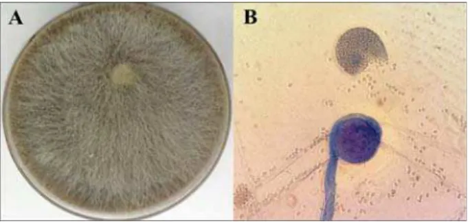
was in accordance with AML M5a. Immunophenotyping with flow cytometry revealed a population of mono-nuclear cells with immunophenotype HLA-DR, cMPO, cLizozim, CD13, CD33, CD15, CD11b, CD11c, CD64, CD14, CD163, CD36 and CD56+, indicating the diagnosis of AML CD56+ with monocytic differentiation. Karyotype was normal 46XY, and molecular analyses for FLT3 and NPM were negative. The patient was ranged in intermedi-ate group I according to Eureopean Leukemia Net classifi-cation. Hemostasis tests showed the following: fibrinogen 6.59 g/L, prothrombin time 70%, partial thromboplastin time 30.7 sec., and D dimer 2.1 μg/L. Biochemical analy-ses of blood showed elevation of lactate dehydrogenase 1,497 U/L, blood urea nitrogen 15.9 mmol/L, creatinin 388 μmol/L, and acidum uricum 917 μmol/L. Liver func-tion tests were within normal limits. Echocardiography showed ejection fraction of 67%, while chest radiography was normal. Abdominal ultrasound showed normal size of the liver and splenomegaly of 195 mm. The patient was treated for five days with hydroxycarbamide 2 × 500 mg/ day including rehydration and xanthine oxidase inhibitors in order to decrease the number of white blood cells. The therapy was continued with cytarabine 2 × 20 mg/sc dur-ing the next ten days. More aggressive treatment was not recommended because of the previous renal failure. After chemotherapy, the patient became neutropenic and febrile. He was treated with broad spectrum antibiotics without effects. Twenty days after the last dose of chemotherapy, the patient felt pain in the right maxillary and zygomatic region, as well as nasal congestion and headache. On the following day, the patient’s right palpebrae started swelling and the pain was spread all over the right half of the face. The radiography of paranasal sinuses revealed opacifica-tion of the right maxillary sinus with central illuminaopacifica-tion. Fungal hyphae was detected in an inducted lavage and aspirate of the right maxillary sinus, while culture isola-tion showed growth of Rhizopusoryzae (Figure 1). X-ray of paranasal sinuses showed filled lumen of cavities with oedematous epithelium. Computed tomography (CT) re-vealed mucosal thickening, hypoattenuation, opacification of sinuses as soft tissue attenuation of the right maxillary, sphenoidal, and both ethmoidal sinuses, as well as partly of the right frontal sinus, but without signs of invasion of endocranium. In the right nasal cavity the thick mucin like material was also present (Figure 2). After positive CT finding and mycological identification of Rhizopusoryzae
in sinonasal lavage and aspirate, the diagnosis of mucor-mycosis of the nose and paranasal cavities was established. Invasive mucormycosis was successfully treated with am-photericin B(5 mg/kg/day), followed by ketoconazole 400 mg/day, according to the results of antimycogram. Symptoms, pain, and changes over the paranasal region were resolved after treatment with antifungal therapy. Control aspirate and lavage of sinuses after two weeks of the last dose of antimycotic drugs were negative. Unfor-tunately, control bone marrow aspirate again showed the presence of 87% of monoblasts. Two months later the pa-tient died due to the primary disease.
DISCUSSION
IFI is a major cause of morbidity and mortality in patients with AL. Despite increased number of diagnosed cases of IFI, due to development of prompt and accurate diagnostic approaches and continual education about fungal diseases, the death rate due to IFI has dropped nearly 50% in the past two decades, from 44% during the 1995–2000 period to 28% during the 2001–2004 period [4]. The most fre-quent causing agents are Aspergillus, Dematiaceous molds such as Bipolaris, Curvularia,and Alternaria species and the Mucormycetes Rhizopus, Mucor,and Lichthemia [6]. Mucoraceae are ubiquitous fungi that are commonly found in soil and particularly in decaying matter [5]. In a study of patients with hematologic malignancies, the most frequent sites of mucormycosis were the lungs (64%) and the or-bito-sinus-facial structures (24%), while cerebral involve-ment and disseminated infection were observed in only 19% and 8% of the cases, respectively [9, 10]. Predispos-ing factors for these infections are neutropenia, especially when neutrophils are lower than 0.5 × 10⁹/L, long-lasting glucocorticosteroid therapy, radiotherapy, malnutrition, diabetes, and other accompanied diseases that have impact on a patient’s immune system [7, 8, 9, 11, 12]. Mucormy-cosis is an aggressive infection that can cause a significant disease in immunocompromised patients. Although many
Figure 2. CT showing the presence of fluid-like content in the right maxillary, sphenoidal and both ethmoidal sinuses, predominantly in the right, as well as partly in the right frontal one, but without invasion of endocranium; in the right nasal cavity, the thick mucin-like material was also noted
659
Srp Arh Celok Lek. 2016 Nov-Dec;144(11-12):657-660
www.srpskiarhiv.rs
patients with rhinocerebral mucormycosis undergo similar treatment, pathogen speciation should not be underap-preciated; it may be imperative to guide antifungal drug selection, as some mucoraceous fungi may exhibit variable resistance to conventional therapy [13].
Mucormycosis due to inhalation of fungal spores be-gins in the nose and paranasal sinuses with spreading to orbital or intracranial structures either by direct invasion or through blood vessels. The mortality rate is high, rang-es 50–85%, while in disseminated and untreated forms it could be 100% [3, 4, 14]. An immediate CT scanning of the paranasal sinuses and an endoscopic examination of nasal passages with biopsies of any suggestive lesions should be performed. Samples from sinuses should be obtained by non-invasive methods, due to condition of patients with neutropenia. Proper samples include inducted sinonasal lavage and aspirate, and should be taken at the onset of the disease before deterioration of symptoms. Clinical samples should be cultured and examined by histology and direct microscopy. Delays in diagnosis and treatment lead to in-creased mortality [11, 12].
We suppose that our patient had previous fungal colo-nization of paranasal sinuses, without symptoms, before diagnosis of AL was established. After worsening of AL, developing aplasia of bone marrow and febrile neutropenia IFI had rapidly progressed due to immunocompromised state. Positive CT finding, absence of response to broad spectrum antibiotics, and deterioration of symptoms were indications that FRS should be considered. CT scan re-vealed spread of the disease over paranasal sinuses and
aspirate and lavage of the sinuses were taken. Rhizopus oryzae was identified and according to antimycogram two antimycotic drugs, amphotericin Band ketoconazole, were successfully applied. Although the mucormycosis was suc-cessfully solved, AL was resistant to chemotherapy and the patient died as a more aggressive therapy could not have been applied because of the previous chronic renal failure.
In hematologic patients with prolonged febrile neutro-penia, headache, painful paranasal sinuses, fungal infec-tion should be seriously taken into considerainfec-tion. An early diagnosis of fungal infections of paranasal sinuses could be relatively easy established nowadays based on the ra-diography, CT scan visualization, and endoscopy with the invasive sampling as biopsy and histology of suspected lesion. New and non-invasive methods for taking samples from sinuses should be developed in order to make early and accurate diagnosis of mucormycosis with the source in the paranasal sinuses and to start early treatment by proper antifungal drug in patients with neutropenia. Clear com-munication between physician and laboratory is critical to ensure correct specimen processing when mucormycosis is suspected clinically.
ACKNOWLEDGEMENT
This work was supported by grant No. III 41004, awarded by the Ministry of Education, Science and Technologi-cal Development of the Republic of Serbia (Project OI 175034).
1. Bow EJ. Considerations in the approach to invasive fungal infection in patients with haematological malignancies. Br J Haematol. 2008; 140(2):133–52. [DOI: 10.1111/j.1365-2141.2007.06906.x]
[PMID: 18173752]
2. Martin GS, Mannino DM, Eaton S, Moss M. The epidemiology of sepsis in the United States from 1979 through 2000. N Engl J Med. 2003; 348(16):1546–54. [DOI: 10.1056/NEJMoa022139]
[PMID: 12700374]
3. Hammond SP, Marty FM, Bryar JM, DeAngelo DJ, Baden LR. Invasive fungal disease in patients treated for newly diagnosed acute leukemia. Am J Hematol. 2010; 85(9):695–9.
[DOI: 10.1002/ajh.21776] [PMID: 20652970]
4. Auberger J, Lass-Florl C, Ulmer H, Nogler-Semenitz E, Clausen J, Gunsilius E, et al. Significant alterations in the epidemiology and treatment outcome of invasive fungal infections in patients with hematological malignancies. Int J Hematol. 2008; 88(5):508–15. [DOI: 10.1007/s12185-008-0184-2] [PMID: 18982251]
5. Kwon-Chung KJ. Taxonomy of fungi causing mucormycosis and entomophthoramycosis (zygomycosis) and nomenclature of the disease: molecular mycologic perspectives. Clin Infect Dis. 2012; 54(1):8–15. [DOI: 10.1093/cid/cir864] [PMID: 22247451] 6. Thompson GR 3rd, Patterson TF. Fungal diseases of the nose and
paranasal sinuses- Current perspectives. J Allergy Clin Immunol. 2012; 321–6. [DOI: 10.1016/j.jaci.2011.11.039] [PMID: 22206776] 7. Taxy JB, El-Zayaty Sh, Langerman Al. Acute fungal sinusitis. Natural
history and the role of frozen section. Am J Clin Pathol. 2009; 132(1):86–93. [DOI: 10.1309/AJCP9HTH9NRPMYCT] [PMID: 19864238]
8. Čolović N, Arsić-Arsenijević V, Suvajdžić N, Đunić I, Tomin D. Povoljan ishod lečenja hepatosplenične kandidijaze kod bolesnice sa akutnom leukemijom. Srp Arh Celok Lek. 2015; 143(5-6):341–5. [DOI: 10.2298/SARH1506341C] [PMID: 26259411]
9. Pagano L, Offidani M, Fianchi L, Nosari A, Candoni A, Piccardi M, et al. Mucormycosis in hematologic patients. Haematologica. 2004; 89(2):207–14. [PMID: 15003897]
10. Vidovic A, Arsic-Arsenijevic V, Tomin D, Djunic I, Jakovic R, Loncar Z, et al. Proven invasive pulmonary mucormycosis successfully treated with amphotericin B and surgery in patient with acute myeloblastic leukemia: a case report. J Med Case Rep. 2013; 7:263. [DOI: 10.1186/1752-1947-7-263] [PMID: 24299522]
11. Parikh SL, Venkatraman G, DelGaudio JM. Invasive fungal sinusitis: a 15 year review from a single institution. Am J Rhinol. 2004; 18(2):75–81. [PMID: 15152871]
12. Lerchenmuller C, Goner M, Buchner T, Berdel WE. Rhinocerebral zygomycosis in a patient with acute lymphoblastic leukemia. Ann Oncol 2001; 12(3):415–9. [PMID: 11332157]
13. Alvarez E, Sutton DA, Cano J, Fothergill AW, Stchigel A, Rinaldi MG, et al. Spectrum of Zygomycete species identified in clinically significant specimens in the United States. J Clin Microbiol. 2009; 47(6):1650–6. [DOI: 10.1128/JCM.00036-09] [PMID: 19386856] 14. Turner JH, Soudry E, Nayak JV, Hwang PH. Survival outcomes in acute
invasive fungal sinusitis: a systemic review and quantitative synthesis of published evidence. Laryngoscope. 2013; 123(5):1112–8. [DOI: 10.1002/lary.23912] [PMID: 23300010]
660
КРАТАК САДРЖАЈ
Увод Инвазивне гљивичне инфекције (IFI) водећи су узрок морбидитета, морталитета и финансијског оптерећења за пацијенте са акутном леукемијом после примене инду-кционе хемотерапије. Неколико последњих деценија ин-циденца IFI се драматично повећала. Лечење IFI је додатно отежано и због повећане учесталости инфекција изазваних не-Aspergillus плеснима (нпр. Mucorales). Пацијенти са не-утропенијом су под високим ризиком за развој инвазивне мукормикозе која има фулминантни ток и високу стопу мор-талитета (35–100%).
Приказ болесника Приказан је случај пацијента старог 72 године, мушкарца, са акутном монобластном леукемијом, који је лечен током пет дана хидроксикарбамидому дози од 2 × 500 mg/дан, праћено цитарабиному дози од 2 × 20 mg/ sc током наредних 10 дана. Пацијенту се појављују симпто-ми главобоље, неутропенија и едем десне половине лица. Компјутеризована томографија (CT) параназалних синуса је открила засенчење синуса, са задебљањем мукозе десних параназалних синуса и са деструкцијом кости. У лавату и
аспирату синуса је показано присуство гљивице Rhizopus oryzae. Инвазиовна мукормикоза је успешно излечена ам-фотерицином Б (5 mg/kg/дан) праћено кетоконазолом (400
mg/дан). Два месеца касније пацијент је егзитирао због примарне болести.
Закључак Код хематолошких пацијената са акутном леуке-мијом код којих постоји присуство аплазије, пролонгиране фебрилне неутропеније и бола у параназалним синусима, потребно је размотрити могућност присуства гљивичне инфекције. Требало би стандардизовати нове неинвазивне методе за узорковање клиничког материјала из синуса, са циљем да се правовремено и тачно успостави дијагноза му-кормикозе, чији извор се налази у параназалним синусима и да се започне рана терапија са одговарајућим антигљи-вичним леком. Добра комуникација између клиничара и миколога је неопходна како би се осигурало правилно и правовремено узорковање лавата и аспирата синуса код сумње на мукормикозу.
Кључне речи: акутна леукемија; неутропенија; мукормико-за; параназални синуси; коинвазивна гљивична инфекција
И
уко
ко
код
п
је т
кут о
јело д о
леуке јо
Наташа Чоловић1,2, Валентина Арсић-Арсенијевић3, Александра Бараћ3, Нада Сувајџић1,2, Данијела Лековић1,2,
Драгица Томин1,2
1Клинички центар Србије, Клиника за хематологију, Београд, Србија; 2Универзитет у Београду, Медицински факултет, Београд, Србија;
3Универзитет у Београду, Медицински факултет, Институт за микробиологију и имунологију, Београд, Србија
Примљен • Received: 02/11/2015 Прихваћен • Accepted:14/12/2015
Čolović N. et al. Mucormycosis of the paranasal sinuses in a patient with acute myeloid leukemia