U
NIVERSIDADE DE
L
ISBOA
F
ACULDADE DE
C
IÊNCIAS
D
EPARTAMENTO DE
B
IOLOGIA
V
EGETAL
T
HE IMPORTANCE OF APOPTOSIS OF
Plasmodium-
INFECTED CELLS IN THE
GENERATION OF IMMUNITY AGAINST
MALARIA INFECTION
Catarina de Almeida Marques
M
ESTRADO EM
M
ICROBIOLOGIA
A
PLICADA
ii
U
NIVERSIDADE DE
L
ISBOA
F
ACULDADE DE
C
IÊNCIAS
D
EPARTAMENTO DE
B
IOLOGIA
V
EGETAL
T
HE IMPORTANCE OF APOPTOSIS OF
Plasmodium-
INFECTED CELLS IN THE
GENERATION OF IMMUNITY AGAINST
MALARIA INFECTION
Dissertação de Mestrado orientada pelo Doutor Miguel Prudêncio, Instituto de Medicina Molecular, Faculdade de Medicina da Universidade de Lisboa, e Prof. Doutor Rui Malhó, Faculdade de Ciências da Universidade de Lisboa.
Catarina de Almeida Marques
M
ESTRADO EM
M
ICROBIOLOGIA
A
PLICADA
iii The present project was presented on the “Young Researchers in Life Sciences 13th Meeting I Joint Edition”, under the form of an oral communication in the scope of the Immunology Session, on the 8th June of 2010.
iv
A
CKNOWLEDGMENTSÀ Maria, por me ter recebido no laboratório, dando-me a excelente oportunidade de conhecer o mundo da ciência por dentro, por todo o entusiasmo, apoio, inspiração e ideias. E por me dar este projecto, que adorei, para desenvolver durante o meu mestrado. Obrigado.
Ao Miguel, por me ter orientado durante dois anos (prémio de melhor orientador EVER!
Ao Prof. Doutor Rui Malhó, por ter aceitado ser Orientador Interno e estar disponível em caso de dúvida.
), por me ensinar tudo e mais alguma coisa, deixar-me meter o nariz em todas as experiências novas, e ter a maior paciência do mundo para me explicar tudo direitinho. Por toda a ajuda nas experiências que me deixavam com os nervos em franja – FLIVO!- e claro, todas as que envolviam a ida ao Lumina. Ainda mais pelos momentos divertidos e por ver que há algo de especial em mim, e confiar em mim.
Ao Prof. Rogério Tenreiro, Coordenador de Mestrado por esclarecer todas as minhas dúvidas, e me ter dado a oportunidade de desenvolver este projecto, mesmo estando ele na “zona cinzenta”. To all Malaria Unit (UMA – you know who you are!), for receiving me so well, for the good moments, all the help whenever I needed, and all the ideas. Um agradecimento especial, claro, às três meninas que tornam a UMA funcional, a Ana Roberto (ai a burocracia…), a Ana Parreira e a Fernanda, porque sem elas não há mosquitos, não há parasita, logo não há experiências e não há mestrado!
To all the Molecular Parasitology Unit (UPAMOL), Gunnar Mair, for the design of the transgenic parasite, and all the enlightenments, to Céline Carret, for everything, specially for the design of the primers to genotype the Casp3KO mice, and to Ana Guerreiro. Em especial, à Ana Rita, colega de curso, de laboratório, e acima de tudo, grande amiga.
A toda a equipa do Biotério do IMM. Especialmente ao Yuri e ao Joel, que sempre tomaram muito bem conta dos meus ratinhos Casp3KO. À Alina, por tudo, e também por me irradiar os parasitas. À Técnica Céu Raimundo, do Serviço de Radiologia do Hospital de Santa Maria, pelas tantas irradiações de parasitas.
Ao Diogo Pereira, da Unidade de Imunobiologia (UIB), tal como ao Dr. Henrique Veiga Fernandes, por tornarem a irradiação dos ratinhos da experiência das quimeras possível.
Aos meus amigos, pelo apoio, e pelos momentos de descontração fora da vida académica.
À minha Família, por partes. À camada sénior, Avô Quim, Avó Zeta, Avô Zé e Avó Laide. Por me apoiarem sempre, sejam quais forem as minhas decisões, e estarem os quatro, muito à frente do “seu tempo”. Aos meus pais, Joca e Nôno, sem quem, claro, não seria quem sou, não teria feito nada do que fiz, nem teria as ambições que tenho. Por tudo. À minha maninha Carol sem quem eu não sou nada. Por me fazer sentir mais leve e me alhear do mundo quando chego a casa.
E claro, ao Zee. E deixaria simplesmente assim, já que não existem palavras para descrever o quanto foi importante para mim tê-lo sempre ao meu lado durante este ano.
A todos, um enorme OBRIGADO (THANKS)!!!!!
v
A
BSTRACTDespite the efforts towards eradication, malaria remains the most deadly parasitic and vector-borne disease, urging for the development of an effective antimalarial vaccine. Recently, a renewed interest in Plasmodium attenuated whole-organism vaccine strategies has emerged, long acknowledged to experimentally induce full and sterile immunity against malaria in mice, monkeys and humans. This approach involves the use of non-replicating but metabolic active sporozoites, either attenuated by exposition to radiation, the Radiation-Attenuated Sporozoites (RAS), or by genetic modification, the Genetically-Attenuated Sporozoites (GAS). These attenuated parasites are able to infect hepatocytes
in vivo as normal sporozoites, but are unable to fully develop in the liver. This arrest in development
has been associated with their apparent failure to prevent host cell apoptosis, which leads to the formation of apoptotic bodies filled with parasite antigens. Several pieces of evidence have suggested that these apoptotic bodies may provide an optimal source of antigens for dendritic cells cross-presentation, and therefore, immune response activation. The aim of this project was to assess whether apoptosis of RAS or GAS infected hepatocytes is involved in the induced protective immunity response against subsequent parasite challenges. Results obtained through the immunization of C57BL/6 background caspase-3 deficient mice with P. berghei RAS or p36p- GAS revealed that these
mice are only partially protected against further infection. These suggest that at least to some extent, caspase-3 mediated apoptosis of infected hepatocytes may be important in generation of immunity by attenuated parasites.
vi
R
ESUMOEm pleno século XXI, e apesar de todas as medidas de controlo implementadas ao longo dos anos, a malária continua a ser uma das doenças mais mortíferas a assolar a humanidade. Só no ano de 2008, a malária foi responsável por 243 milhões de casos de doença e quase um milhão de mortes, maioritariamente no continente Africano.
A malária, ou paludismo, é causada por parasitas pertencentes ao género Plasmodium, um grupo de protozoários capazes de infectar grupos diversificados de animais vertebrados como mamíferos, aves e répteis. Em humanos, a malária é causada maioritariamente por quatro espécies, P.
falciparum, P. vivax, P. ovale e P. malariae, e é transmitida através da picada de mosquitos fêmea
infectados do género Anopheles. Uma vez inoculado no hospedeiro mamífero, o parasita, sob a forma de esporozoíto, inicia a fase assexuada do seu ciclo de vida, desenvolvendo-se inicialmente assintomaticamente no fígado, onde milhares de merozoítos, a forma do parasita capaz de infectar eritrócitos, são originados. Uma vez libertados na corrente sanguínea, os merozoítos rapidamente infectam eritrócitos, dando origem a mais merozoítos, e iniciando a fase eritrocitária do seu ciclo de vida e sintomática da doença. Contudo, ao fim de alguns ciclos de multiplicação em eritrócitos, alguns merozoítos acabam por gerar gametócitos, as formas sexuais do parasita, que são ingeridas pelo mosquito aquando da sua refeição de sangue. No mosquito, os gametócitos dão início à fase sexuada do ciclo de vida do parasita, que culmina com a formação de esporozoítos capazes de infectar novos hospedeiros mamíferos.
Durante décadas, muitos esforços têm sido empregues com o intuito de erradicar a malária. Contudo, estes têm-se revelado infrutíferos, e tanto parasitas como mosquitos resistentes às medidas de controlo empregues têm emergido nos últimos anos. Perante este cenário, tem-se sugerido que o desenvolvimento de terapias capazes de bloquear a transmissão do parasita do Homem para o mosquito, nomeadamente o de uma vacina eficaz contra a malária, é urgentemente necessário e crucial para se alcançar a erradicação da doença.
Há muito que se tenta desenvolver uma vacina anti-malárica eficaz. No entanto, tal tem-se revelado um dos maiores desafios da medicina, principalmente devido à extraordinária complexidade do ciclo de vida do parasita, durante o qual este se desenvolve e diferencia em formas que apresentam uma morfologia, metabolismo e conteúdo antigénico completamente distintos consoante a fase em que o parasita se encontra. A fase hepática constitui um alvo ideal para o desenvolvimento de uma vacina, uma vez que o desenvolvimento de uma vacina pré-eritrocitária eficaz teria simultaneamente capacidade profiláctica e de bloqueio da transmissão: a eliminação das formas pré-eritrocitárias do parasita (o esporozoíto e a forma exo-eritrocitária em desenvolvimento – EEF - dentro dos hepatócitos), permitiria impedir o aparecimento dos sintomas da infecção e, consequentemente, a transmissão do parasita ao mosquito. Contudo, uma vacina pré-eritrocitária tem que ser 100% eficaz em eliminar o parasita: basta que um esporozoíto se desenvolva completamente no fígado para que ocorra doença e, consequentemente, transmissão.
Vários estudos demonstram que a administração de esporozoítos atenuados mas metabolicamente activos é capaz de induzir uma resposta imune eficiente de longa duração em ratinhos, macacos e humanos, contra futuras infecções por esporozoítos. De facto, a desilusão relativa à eficácia de vacinas baseadas apenas em partes recombinantes de antigénios do parasita,
vii tem despertado um grande interesse para o desenvolvimento de uma vacina usando esporozoítos vivos atenuados que, em última análise, constitui uma vacina multi-antigénica e em tudo semelhante ao parasita infeccioso.
Os esporozoítos podem ser eficientemente atenuados através da sua exposição a uma determinada dose específica de radiação ionizante (X ou γ) (radiation attenuated sporozoites - RAS), ou da sua manipulação genética, em que genes essenciais para o desenvolvimento das formas pré-eritrocitárias do parasita são danificados (genetically attenuated sporozoites - GAS). Em ambas as estratégias de atenuação, os esporozoítos atenuados mantêm a sua capacidade de invadir hepatócitos, tal como o parasita selvagem, mas demonstram uma grave deficiência no seu desenvolvimento uma vez dentro da célula hospedeira, que os impede de produzir merozoítos e assim, causar doença. Esta deficiência em completar o seu desenvolvimento dentro do hepatócito tem sido associada com a incapacidade que os parasitas atenuados têm de inibir a apoptose da célula hospedeira.
Ao longo da evolução, os microrganismos intracelulares desenvolveram mecanismos que lhes permitem manipular os processos biológicos da célula hospedeira para seu benefício. Um dos processos que mais pressão selectiva tem imposto aos microrganismos intracelulares tem sido a morte das células hospedeiras por apoptose. Apesar de a apoptose poder ser desencadeada através de diversos estímulos e vias de sinalização, a via extrínseca, accionada por ligandos extracelulares, e a via intrínseca, desencadeada por sinais internos como o stress, são as mais comuns em células de mamíferos. Embora sejam despoletadas por diferentes estímulos, as duas vias apoptóticas convergem na activação de efectores comuns, as proteínas caspase, nomeadamente a da caspase-3, que aquando da sua activação, leva em última instância à morte celular e à resultante formação de corpos apoptóticos. Tal como em outros microrganismos intracelulares, tem sido demonstrado que os estádios pré-eritrocitários de Plasmodium possuem a capacidade de inibir a apoptose dos hepatócitos infectados, tanto aquando do processo de invasão, como durante os estádios iniciais e tardios do seu desenvolvimento. Aparentemente, a protecção da célula hospedeira à apoptose durante o desenvolvimento do parasita requer que este se mantenha vivo, e possivelmente, em processo de desenvolvimento. Várias observações sugerem que os parasitas atenuados, tanto RAS como GAS, são incapazes de evitar que a célula infectada entre em processo de apoptose e que, de facto, a imunidade gerada aquando da imunização com os parasitas atenuados resulta da sua incapacidade em proteger a célula hospedeira de entrar em apoptose aquando da infecção.
Quando a célula infectada sofre apoptose, formam-se corpos apoptóticos carregados com antigénios celulares e do parasita. Estes podem depois ser fagocitados por células apresentadoras de antigénios, tais como macrófagos e células dendríticas. Posteriormente, estas células podem fazer a apresentação destes antigénios ao sistema imunitário, desencadeando respostas imunes específicas contra o parasita. Vários estudos têm associado as células dendríticas com o estabelecimento da imunidade protectora induzida pelos parasitas atenuados, apontando-as como o veículo entre as células infectadas em apoptose e o sistema imunitário. De facto, as células dendríticas encontram-se associadas com a apresentação de antigénios exógenos através de moléculas de histocompatibilidade de classe I a células T CD8+, os principais elementos da resposta imune celular gerada pelos parasitas atenuados.
viii O presente trabalho pretende testar a hipótese de que a apoptose das células infectadas com os parasitas atenuados se encontra intimamente relacionada com o estabelecimento bem sucedido da resposta imune gerada por estes parasitas contra posteriores infecções por esporozoítos infecciosos.
Recorrendo a ratinhos da linhagem C57BL/6 deficientes em caspase-3 (Casp3KO), um elemento-chave em diversas vias da apoptose, vários ensaios de imunização com parasitas P. berghei ANKA atenuados, quer por a radiação γ (Pb-RAS), ou por manipulação genética (Pbp36p
-), foram realizados. Em cada ensaio, a posterior infecção foi efectuada com esporozoítos P. berghei ANKA que expressam constitutivamente a proteína de fluorescência verde (PbGFP) ou luciferase (PbLuci). Estes parasitas permitem, respectivamente através da análise por PCR quantitativo em tempo real (qRT-PCR) ou imagiologia in vivo em tempo real, inferir o nível de infecção no fígado e assim, a eficiência do regime de imunização. Contudo, o estabelecimento de uma resposta imunitária completamente eficiente contra a infecção foi apenas irrefutavelmente depreendida através da vigilância e seguimento do aparecimento de parasitas no sangue dos animais. Estes ensaios permitiram-nos verificar que os ratinhos Casp3KO encontram-se apenas parcialmente protegidos contra a infecção com esporozoítos infecciosos, sugerindo que as vias apoptóticas dependentes de caspase-3 se encontram possivelmente envolvidas no estabelecimento da imunidade induzida pelos parasitas atenuados.
De modo a excluir a possibilidade de que a imunidade parcial observada nos ratinhos Casp3KO se deve a uma deficiência ao nível do sistema imunitário destes animais, uma vez que se pensa dever à diminuição do nível de apoptose das células infectadas no fígado, a medula óssea de ratinhos Casp3KO foi substituída pela de ratinhos C57BL/6 normais, e vice-versa. Os ratinhos quimera resultantes foram imunizados com Pb-RAS, e posteriormente infectados com PbLuci. Infelizmente, este ensaio não nos permitiu retirar quaisquer conclusões concretas, apesar de sugerir que o fenótipo observado nos ratinhos Casp3KO imunizados se deve à sua maior resistência à apoptose nas células hepáticas infectadas com os parasitas atenuados.
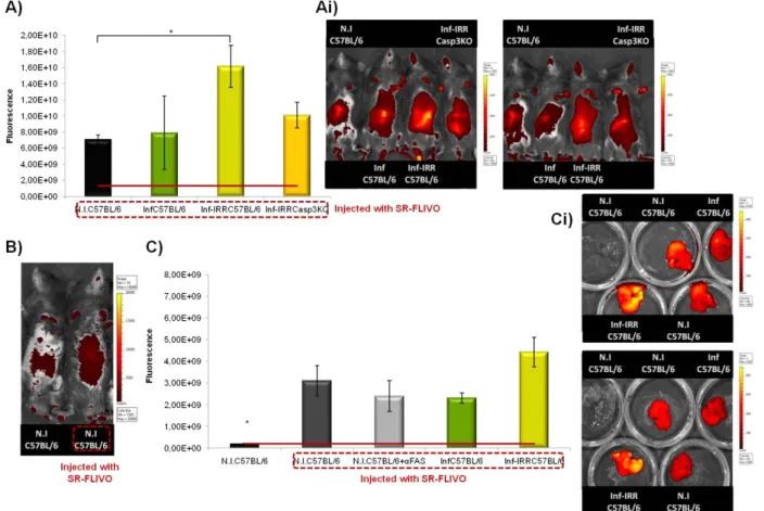
Através de uma sonda fluorescente que permite detectar níveis de apoptose in vivo, foi-nos possível inferir que há um aumento do nível de apoptose no fígado de ratinhos C57BL/6 infectados com PbRAS, que é aparentemente superior ao observado em ratinhos Casp3KO infectados com os mesmos parasitas, e ratinhos C57BL/6 infectados com parasitas infecciosos. Estes resultados permitem-nos de certo modo confirmar as observações de que os parasitas atenuados por radiação são incapazes de proteger a célula hospedeira de sofrer apoptose.
Em conjunto, os resultados aqui apresentados aparentam suportar a hipótese proposta, de que a apoptose das células infectadas com parasitas atenuados é relevante para o estabelecimento da resposta imune efectiva induzida por estes. Perante este panorama, planeámos construir um parasita
P. berghei ANKA transgénico que expressa e exporta para o citoplasma das células hospedeiras
caspase-2, um factor pró-apoptótico, com o intuito de induzir a apoptose das células infectadas e inferir se a infecção de ratinhos com este parasita é capaz de induzir uma resposta imune eficaz contra infecções posteriores com esporozoítos infecciosos P. berghei ANKA.
ix
M
OST USEDA
BBREVIATIONSBS – Blocking Solution.
Casp3KO mice - C57BL/6 background caspase-3 deficient mice.
CD8+ TCM – central memory CD8+ T cells.
CD8+ T
EM – effector memory CD8+ T cells.
ChimC57BL/6 – C57BL/6 mice reconstituted with Casp3KO mice bone marrow.
ChimCasp3KO - Casp3KO mice reconstituted with C57BL/6 mice bone marrow.
CM – cerebral malaria.
CSP– circumsporozoite protein.
DC– Dendritic Cell.
EEF– Exoerythrocytic form.
ELISA – enzyme-linked immunosorbent assay.
ELISPOT – enzyme-linked immunosorbent spot assay.
GAS – Genetically Attenuated Sporozoites.
i.p. – Intra-peritoneal.
i.v. – intravenously.
ImmC57BL/6 – group of C57BL/6 mice that were
immunized, either with PbRAS or PbP36p- sporozoites,
and subsequently infected (challenged) with WT PbGFP sporozoites.
ImmCasp3KO – group of Casp3KO mice that were
immunized, either with PbRAS or PbP36p- sporozoites,
and subsequently infected (challenged) with WT PbGFP sporozoites.
InfC57BL/6 – group of C57BL/6 mice that were only
infected.
InfCasp3KO – group of Casp3KO mice that were only infected.
Inf-IrrC57BL/6 – C57BL/6 mice infected with PbGFP-RAS.
Inf-IrrCasp3KO – Casp3KO mice infected with PbGFP-RAS.
N.I.C57BL/6 – non-infected C57BL/6 mice.
NIrr – Non-irradiated.
p.i. – post infection.
PbGFP – green fluorescent protein (GFP) – expressing P.
berghei ANKA.
PbGFP-Irr Hosp – PbGFP parasites that were exposed to
a γ-radiation dose of 16 Krad in a Compagnie Oris
Industrie IBL 437C irradiator.
PbGFP-Irr IMM – PbGFP parasites that were exposed to a γ-radiation dose of 16 Krad in a MDS Gamma Cell 3000
Elan irradiator.
PbGFP-RAS – Radiation attenuated PbGFP sporozoites.
PbLuci – luciferase – expressing P. berghei ANKA.
Pbp36p- - P36p deficient GFP – expressing Plasmodium berghei ANKA.
PbRAS – Plasmodium berghei radiation attenuated
sporozoites.
PV and PVM – parasitophorous vacuole and
parasitophorous vacuole membrane.
qRT-PCR - Quantitative Real-Time Reverse Transcription PCR.
RAS – Radiation Attenuated Sporozoites.
RBM – Roll Back Malaria partnership.
RT – Room temperature.
WHO – World Health Organization.
x
T
ABLE OFC
ONTENTSAcknowledgements iv
Abstract v
Resumo vi
Most used Abbreviations ix
Table of Contents x
Introduction 1
Malaria 1
Plasmodium life cycle 2
Malaria Eradication Efforts and Control Measures 4
Malaria Vaccination Strategies 5
The Subunit Vaccine Approach 5
The Live Attenuated Whole Organism Vaccine Approach – RAS & GAS 6
Life as an intracellular Pathogen – Modulation of Host Cell Apoptosis 10
Immune Responses induced by RAS and GAS 12
How are RAS and GAS Immune Responses Triggered? – DCs come into Play 14
Aim 15
Materials and Methods 17
Mice 17
Parasites 17
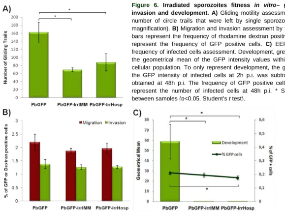
PbGFP-RAS fitness – in vitro gliding, migration, invasion and development assessment 17
P. berghei infection progression in Casp3KO mice 18
Immunization with PbGFP-RAS or PbP36p-GAS and parasite challenge experiments 18
Quantification of PbGFP liver infection by qRT-PCR 18
Quantification of PbLuci liver infection by Real-Time in vivo imaging 19
Caspase 3 KO bone marrow chimeric mice 19
In vivo detection of apoptosis in the liver 20
Transgenic Parasite – Pro-Caspase-2 expressing and exporting P. berghei 20
Statistical Analysis 22
Results and Discussion 23
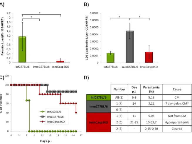
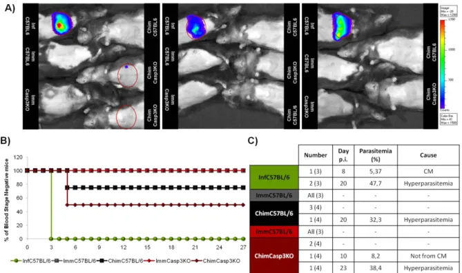
Infection with P. berghei ANKA – a different and variable outcome for Casp3KO mice 23
P. berghei ANKA RAS –the expected phenotype 25
Immunization with attenuated sporozoites – RAS and GAS 27
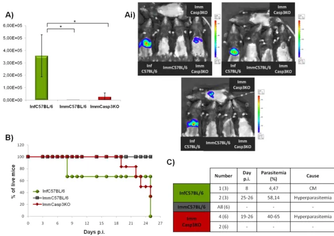
Immunization of chimeric mice with PbRAS 31
Detection of Apoptosis in vivo – mice infected with PbRAS have higher levels of apoptosis in the liver 33
Transgenic Parasite – a P. berghei ANKA parasite expressing and exporting to the host cytoplasm a pro-apoptotic factor 35
Conclusion and Future Perspectives 38
I
NT RO DUC T IO N 1I
NTRODUCTION
M
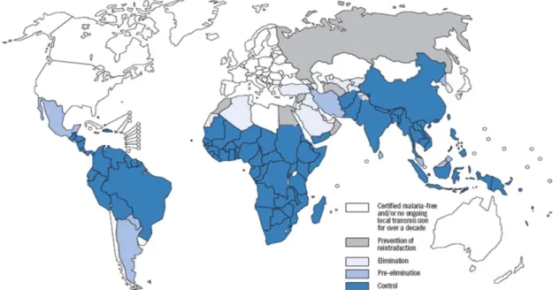
ALARIAMalaria is a disease that, despite all the efforts towards its eradication, and all the scientific breakthroughs, remains, in the 21st century, the world’s most deadly and morbid parasitic and vector-borne infectious disease. Presently, malaria is a life-threatening risk for 3,3 billion people over 109 countries, in the African, Asian and South American continents (Figure 1.). In 2008 alone, it was responsible for as many as 243 million cases of disease and 863.000 deaths, 85% of the losses being African children under the age of 5 (WHO, 2009). Moreover, malaria has an overwhelming impact on the affected countries’ economy, being annually accountable for a decrease in the gross domestic product (GDP) of as much as 1.3% in countries with high levels of transmission, and has consequently lead to economical and social disparities between countries with and without malaria (WHO, 2010).
Malaria is caused by protozoan parasites from the Plasmodium genus, and a member of the Apicomplexa phylum, to which other human pathogens, such as Toxoplasma gondii and
Cryptosporidium parvum, also belong (Ejigiri and Sinnis, 2009). By 2008 there were 199 Plasmodium
described species that, as a group, are able to infect a broad range of vertebrate hosts such as mammals, birds, and squamat reptiles (Martinsen et al., 2008), although each Plasmodium species has a remarkably restricted host range, and typically only infects few closely related vertebrate species (Matuschewski, 2006). In humans, malaria is mainly caused by four Plasmodium species, P.
falciparum, P. vivax, P. ovale and P. malariae, although a fifth species, P. knowlesi, a natural parasite
of macaque monkeys in south-east Asia, has been recently described to have occasionally infected and caused disease in humans (WHO, 2010). Together with P. vivax, P. falciparum, which is responsible for most of the severe and deadly malaria cases, accounts for the vast majority (over 93%) of the clinical cases notified each year (WHO, 2009), while P. ovale and P. malariae contribute to a lesser number of malaria infections (RBM, 2008). Malaria is transmitted to the vertebrate host by the bite of infected female mosquitoes, members of genera that comprise the most ubiquitous and
Figure 1. Global map of malaria-free countries and malaria-endemic countries in phases of control, pre-elimination, elimination and prevention of reintroduction by the end of 2008 (WHO, 2009).
I
NT RO DUC T IO N 2 abundant arthropod populations in most parts of the world: Anopheles in the case of mammalian malaria, and both Culex or Aedes in the case of avian malaria (Matuschewski, 2006). In fact, the use of such vectors constitutes one of the main reasons for malaria’s success.Plasmodium
L
IFEC
YCLEPlasmodium parasites possess a complex life cycle, divided into two obligatory hosts where
asexual, in the vertebrate, and sexual, in the mosquito, stages take place. During both, the parasite follows a developmental programme where it changes from extracellular motile invasive stages, to intracellular replicative ones (Matuschewski, 2006).
While taking its blood meal from a mammal host, the infected female Anopheles mosquito deposits sporozoites, a parasite infective form that is unable to infect erythrocytes, into the host avascular dermal tissue (Amino et al., 2006) (Figure 2. A). Here, sporozoites persist for some time, 1-3 hours (Amino et al., 2006; Yamaushi et al., 2007), apparently moving in a random fashion by gliding motility, an active movement pattern characteristic of Plasmodium spp. This is accompanied by active migration through the host cells with membrane disruption, and is required for sporozoite exit from the dermis. Migration, by traversing cells, allows the sporozoites to penetrate cell barriers, to escape destruction by phagocytic cells in the dermis, and to reach successfully the bloodstream (Amino et al., 2008). However, only a proportion of the initially inoculated sporozoites leaves the dermis, penetrates a blood vessel, and is carried within the bloodstream. Some sporozoites remain in the dermis, where they are probably eliminated by recruited phagocytes, while others penetrate a lymphatic vessel and reach the lymph node. Here, sporozoites are either phagocytosed by dendritic cells (DCs) or partially develop into early small exoerythrocytic forms (EEFs), before being degraded (Amino et al., 2006). Once in the bloodstream, sporozoites rapidly home to the liver, starting an obligatory and clinically silent liver, or pre-erythrocytic, stage of infection that, in humans, can last for 6 to 40 days, depending on the Plasmodium species (Murray et al., 2005). In the liver, sporozoites glide freely along the sinusoidal epithelium until they get sequestered. Here, they invade Kupffer cells, the liver’s resident macrophages, and gain access to the liver parenchyma (Frevert et al., 2005) (Figure 2. B). Sporozoites then migrate through several hepatocytes, before productively invading a single final one with the formation of a parasitophorous vacuole (PV) (Mota et al., 2001) (Figure 2. C). Afterwards, each invasive sporozoite dedifferentiates and develops into an EEF that grows and increases dramatically in volume without annihilating the host cell (reviewed in Vaughan et al., 2008) (Figure 2. D). During its growth, the EEF multiplies by schizogony (a process in which karyokinesis is temporally separated from cytokinesis, and a hallmark of Plasmodium spp. replication stages; Aly et al., 2009) into thousands of merozoites, the parasite’s next infective form capable to infect erythrocytes, that stay enclosed within the PV membrane (PVM) (Sturm et al., 2006). When EEF development is completed, merozoites are released from the PVM freely in the host cell cytoplasm, after which merozoite-filled vesicles from host cell membrane origin, the merosomes, are formed and released into the liver sinusoid (Sturm et al., 2006) (Figure 2. E).
Once in the liver sinusoids, the merosomes enter the bloodstream and accumulate in the lung’s microvasculature, where they finally break up releasing merozoites into the bloodstream (Baer et al., 2007; Strum et al., 2006) (Figure 2. F), and starting the symptomatic blood, or erythrocytic, stage of infection. Merozoites then rapidly attach to the erythrocytes, further penetrating them with the
I
NT RO DUC T IO N 3 formation of a PV. Subsequently, the invasive merozoite converts into a ring stage form, which develops into a trophozoite stage. The trophozoite then starts to multiply by schizogony, transforming itself into an erythrocyte schizont stage, which ultimately originates newly formed merozoites. When fully developed, merozoites egress from the erythrocyte, by PV and erythrocyte membrane rupture, after which they rapidly infect other erythrocytes, initiating a new erythrocytic cycle (reviewed in Silvieet al., 2008) (Figure 2. F). It is when the erythrocytes release merozoites that the typical pattern of
malaria symptoms appears: the paroxysms, consisting of chills, high fever and malarial rigors (Murray
et al., 2005). These usually appear periodically, the time interval depending on the length of the Plasmodium species erythrocytic cycle (in humans can be of 24-72 hours), and may remain relatively
mild, or progress to severe attacks with hours of sweating, chills, persistent high fever and exhaustion (Murray et al., 2005). If not treated, P. falciparum malaria evolves to a severe, and maybe fatal, disease with involvement of the brain, cerebral malaria, due to the accumulation of parasitized cells and cellular debris. After several erythrocytic cycles, some merozoites give origin to the sexual stages, the male and female gametocytes (Figure 2. G).
When a mosquito takes its blood meal from an infected individual, gametocytes are ingested, starting the sexual part of the parasite’s life cycle (Figure 2. H). Here, the midgut lumen environment triggers gametocytes to rapidly undergo gametogenesis (Bilker et al., 1997; Bilker et al., 1998), originating the male and female gametes that subsequently fuse to form the zygote, the only diploid form during the parasite’s life cycle. Soon after its formation, the zygote endures meiosis and genetic recombination, converting itself into an ookinete, a motile form of the parasite. Thereafter, the ookinete breaches the peritrophic matrix that encloses the blood meal and penetrates the mosquito midgut epithelium, where it traverses several epithelial cells. Subsequently, it exits the epithelium, and initiates its transformation into a sessile oocyst, the only extracellular developmental stage of the
Figure 2. Plasmodium spp. life cycle. Detailed description in the text. (A) The infected mosquito deposits sporozoites in the host’s skin where they gain access to a blood vessel. (B) In the liver sinusoids, sporozoites invade Kupffer cells and get access to the liver parenchyma. (C) Sporozoites migrate through several hepatocytes, invading a final one with formation of a PV. (D) Sporozoites then develop into EEFs, giving rise to thousands of merozoites. (E) Merosomes are release from the infected hepatocyte into the sinusoid lumen. (F) Erythrocytic cycle. (G) Some merozoites originate the male and female gametocytes. (H) During a blood meal, the mosquito uptakes the gametocytes, starting the parasite’s sexual part of the life cycle that ends with the accumulation of thousands of sporozoites in the salivary glands. (I) The mosquito takes a blood meal, and the cycle restarts. Adapted from Plasmodium life cycle from Malaria Unit of IMM.
I
NT RO DUC T IO N 4 parasite (reviewed in Aly et al., 2009). Afterwards, the oocyst develops, undergoing several mitotic divisions to originate sporoblasts, from which sporozoites start budding. When fully developed, thousands of mature sporozoites actively egress the oocyst and enter the hemocoel, where are carried through all mosquito tissues by the circulatory system, the hemolymph. While passing through the salivary glands, sporozoites adhere to the basal lamina, breach it, and invade the salivary gland secretory acinar cells, getting access to the salivary duct, where they accumulate and finish their maturation (reviewed in Aly et al., 2009). When the mosquito takes its next blood meal, sporozoites are injected into the mammalian host, starting the asexual part of the parasite’s life cycle (Figure 2. I).M
ALARIAE
RADICATIONE
FFORTS ANDC
ONTROLM
EASURESThroughout time, many have been the efforts to control, eliminate and, ultimately, eradicate malaria. However, these have not been completely successful, although malaria elimination was effectively achieved in Europe, North America and some countries in the North of Africa (Figure 1.). In 1959, the World Health Organization (WHO) launched the first attempt to eradicate malaria, the Global Programme for Malaria Eradication, which was based on mosquito control through indoor residual spraying (IRS) with dichloro-diphenyl-trichloroethane (DDT), and treatment of the infected populations with chloroquine (Greenwood, 2009). Sadly, the ambitious programme did not succeed in its main goal, and was abandoned in 1969. Nevertheless, it allowed malaria elimination in over 30 countries, a decrease of clinical burden in several endemic countries outside the African continent, and the removal of malaria risk from over one billion people (Greenwood, 2008; Greenwood, 2009). After the programme was abandoned, for some time no other attempts were made to eradicate malaria, and the efforts were directed to regional control and elimination, as has been done previously to the programme for years, back then, with limited success (Greenwood, 2009). In 2007, the Melinda and Bill Gates Foundation, the WHO, and the Roll Back Malaria Partnership (RBM) joint efforts to accomplish malaria eradication. This time, eradication will be endorsed by promoting national and regional gradual elimination using combinations of tools that are currently available: the artemisinin combination therapies (ACTs), the chemoprophylaxis or intermittent preventive treatment (IPT), IRS, and insecticide–treated bed nets (ITNs) or long-lasting ITNs (LLITNs) (Greenwood, 2008; Greenwood, 2009). It is believed that in many epidemiological situations the correct widespread and combination of these tools will be able to achieve up to 90% reduction in malaria clinical burden regionally. However, it is doubtful that in medium or high transmission areas these will be enough to accomplish malaria elimination and ultimately, eradication (Greenwood and Targett, 2009). Moreover, the emergence of resistance against several antimalarials by the parasite, and against several insecticides by the mosquito, threaten their efficacy. Therefore, it is argued that tools efficient in blocking parasite transmission, such as new gametocytocidal drugs, safer than primaquine, along with an efficient antimalarial vaccine, are essential and crucial for ultimate malaria eradication (reviewed in Greenwood, 2008).
I
NT RO DUC T IO N 5M
ALARIAV
ACCINATIONS
TRATEGIESSince the early 19th century, researchers have struggled to develop an effective antimalarial vaccine, long acknowledged as one of the greatest challenges of medicine (Luke and Hoffman, 2003). The main reason for this fruitless quest relies on the complexity of the parasite’s life cycle, comprising three distinct stages, each with a different gene expression pattern and consequently, different morphology, metabolism and antigen content (Vaughan et al., 2010). The pre-erythrocytic stages of the parasite, the sporozoite and the developing EEF, constitute attractive targets for vaccine development: they are present in lower numbers than the erythrocytic stages, are completely asymptomatic, and provide a relatively large window of opportunity for an effective immune response to eliminate the parasite (Vaughan et al., 2010), given that parasite development in the human liver lasts for several days. Moreover, pre-erythrocytic stages appear to not exhibit substantial antigenic variation or antigenic polymorphisms (Vaughan et al., 2010), conferring to a pre-erythrocytic vaccine the potential of being effective against heterologous parasite strains. If completely efficient, a pre-erythrocytic vaccine would promote the elimination of the parasite in the liver, therefore preventing disease onset, and consequently, parasite transmission. In fact, the major challenge for a successful pre-erythrocytic vaccine is that it is an all-or-none vaccination strategy, as it must eliminate every single parasite in the liver because, if even just one is able to fully develop, the parasite will proceed to the blood, cause disease and consequently, be transmitted (Vaughan et al., 2010).
The development of an effective pre-erythrocytic vaccine has relied mainly on two strategies, the subunit and the live attenuated whole organism approaches.
T
HES
UBUNITV
ACCINEA
PPROACHMost of the efforts to develop an effective vaccine against malaria have focused on the recombinant subunit approach. Several vaccine candidates have emerged through time, but the majority proved to be inefficient in inducing protective immunity in humans and were early abandoned. However, two strong candidates have reached human clinical trialsa
Sf66 was the first vaccine candidate to incorporate peptide sequences derived from both pre- and erythrocytic stages of the parasite life cycle. Sf66 was based on a synthetic peptide unit that, besides comprising the immunodominant unit NANP (N, asparagines; A, alanine; P, proline) of the CSP, also incorporated a fragment of the merozoite surface protein 1 (PfMSP-1, involved in the merozoite initial attachment to the erythrocyte upon infection) (reviewed in Silvie et al., 2008), and two uncharacterized peptides of 55 and 35KDa (Snounou and Rénia, 2007). Initially, Sf66 showed strong evidence of conferring protection against P. falciparum blood-stage challenge in monkeys and human volunteers. Facing such results, Sf66 was further tested, between 1992 and 1999, in several independent clinical , the Sf66 and the RTS,S vaccines, both comprising parts of the P. falciparum circumsporozoite protein (CSP), which is only expressed in the pre-erythrocytic stages of the parasite, both in the sporozoite and the EEF (Greenwood and Targett, 2009).
a
Steps in malaria vaccine development. (1) Research and preclinical development – identification of antigens and creation of the vaccine concept, preclinical evaluation in animal models and development of the manufacturing process. (2) Phase I
Clinical Trials (CT) – safety profile and immune response preliminary evaluation in naïve and exposed populations. (3) Phase II CT – safety and potential side effects monitoring, immune response measurement, efficacy against infection and clinical
disease evaluation and determination of the optimum dosage and schedule. (4) Phase III CT – further safety and potential side effects monitoring and efficacy evaluation on a large scale. (5) Submission to regulatory authorities. (6) Introduction in the
market. (7) Phase IV CT – post-marketing safety monitoring, protection duration measurement and vaccine compliance
I
NT RO DUC T IO N 6 trials in endemic countries. Unfortunately, it showed a reduced efficacy against infection, and was abandoned after being considered of no use in malaria control (Snounou and Rénia, 2007).The RTS,S vaccine is the most clinically advanced antimalarial vaccine candidate so far. It is based on the hepatitis B surface antigen (HBsAg or S antigen) virus-like particle platform, genetically engineered to harbour a P. falciparum CSP construct comprising 19 NANP repeats of the protein central region, known to induce B cell responses, and its entire C-terminal flanking region, known to induce specific T cell responses (Casares et al., 2010). To enhance its immunogenicity, the RTS,S construct was formulated in association with several adjuvant systems (AS), shown to induce strong humoral and cellular immune responses. Two formulations, RTS,S/AS02 and RTS,S/AS01 have shown promise results in studies with human volunteers, and were further tested in phase II clinical trials in endemic countries (GSK, 2010). In these clinical trials both formulas were shown to be safe to use as a vaccine in young children, with a strain unspecific efficacy against infection of about 45%, sadly far from ideal. Unexpectedly, since RTS,S was designed to act on pre-erythrocytic stages by only incorporating CSP as an immunogen, RTS,S formulations have also shown to be effective against clinical malaria, with around 44% efficacy, and against severe malaria development, with nearly 49% efficacy (Vekemans et al, 2009; Casares et al., 2010). Unfortunately, the efficiency of both formulas tended to decline with time. Altogether, RTS,S/AS01 showed a higher efficiency and more favourable safety data than RTS,S/AS02, and was selected for further evaluation in a phase III pre-licensure pivotal efficiency trial in several endemic countries, meaning that, a partially effective antimalarial vaccine will, hopefully, be available in a near future (Vekemans et al, 2009). Nonetheless, giving that the RTS,S has an efficacy lower that 50% against infection, it is arguable whether RTS,S will be useful when added to the other control measures in the quest to eradicate malaria (90% with the current tolls compared to 93% when combined with the vaccine). Moreover, the fact that it is only based on one P. falciparum protein raises the concern of whether a single protein based vaccine will be adequate to sustain malaria control, especially because it may bear the risk of emergence of resistant parasites (Greenwood and Targett, 2009; Luke and Hoffman, 2003).
T
HEL
IVEA
TTENUATEDW
HOLEO
RGANISMV
ACCINEA
PPROACH–
RAS
&
GAS
The observation that subunit vaccines fail to provide any significant and long lasting protective immunity has stimulated recent attempts to produce a vaccine based on an alternative approach, in which non-replicating but metabolically active live attenuated whole organisms are used. In fact, attenuated whole organism vaccines represent 75% of the currently licensed viral and anti-bacterial vaccines (Good, 2005), as it is the case of the ones against poliovirus, measles, typhoid fever, and tuberculosis (reviewed in Hoffman et al., 2010). Throughout the years, it has been shown that this approach can, at least in theory, be applied to the development of a highly effective antimalarial vaccine.
In 1967, it was demonstrated that immunization of mice with P. berghei, a rodent malaria parasite, radiation-attenuated sporozoites (RAS) was able to completely abrogate the onset of blood stage infection after challenge with infectious P. berghei sporozoites (100% efficacy) (Nussenzweig et al., 1967). Subsequently, the same level of sterile protection against infection was also induced in monkeys (Collins and Contacos, 1972) and humans (Clyde et al., 1973), immunized with P. cynomolgi RAS and P. falciparum RAS, respectively. Moreover, the protection conferred by immunizations with
I
NT RO DUC T IO N 7 RAS was shown to be long-lasting either in mice, monkeys or humans (Nussenzweig et al., 1967; Clyde et al., 1973; Hoffman et al., 2002). Soon, it became evident that the safety and efficacy of the RAS parasites relied on a precise and adequate radiation dose (ionizing radiation, either X- or γ- radiation). The proper irradiation of the sporozoites introduces random mutations and breaks into the DNA, to an extent that allows them to survive and keep their ability to successfully invade hepatocytes, although their capacity to fully develop is completely abrogated, and therefore, infection with these RAS does not cause disease and parasite transmission (Figure 3. B) (Vaughan et al., 2010). Conversely, under-irradiation of sporozoites allows them to remain infectious and to fully mature in the liver, therefore causing malaria, while over-irradiation completely inactivates or kills the sporozoites, that consequently are not able to infect hepatocytes (Vanderberg et al., 1968). In fact, over-irradiated sporozoites fail to confer any significant protection, suggesting that antigens expressed by persistent, partially developed liver stage parasites may be critical for the induction and maintenance of protective immunity, thereby indicating that RAS must invade liver cells and persist as at least partially developed EEFs to induce an immune response (Ballou, 2007). Because RAScomprehend the entire parasite, they act as a complete multi-antigen vaccine, possessing all the antigens of the infective non-irradiated sporozoites (N-Irr). Therefore, the resulting immune response is quite complex, involving both cellular and humoral immune responses against infected hepatocytes and surface antigens present in the sporozoite surface, like the CSP (reviewed in Ballou, 2007).
Despite all the promising results of obtaining 90-100% efficacy of protection against infection in humans with RAS (Hoffman et al., 2002), for decades it was considered impossible to develop, license and commercialize a live attenuated whole organism vaccine for P. falciparum (Ballou, 2007). The main obstacles to its development were mainly associated with the processes of producing, collecting and purifying the sporozoites, which present a high risk of contamination with other potentially
Figure 3. The fate of wild-type and attenuated parasites once in the mouse liver. (A) Normal infection by a wild type
sporozoite. (B) Infection by RAS. (C) Infection by GAS. (B) and (C) both successfully home to the liver and infect hepatocytes, but do not fully develop to generate merozoites.
(D) Different levels of parasite development inside the
hepatocyte by several GAS. Adapted from Waters et al., 2005, and Vaughan et al., 2010.
I
NT RO DUC T IO N 8 pathogenic transmissible agents, from both human and mosquito origin, as well as with allergenic mosquito proteins. These concerns come from the fact that sporozoite production involves feeding the mosquitoes with gametocyte-infected human blood (always considered to carry an inherent non-zero risk of infectious agents), and further sporozoite collection and purification, involving the removal and disruption of the mosquito’s salivary glands. Moreover, the capability of scaling up these processes has also been a great limiting factor, as well as how this type of vaccine would be preserved and taken to the endemic areas (Ballou, 2007). In 2003, Stephen Hoffman, founder of the Sanaria Inc., and co-workers reappraised the potential of an attenuated whole organism vaccine against malaria, as well as the obstacles to produce it, and took the challenge to develop such a vaccine (Luke and Hoffman, 2003). Presently, Sanaria Inc. possesses the world’s first licensed facility for manufacturing a live attenuated malaria vaccine, where an entire single coordinated process of producing and preserving the first generation attenuated whole organism vaccine against P. falciparum malaria, the SanariaTM PfSPZ vaccine, has been developed (Hoffman et al., 2010). This tightly controlled process has enabled Sanaria Inc. to overcome the risks and limitations of contaminations, improper radiation of the sporozoites, as well as their further preservation and delivery to endemic countries, by cryopreservation, storage and distribution of the RAS in liquid nitrogen vapor phase (LNVP), an electricity-independent platform (Hoffman et al., 2010). Currently, the SanariaTM PfSPZ vaccine, given by intradermal (ID) or subcutaneous inoculation (SC) is being tested in phase I clinical trials, in experimentally challenged malaria naïve volunteers to assess its safety, immunogenicity, adequate dose, and protective efficacy (Vaughan et al., 2010). Ultimately, during clinical trials SanariaTM PfSPZ must demonstrate that it is aseptic (free from contaminating pathogens), pure (free of contaminating mosquito material), non-replicating (arrests during EEF development and is thus unable to cause malaria), and potent (capable of inducing protective immune response), in order to be introduced in the market (Hoffman et al., 2010). If it develops favourably, after all these years of research, an attenuated whole organism vaccine against malaria, with a high efficacy of protection, may finally be available in a relatively near future, and become an important tool to achieve malaria eradication.Although the efforts and breakthroughs accomplished by Sanaria Inc. appear to finally make feasible the development of a live attenuated whole organism vaccine using RAS, there will always be reservations regarding the safety and reproducibility of the radiation process (Ballou, 2007), as it is unacceptable that a vaccine itself may cause disease if it is improperly attenuated. Fortunately, advances in genetic engineering may have allowed researchers to overcome this problem. The availability of genome sequences for a number of Plasmodium species, including of P. falciparum, the generation of stage-specific gene expression data, and the capacity to genetically manipulate the parasite, has allowed the search for essential genes involved in parasite survival at different stages of its life cycle. Together, these have enabled the informed genetic manipulation of the parasite to produce genetically-attenuated sporozoites (GAS), that can be designed to exhibit distinct biological and antigenic features, therefore allowing the creation of strains that are safe and elicit complete protection, with low dose immunizations (Vaughan et al., 2010).
It has been demonstrated that the gain of infectivity by sporozoites (from non-infective sporozoites in the midgut to infective sporozoites inside the salivary glands) is accompanied by an extensive differential upregulated expression of the upregulated in infectious sporozoites (UIS) genes
I
NT RO DUC T IO N 9 (Matuschewski et al., 2002). Disruption of several of UIS genes, which the parasite lacks the ability to compensate for their loss, has been done in rodent parasites like P. berghei and P. yoelii, and allowed researchers to test whether it is feasible to develop an efficient genetically-attenuated antimalarial vaccine. In P. berghei, disruption of the UIS3 and UIS4 genes, both encoding transmembrane proteins present in the EEF PVM, as well as of the P36p gene (also termed P52), encoding for a putative glycosylphosphatidylinositol (GPI)-anchored protein, member of the 6-Cys domain protein family, has been successfully achieved by double cross-over recombination gene replacement. The resulting parasites, uis3-, uis4- and p36p-, have revealed complete arrest in EEF development soon after hepatocyte invasion (Figure 3. D), although all three have shown, as expected, to present a phenotype comparable to the one of the wild type (WT) parasite regarding the blood stage and mosquito stage infections, as well as hepatocyte migration and invasion processes (Mueller et al., 2005a; Mueller et al., 2005b; van Djik et al., 2005). However, in the case of the p36p- parasites, a consensus regarding the later processes has not been established. In two independent studies wherep36p- parasites were generated using similar strategies, one reported an increase in migration and a decrease in invasion with PV formation (Ishino et al., 2005), while the other described that both processes are comparable to those of the WT parasite, although invasion occurred without the formation of a PV (van Djik et al., 2005). Nevertheless, when mice were immunized with uis3-, uis4- or
p36p- parasites, complete and long-lasting immunity was generated against further challenges with WT sporozoites (Mueller et al., 2005a; Mueller et al., 2005b; Ishino et al., 2005; van Djik et al., 2005), demonstrating that rodent model GAS are highly effective vaccines. Unfortunately, when given to mice in high doses, uis4- and p36p- parasites (from both studies) were reported to cause breakthrough infections, although with a delayed appearance of parasites in the blood (Mueller et al., 2005b; Ishino
et al., 2005; van Djik et al., 2005), which may surrender their potential to be used as vaccines.
Recently, successful induction of complete long-lasting immunity was also reported to be achieved by disruption of other sporozoite specific gene, the SAP-1 (sporozoite asparagine-rich protein-1) in P.
yoelii (Aly et al., 2008). The sap1- parasites showed a phenotype comparable to the one of the WT regarding blood stage and mosquito infections, as well as migration and invasion of hepatocytes. Resembling what was described for the three aforementioned GAS, sap1- parasites showed complete early EEF development arrest (Figure 3. D) and, like the uis3- parasites, no breakthrough infections
occurred, even when extremely high doses of sap1- sporozoites were injected into the mice. The sap1 -parasites were shown to lack the expression of essential genes such as the UIS3, UIS4 and P36p genes, suggesting SAP-1 may be involved in a post-transcriptional mechanism of gene expression control, and that actually, more genes may be also abrogated in this parasite (Aly et al., 2008). This lack of several genes expression turns the sap1- parasite into an attractive GAS vaccine candidate due to its quasi-multi-loci attenuation.
Altogether, the data obtained with rodent models GAS, especially of uis3- and sap1- parasites, have shown that safe and highly protective live attenuated malaria parasites can be created by genetic manipulation. Moreover, these studies may allow researchers to transfer the knowledge acquired to the development of a P. falciparum GAS, since all the aforementioned genes possess orthologues in other Plasmodium species, including P. falciparum (Mueller et al., 2005a; Mueller et al., 2005b; Ishino
I
NT RO DUC T IO N 10 Recently, to assess the potential to create a GAS vaccine for human malaria, the P36p and theP36, a non-UIS gene encoding a putative secreted protein member of the 6-Cys domain protein
family, genes were simultaneously disrupted in P. falciparum by double cross-over recombination gene replacement, to avoid genetic reversion and reconstitution of the WT locus (VanBuskirk et al. 2009). The resulting p36p-/p36- parasites presented a phenotype comparable to the one of the WT
parasite regarding the blood stage and mosquito stage infections, as well as hepatocyte migration and invasion processes. Moreover, P. falciparum p36p-/p36- parasites exhibited a profound EEF
development arrest, and were not able to survive beyond the third day post infection (p.i.), both in vitro (using a human hepatic cell line) and in vivo (using chimeric mice carrying human hepatocyte transplant), and therefore, were unable to persist in a growth-arrested state (VanBuskirk et al. 2009). These results were in accordance with the ones obtained in the P. yoelii model in which P36p and the
P36 orthologues were simultaneously disrupted. The P. yoelii p36p-/p36- parasite showed complete
arrest of the EEF development after invasion without PV formation (Figure 3. D) and no breakthrough infections were reported. Besides, complete immunization against further challenges with WT sporozoites was achieved (Labaied et al.2007). Facing such a strong phenotype of EEF development arrest, as well as the promising results obtained using a rodent model, the P. falciparum p36p-/p36
-parasite has been selected to advance into phase I clinical trials in human volunteers (Vaughan et al., 2010).
Altogether, these results, along with the technologies developed by Sanaria Inc., which can be adapted to GAS, indicate that it is feasible, in a somewhat near future, to generate a safe and efficient
P. falciparum GAS vaccine that offers the advantages of genetic homogeneity, standardization,
batch-to-batch consistency and testable identity over the RAS, and even, the subunit candidates.
L
IFE AS AN INTRACELLULARP
ATHOGEN–
M
ODULATION OFH
OSTC
ELLA
POPTOSISAttenuated Plasmodium parasites, both RAS and GAS, are unable to fully develop inside the infected hepatocyte, arresting their development at early EEF stages. Several pieces of evidence have suggested that this may be due to their inability to prevent host cell apoptosis during development inside the hepatocyte.
By living and developing within a host cell, intracellular pathogens are protected from immediate attacks by mechanisms of the host immune system. To achieve a successful intracellular life style, these pathogens have developed powerful methods to manipulate host cell functions in their own advantage, such as those involved in normal cell proliferation, cellular development and, specially, cell death by apoptosis, which is activated upon external or internal stimuli (van de Sand et al., 2005). Apoptosis is an active, highly specific and regulated, physiological process that plays an important role in tissue development and homeostasis, regulation and termination of immune responses and, importantly, in the removal of damaged or infected cells (reviewed in Carmen and Sinai, 2007).
There are two main pathways leading to apoptosis in mammalian cells, the intrinsic and the extrinsic pathways, both converging on common effectors, the caspases (cysteine proteases that cleave after aspartate residues). Briefly, the extrinsic pathway is triggered by the activation of a death receptor on the cell plasma membrane surface (e.g. FasL) following the binding of an external ligand (e.g. Fas). This leads to the formation of the death-inducing signalling complex (DISC) that activates the initiator caspase, caspase 8 that further activates an effector one, the caspase 3. Once activated,
I
NT RO DUC T IO N 11 caspase 3 ultimately leads to cell death, by being responsible for multiple proteins activation. Meanwhile, the intrinsic pathway is initiated by internal signals like DNA damage or stress, and relies on the release of cytochrome c from the mitochondria. Once in the cytosol, cytochrome c interacts with the apoptosis activating factor 1 (Apaf-1) that oligomerizes to form an apoptosome, which in turn, recruits and promotes the activation of the initiator caspase 9, further activation of caspase 3 and, consequently, cell death (reviewed in Carmen and Sinai, 2007; James and Green, 2004). Both pathways are tightly regulated by multiple inhibitory or survival mechanisms, such as the pro-survival responses mediated by the phosphatidylinositol 3-kinase (PI3-K) pathway, known to interfere and inhibit apoptosis (reviewed in Carmen and Sinai, 2007).Although no inflammatory response is elicited when infected cells undergo apoptosis, phagocytes are attracted to the site, where they efficiently recognize and eliminate the apoptotic cell harbouring the intracellular pathogen. Throughout evolution, host cell apoptosis has therefore placed a strong selective pressure on intracellular pathogens, either virus, bacteria and parasites, to inhibit the host cell apoptotic machinery (Heussler et al., 2001). Among protozoans, Toxoplasma gondii, Theileria spp., Trypanosoma cruzi and Leishmania spp. are known to confer resistance to host cell apoptosis, and Plasmodium spp. is not an exception (reviewed in Carmen and Sinai, 2007).
Once in the liver parenchyma, sporozoites traverse several hepatocytes before productively invading a final one with the formation of a PV (Figure 1. C). Upon traversal, wounded hepatocytes secrete hepatocyte growth factor (HGF), probably as a result of transient loss of their membrane integrity. HGF then binds to its tyrosine kinase receptor c-mesenchymal-epithelial transition factor (c-MET) on the surface of neighbour hepatocytes (Carrolo et al., 2003), leading to the activation of the HGF/c-MET signalling. This then mediates the coordinated execution of multiple cellular processes that both render the hepatocytes more susceptible for infection (Carrolo et al., 2003) and lead to the activation of the pro-survival PI3-K pathway (Leirião et al., 2005a). Activation of the PI3-K pathway by HGF/c-MET signalling has been shown, both in vitro and in vivo, to prevent host cell apoptosis during the initial phases of hepatocyte infection (Leirião et al., 2005a). However, this has not been observed at later stages of EEF development, since HGF/c-MET signalling requirement during Plasmodium infection of hepatocytes occurs at the early stages of establishment and development of sporozoites within the host cell (Carrolo et al., 2003). Nevertheless, prevention of apoptosis during infection mediated by the PI3-K pathway may also be induced by other signalling pathways, or even be mediated by other pathways. This is supported by the report that sporozoite microneme protein essential for cell traversal (SPECT) disrupted P. berghei parasites, which lack the ability to migrate through hepatocytes, and therefore, to induce HGF secretion, are able to infect hepatocytes in vitro comparably to the WT parasites (Ishinho et al., 2004). At later phases of EEF development, the parasite grows and develops far beyond the normal size of the host cell (Figure 1. D), exerting an important stress factor on the infected hepatocyte. However, infected hepatocytes were reported to not exhibit signs of cell death and to be protected against apoptosis induced by several stimuli, both in
vitro and in vivo (van de Sand et al., 2005). Actually, resistance to apoptosis seems to become more
pronounced during the EEF development, and to be independent from the PI3-K pathway. It is more likely that at later stages of hepatocyte infection the parasite blocks potential pro-apoptotic factors,
I
NT RO DUC T IO N 12 most probably by secretion of parasite molecules, rather than stimulating host cell survival pathways(van de Sand et al., 2005). In the meantime, these mechanisms remain to be elucidated.
Apparently, host cell protection from apoptosis requires that the parasite remains alive. There are indications that P. berghei RAS and P. berghei p36p- infected hepatocytes show evidence of apoptosis, like caspase-3 activation, shortly after invasion and early developmental phases. This suggests that these attenuated parasites, which are deficient in completing their development within the hepatocyte, fail to protect the host cell from entering in the apoptotic process (Leirião et al., 2005b; van Dijk et al., 2005). Interestingly, at the same time point, p36p- parasite-infected hepatocytes presented a significantly higher level of apoptosis than those infected with RAS, a difference that may be explained by the observed longer survival and development time of RAS within the host cell (Figure 3. B and D) (van Dijk et al., 2005). It appears that, the longer the attenuated parasite develops and survives within the host cell, the longer it is protected from apoptosis, maybe because the parasite, by arresting at an early stage of EEF development, lacks the capability of expressing factors that are essential for host cell apoptosis prevention, or by not developing further, ends up degenerating and subsequently, stops expressing the factors responsible for the protection. Surprisingly, it seems that within an infectious dose of WT parasites, some fail to prevent host cell apoptosis, maybe because they are deficient in some stage of their development, and end up degenerating (Leirião et al., 2005b; van Dijk et al., 2005). Nevertheless, this appears to be normal, occurring to a low percentage of WT parasites within an infectious dose.
I
MMUNER
ESPONSES INDUCED BYRAS
ANDGAS
As previously mentioned, immunization with live attenuated parasites elicits robust and long-lasting immune responses that completely protect against further infections. However, the nature of these immune responses is still not fully understood, although it appears that these may precisely be generated as a result from the fact that both RAS and GAS parasites fail to prevent host cell apoptosis.
For practical and ethical reasons, the vast majority of the studies aiming to understand the immune responses triggered by RAS and GAS immunization protocols have been performed using
Plasmodium parasite rodent model systems, mainly P. berghei and P. yoelii with laboratory mice from
two major backgrounds, BALB/c and C57BL/6. Collectively, results indicate that the sterile immunity generated by RAS is of a multi-factorial nature, involving both humoral and cellular immune responses, respectively targeting the sporozoite and the infected hepatocyte (Jobe et al., 2009). Although it includes both CD4+ and CD8+ T cells responses, the protective immune response was reported to be primarily mediated by intrahepatic interferon-γ (IFN-γ) producing effector memory CD8+ T cells (TEM), accompanied by the presence of central memory CD8
+
T cells (TCM) that endure homeostatic proliferation, forming a reservoir of memory T cells (Berezon et al., 2003). The importance of CD8+ T cell response is supported by the fact that immune protection by RAS is major histocompatibility class 1 (MHC-I)-dependent (White et al., 1996).
It has been argued that the success of the protection conferred by RAS and GAS, is owed to the fact that they elicit immune responses that are broad-based against multiple antigen targets, both from the sporozoite and early EEF developmental stages (Hoffman and Doolan, 2000). Nevertheless, evidence that the antibody responses induced were predominantly directed against the antigenic
I
NT RO DUC T IO N 13 repetitive central domain of the CSP (Zavala et al., 1986), as well as that adoptive transfer ofCSP-specific CD8+ or CD4+ T cell clones was able to confer full protection against WT parasite sporozoite challenge (Romero et al., 1989; Rodrigues et al., 1991; Renia et al., 1993), led to the hypothesis that RAS-induced immune responses might be mediated by few immunodominant antigens, such as the CSP. In fact, this has prompted the development of subunit vaccines essentially based on CSP, such as the RTS,S and Sf66 vaccines (see above). However, the modest efficacies obtained by these vaccines, along with incongruous results obtained recently in two independent reports (Kumar et al., 2006; Grüner et al., 2007), question the importance of immune responses against CSP in the induction and acquisition of sterile protection.
Using BALB/c mice tolerant to P. yoelii CSP, therefore devoid of T cell dependent responses against CSP, Kumar et al., 2006 reported that CSP is a crucial antigen for the efficacy of RAS immunization. However, Grüner et al., 2007 demonstrated that sterile protection against P. berghei malaria can be induced independently of specific T cell immune responses to CSP. They have shown that BALB/c mice could be fully immunized by P. berghei RAS whose endogenous CSP had been replaced by the P. falciparum CSP, which does not elicit minimal T cells responses in those mice. Moreover, Mauduit et al., 2009 corroborated these later results, and actually proposed that this disparity could be due to the fact that immune responses against CSP and non-CSP antigens are induced differently in the two rodent malaria species. They have suggested that in P. berghei immune responses against non-CSP antigens, responsible for the sterile protection, are rapidly induced after RAS immunization, while in P. yoelii the responses arising rapidly are against CSP, being the ones targeting non-CSP antigens only achieved after several boosts with RAS. Nevertheless, it is still assumed that CSP is indeed the immunodominant antigen regarding the humoral responses, being the anti-CSP antibodies, generated after P. yoelii RAS, uis3- or uis4- parasite immunizations, able to neutralize infective sporozoites (Kumar et al, 2009).
As mentioned, RAS-induced protective immunity is mainly mediated by IFN-γ producing CD8+ TEM cells. CD8+ T cells eliminate target cells mostly by engaging the perforin/granzyme B pathway to induce targeted cell apoptosis (cytotolytic activity). Here, through a pore formed by perforin, granzyme B is delivered to the targeted cell cytoplasm where it activates pro-apoptotic factors that ultimately lead to caspase-3 activation, or directly activates caspase-3 (James and Green, 2004). Surprisingly, protection against infection was successfully achieved in perforin as well as in granzyme B deficient mice after RAS immunization, suggesting that CD8+ T cells were primarily killing infected hepatocytes through other mechanism (reviewed in Vaughan et al., 2010). This scenario is actually logical if we take in consideration that infected hepatocyte apoptosis is inhibited by the developing parasite, and that this is one possible reason for the development of IFN-γ mediated CD8+ T cell immune response against infected cells, since IFN-γ affects the parasite indirectly, by inducing nitric oxide (NO) production in the infected cell. Nevertheless, cytolytic elimination of infected hepatocytes has also been reported, but appears to be less predominant than the IFN-γ mediated CD8+ T cell response (van de Sand et al., 2005).