i Delivery of methotrexate using fucoidan/chitosan pH sensitive nanoparticles
Ana Isabel Ferreira Barbosa
Dissertação do 2º Ciclo de Estudos Conducente ao Grau de Mestre em Química Farmacêutica
Trabalho realizado sob a orientação da Doutora Sofia Teresa Antunes Costa Lima e da Professora Doutora Maria de La Salette de Freitas Fernandes Hipólito Reis Dias
Rodrigues
ii DE ACORDO COM A LEGISLAÇÃO EM VIGOR, NÃO É PERMITIDA A REPRODUÇÃO
iii _____________________________________________________________
(Orientadora: Doutora Sofia Teresa Antunes Costa Lima)
_____________________________________________________________ (Coorientadora: Professora Doutora Maria de La Salette de Freitas Fernandes Hipólito
Reis Dias Rodrigues)
_____________________________________________________________ (Diretora do Mestrado em Química Farmacêutica:
iv - This page is intentionally left blank. -
v ABSTRACT
Nature has always been a source of inspiration to answer many questions in science matters. In fact, natural products are very useful as starting points in drug discovery, revealing active compounds whose structure can be a promising scaffold to find or synthesise new products. Speaking about the marine environment, their immense biodiversity can be a promising database to find interesting compounds with potential bioactivity or with characteristics that can be valuable in different subjects. The interest in marine polysaccharides has been increasing in the last years, due to their biological activities and low toxicity. Moreover, these interesting structures can be easily modified and applied to nanotechnology. Nanotechnology has proven to be a valuable tool in medicine, especially allowing the creation of drug delivery systems whose properties improved the efficacy and safety of drugs administered to treat diseases. Drug delivery systems may be designed to respond to stimulus, like the pH conditions in the whole organism, and rely on their specific chemical properties to release the drug in the intended site of action.
In this work, two marine polysaccharides (chitosan and fucoidan) were explored as pH-sensitive nanoparticles for drug delivery. The marine polysaccharides fucoidan and chitosan were mixed in ratios of 1:1, 3:1 and 5:1, respectively, to evaluate the integrity in different values of pH. The fucoidan/chitosan nanoparticles were characterized in terms of size, polydispersity, zeta potential, FTIR analysis, morphology (TEM and Cryo-SEM) and their pH sensitiveness.
Methotrexate is an antimetabolite immunosuppressive drug widely used in cancer therapy and other inflammatory diseases like rheumatoid arthritis and psoriasis. Despite its efficacy, methotrexate has associated severe side effects. As a solution to overcome these significant side effects, it was thought to entrap the methotrexate in the produced fucoidan/chitosan nanoparticles to reduce methotrexate non-specific interactions, and take advantage of the lower pH in the tumor microenvironment and inflamed tissue areas as a strategy to successfully deliver the drug in that area. In a general way, the nanoparticles were stable for storage at room temperature over 10 weeks.
The methotrexate loaded fucoidan/chitosan nanoparticles were also characterized in terms of size, polydispersity, zeta potential and pH sensitiveness. The size of the nanoparticles ranged from 300 to 400 nm, the zeta potential value was dependent on the ratio of fucoidan/chitosan, but revealed a stable surface potential without tendency to aggregate. In terms of pH sensitiveness, the 1F/1C ratio was the most responsive to different pH conditions, and both 3F/1C and 5F/1C maintained their integrity in different pH environments (2.5, 6.0, 7.0 and 7.4). When stored at room temperature the
MTX-vi loaded 3F/1C and 5F/1C nanoparticles loss their drug content shortly. The 1F/1C nanoparticles were able to retain the entrapped MTX up to 16 weeks.
All formulations were tested for in vitro drug release in biorelevant media to assess their oral delivery but also topical environment. Cell viability studies were also performed to all formulations. The 1F/1C has showed cytotoxicity at the highest tested concentration, in fibroblast and keratinocyte cell lines due to their surface potential, but 3F/1C and 5F/1C revealed biocompatibility.
Moreover, the in vitro skin permeation was studied, and particularly the ratio of 5F/1C showed an enhanced permeability when compared to free methotrexate.
From the overall obtained results, the use of pH sensitive fucoidan/chitosan nanoparticles for the delivery of methotrexate might be considered as a strategy to find solutions in cancer and inflammatory diseases.
vii RESUMO
A natureza sempre foi uma fonte de inspiração para responder a muitas questões científicas. Na verdade, os produtos naturais são muito úteis como pontos de partida na descoberta de fármacos, revelando compostos ativos cuja estrutura pode ser um molde promissor para encontrar ou sintetizar novos produtos. Falando sobre o meio ambiente marinho, a sua imensa biodiversidade pode ser uma fonte de dados promissora para encontrar compostos interessantes com potencial bioatividade ou com características que podem ser valiosas em diferentes disciplinas. O interesse em polissacarídeos marinhos tem aumentado nos últimos anos, devido às suas atividades biológicas e baixa toxicidade. Além disso, essas estruturas interessantes podem ser facilmente modificadas e aplicadas à nanotecnologia. A nanotecnologia provou ser uma ferramenta valiosa na medicina, especialmente permitindo a criação de sistemas de administração de medicamentos cujas propriedades melhoraram a eficácia e a segurança dos fármacos administrados para tratar doenças. Os sistemas de administração de medicamentos podem ser projetados para responder a um estímulo, como as condições de pH em todo o organismo, e dependem de suas propriedades químicas específicas para liberar o medicamento no local de ação pretendido.
Neste trabalho, dois polissacarídeos marinhos (quitosano e fucoidan) foram explorados como nanopartículas sensíveis ao pH para entrega de fármacos. Os polissacarídeos marinhos fucoidan e quitosano foram misturados em razões de 1:1, 3:1 e 5:1, respetivamente, para avaliar a integridade em diferentes valores de pH. As nanopartículas de fucoidan/quitosano foram caracterizadas em termos de tamanho, polidispersão, potencial zeta, análise FTIR, morfologia (TEM e Cryo-SEM) e a sua sensibilidade ao pH.
O metotrexato é um fármaco antimetabolito imunossupressor amplamente utilizado na terapia do cancro e outras doenças inflamatórias, como artrite reumatóide e psoríase. Apesar da sua eficácia, o metotrexato apresenta efeitos secundários graves. Como uma solução para superar esses efeitos secundários significativos, pensou-se em encapsular o metotrexato nas nanopartículas de fucoidan/ quitosano produzidas para reduzir as interações não específicas do metotrexato e aproveitar o menor pH no microambiente do tumor e áreas de tecido inflamado como estratégia para entregar com sucesso o fármaco nessa área. De forma geral, as nanopartículas foram estáveis para um armazenamento à temperatura ambiente durante 10 semanas.
As nanopartículas de fucoidan/ quitosano carregadas com metotrexato também foram caracterizadas em termos de tamanho, polidispersão, potencial zeta e sensibilidade ao pH. O tamanho das nanopartículas variou de 300 a 400 nm, o valor do
viii potencial zeta dependia do rácio de fucoidan/ quitosano, mas revelou um potencial de superfície estável sem tendência a agregação. Em termos de sensibilidade ao pH, o rácio 1F/1C foi a mais sensível a diferentes condições de pH, e tanto os rácios 3F/1C como 5F/1C mantiveram sua integridade em diferentes ambientes de pH (2.5, 6.0, 7.0 e 7.4). Quando armazenados à temperatura ambiente, as nanopartículas 3F/1C e 5F/1C carregadas com MTX perdem seu conteúdo em fármaco num curto intervalo de tempo. As nanopartículas 1F/1C conseguiram reter o MTX encapsulado até 16 semanas.
Todas as formulações foram testadas para liberação de fármaco in vitro em meios biorrelevantes para avaliar a sua administração oral, mas também o ambiente tópico. Estudos de viabilidade celular também foram realizados para todas as formulações. O rácio 1F/1C mostrou citotoxicidade na maior concentração testada, em linhas celulares de fibroblastos e queratinócitos devido ao seu potencial de superfície, mas os rácios 3F/1C e 5F/1C revelaram biocompabilidade.
Além disso, a permeação da pele in vitro foi estudada, e particularmente o rácio de 5F/1C mostrou uma permeabilidade aumentada quando comparada com o metotrexato livre.
Globalmente, a partir dos resultados obtidos, o uso de nanopartículas de fucoidan/ quitosano sensíveis ao pH para a distribuição de metotrexato pode ser considerado como uma estratégia para encontrar soluções no tratamento de cancro e doenças inflamatórias.
Palavras-chave: quitosano, entrega de fármacos, fucoidan, metotrexato, nanopartículas sensíveis ao pH
ix ACKNOWLEDGEMENTS
I am sincerely grateful to Professor Salette Reis, for giving me the unforgettable opportunity to work in her research group, for all the words of encouragement and support to always take the next step through these months. Her presence in this work always inspired me to give the best I could.
I would like to express my deepest gratitude to Dr. Sofia Lima for making this dream come true. Everyday beside her was a journey of knowledge, so I am thankful for always putting everything on the right pathway, giving me the confidence to look forward and giving so much attention to all the things I needed. This work wouldn’t be possible without her valuable help and guidance.
I would like to thank all my professors in this Master’s degree, in particular to Professor Madalena Pinto, for giving me the tools and knowledge to continue this study.
I am thankful to all members of Laboratório de Química Aplicada, especially to my colleges at Molecular Biophysics and Biotechnology, for accepting me in their family and for always helping me along these months.
I am thankful to Professor Marcela Segundo, Dr. Luísa Barreiros and MSc Sara Fernandes for the valuable help in the HPLC determinations.
I would also like to thank Eng. Manuela Barros and Vânia Dias, for always helping gathering all the materials to prepare the procedures in this work.
To my family and friends, I am lucky to have them beside me unconditionally, and I thank them for always believing in me and supporting every choice I made.
This research was partially supported through national funds provided by FCT – Foundation for Science and Technology and European Regional Development Fund (ERDF) and COMPETE under the projects PEst-C/MAR/LA0015/2013, PTDC/MAR-BIO/4694/2014, PT2020 UID/MULTI/04378/2013 and INNOVMAR - Innovation and Sustainability in the Management and Exploitation of Marine Resources, reference NORTE-01-0145-FEDER-000035, Research Line NOVELMAR. This work also received financial support from the European Union (FEDER funds through COMPETE POCI-01-0145-FEDER-016790).
x - This page is intentionally left blank. –
xi INDEX Abstract ... v Resumo ... vii Acknowledgements ... ix Index ... xi
List of Figures ... xiv
List of Tables ... xvi
List of Abbreviations ... xvii
1. Introduction ... 1
1.1. Exploitation of marine environment in the quest for new compounds ... 1
1.2. Drug Delivery Systems ... 2
1.2.1. pH sensitive drug delivery systems ... 4
1.3. Marine polysaccharides as drug delivery systems ... 5
1.3.1. Fucoidan ... 7
1.3.2. Chitosan ... 9
1.3.3. Fucoidan/Chitosan Nanoparticles ... 10
1.4. Methotrexate ... 11
1.5. Aim and Strategy ... 12
2. Materials and Methods ... 13
2.1. Materials ... 13
2.2. Methods ... 13
2.2.1. Preparation of Fucoidan/Chitosan Nanoparticles ... 13
2.2.2. Characterization of Fucoidan/ Chitosan Nanoparticles ... 14
2.2.2.1. Particle Size and Polydispersity Index ... 14
2.2.2.2. Zeta Potential ... 14
2.2.2.3. Entrapment Efficiency and Loading Capacity ... 14
2.2.2.4. Fourier Transform Infrared Spectroscopy ... 15
xii
2.2.2.6. Cryo-Scanning Electron Microscopy ... 16
2.2.2.7. pH Responsiveness ... 16
2.2.2.8. Lyophilization ... 16
2.2.2.9. Stability Studies ... 17
2.2.2.10. In vitro Drug Release ... 17
2.2.2.11. In vitro Skin Permeation Using Franz Diffusion Cell ... 18
2.2.3. Cellular Studies... 19
2.2.3.1. Cell Culture ... 19
2.2.3.2. Cell Viability Assay ... 19
2.3. Statistical Analysis ... 20
2.4. Design of chemical structures ... 20
3. Results and Discussions ... 21
3.1. Preparation of Fucoidan/Chitosan Nanoparticles ... 21
3.1.1. Determination of size, polydispersity and surface potential of the nanoparticles ... 22
3.1.2. Fourier Transform Infrared Spectroscopy Studies ... 23
3.1.3. Transmission Electron Microscopy Analysis ... 25
3.1.4. Cryo-Scanning Electron Microscopy Analysis ... 26
3.1.5. Characterization of the MTX-loaded nanoparticles ... 28
3.1.6. pH sensitive assessment ... 29
3.2. Storage Stability ... 30
3.2.1. Storage stability at room temperature ... 31
3.2.2. Storage stability at 4ºC ... 33
3.2.3. Freeze-drying optimization ... 36
3.3. In vitro MTX release ... 37
3.3.1. In vitro release assay in biorelevant media to simulate gastrointestinal tract 37 3.3.2. In vitro release assay under intestinal and physiological conditions ... 39
xiii
3.3.4. In vitro release assay to simulate skin release ... 41
3.4. Cell viability ... 44
3.4.1. Cell viability study in L292 cell line ... 44
3.4.2. Cell viability study in HaCaT cell line ... 45
3.4.3. In vitro skin permeation assay ... 46
4. Conclusions and Future Perspectives ... 48
xiv LIST OF FIGURES
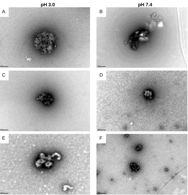
Figure 1. Ideal characteristics of a nanodelivery system. Adapted from Jain14. ... 3 Figure 2. Marine origin polysaccharides structures grouped according to their electrostatic nature. Adapted from Cardoso.34 ... 6 Figure 3. Chemical structure of fucoidan unit from Fucus vesiculosus. ... 7 Figure 4. Structure of chitosan. ... 9 Figure 5. Chemical structures of methotrexate (1) and folic acid (2). Adapted from Wong.75 ... 11 Figure 6. Scheme of fucoidan/chitosan nanoparticles’ preparation. ... 22 Figure 7. FTIR spectra of chitosan, fucoidan and nanoparticles 1F/1C, 3F/1C and 5F/1C. ... 24 Figure 8. TEM images obtained for the designed nanoparticles. Freshly prepared the F/C nanoparticles are in pH 3.0 (A, C and E) and under physiological conditions the pH would be 7.4 (B, D and F). The nanoparticles were observed in both pH conditions: 1F/1C (A, B), 3F/1C (C, D) and 5F/1C (E, F). Scale bar: 200 nm. ... 25 Figure 9. Cryo-SEM images obtained for studied nanoparticles: A) 1F/1C, B) 3F/1C and C) 5F/1C. Scale bar: A, B) 2 μm; C) 3 μm. ... 27 Figure 10. Storage stability of drug-free formulations at room temperature. Parameters size, PDI and zeta potential were determined for 1F/1C (A, B and C), 3F/1C (D, E and F) and 5F/1C (G, H and I). Each result represents the mean ± standard deviation for n=6 measurements. *P<0.05, **P<0.01, ***P<0.001 ... 31 Figure 11. Storage stability of MTX-loaded formulations at room temperature. Parameters size, PDI and zeta potential were determined for MTX-loaded 1F/1C (A, B and C), 3F/1C (D, E and F) and 5F/1C (G, H and I). Each result represents the mean ± standard deviation for n=6 measurements. *P<0.05, **P<0.01, ***P<0.001 ... 32 Figure 12. Storage stability of MTX-loaded formulations at room temperature. Parameters for drug content in A) 1F/1C, B) 3F/1C and C) 5F/1C. Each result represents the mean ± standard deviation for n=3 measurements. **P<0.01, ***P<0.001 ... 33 Figure 13. Storage stability of drug-free formulations at 4ºC temperature. Parameters size, PDI and zeta potential were determined for 1F/1C (A, B and C), 3F/1C (D, E and F) and 5F/1C (G, H and I). Each result represents the mean ± standard deviation for n=6 measurements. *P<0.05, **P<0.01, ***P<0.001 ... 34 Figure 14. Storage stability of MTX-loaded formulations at 4ºC. Parameters size, PDI and zeta potential were determined for MTX-loaded 1F/1C (A, B and C), 3F/1C (D, E and F) and 5F/1C (G, H and I). Each result represents the mean ± standard deviation for n=6 measurements. ... 35
xv Figure 15. Storage stability of MTX-loaded formulations at 4ºC temperature. Parameters for drug content in A) 1F/1C, B) 3F/1C and C) 5F/1C. Each result represents the mean ± standard deviation for n=3 measurements. **P<0.01, ***P<0.001 ... 35 Figure 16. In vitro release of MTX loaded in 1F/1C (A), 3F1C (B) and 5F1C (C) nanoparticles under simulated gastrointestinal conditions using FaSSGF, FaSSIF and PBS pH 7.4 at physiological temperature (37ºC). Data points correspond to the mean ± standard deviation for n=3 replicates (n=3 measurements of MTX release per replicate).38 Figure 17. In vitro release of MTX loaded in 1F/1C (A), 3F1C (B) and 5F1C (C) nanoparticles under simulated intestinal conditions using FaSSIF at physiological temperature (37ºC). Data points correspond to the mean ± standard deviation for n=3 replicates. ... 39 Figure 18. In vitro release of MTX from 1F/1C, 3F/1C and 5F/1C NPs in PBS pH 6.5 receptor media (80 mL) at 37ºC. Data points correspond to the mean ± standard deviation for n=2 replicates (n=3 measurements of MTX release per replicate). ... 40 Figure 19. In vitro release of MTX from 1F/1C, 3F/1C and 5F/1C NPs in PBS pH 7.4 receptor media (80 mL) at 37ºC. Data points correspond to the mean ± standard deviation for n=2 replicates (n=3 measurements of MTX release per replicate). ... 41 Figure 20. In vitro release of MTX from 1F/1C NPs in PBS pH 5.5 receptor media (80 mL) at 32ºC. Data points correspond to the mean ± standard deviation for n=3 replicates (n=3 measurements of MTX release per replicate). ... 42 Figure 21. In vitro release of MTX from 3F/1C NPs in PBS pH 5.5 receptor media (80 mL) at 32ºC. Data points correspond to the mean ± standard deviation for n=3 replicates (n=3 measurements of MTX release per replicate). ... 42 Figure 22. In vitro release of MTX from 5F/1C NPs in PBS pH 5.5 receptor media (80 mL) at 32ºC. Data points correspond to the mean ± standard deviation for n=3 replicates (n=3 measurements of MTX release per replicate). ... 43 Figure 23. Cytotoxicity of unloaded (A) and drug (B) loaded F/ C nanoparticles. Free MTX and MTX-loaded F/C range of concentrations from 12.5 to 200.0 μg mL-1
in L929 cell line. Each result represents the mean ± standard deviation for n=4 replicates of 2 assays. .... 44 Figure 24. Cytotoxicity of unloaded (A) and drug (B) loaded F/C nanoparticles. Free MTX and MTX-loaded F/C nanoparticles were assessed within the range of concentrations from 12.5 to 200.0 μg mL-1 in HaCaT keratinocytes cell line. Each result represents the mean ± standard deviation for n=4 replicates of 2 assays. ... 45 Figure 25. MTX skin permeation. In vitro amount of MTX permeated in relation to the initially added, over time in pig ear skin as free drug (black squares) and MTX-loaded within (A) 3F/1C (open grey circles) and (B) 5F/1C (grey squares) nanoparticles. Each value represents the mean ± SD (n ≤ 3). **P<0.01, ***P<0.001 ... 46
xvi LIST OF TABLES
Table 1. Examples of different pH-sensitive nanocarriers ... 5
Table 2. Effect of fucoidan on tumor growth in tumor-bearing mice ... 8
Table 3. Ratios of Fucoidan/Chitosan (F/C) nanoparticles ... 21
Table 4. Characterization of nanoparticles ... 22
Table 5. List of the characteristic peak of the compounds and nanoparticles determined by FTIR... 24
Table 6. Mass of methotrexate added in each formulation ... 27
Table 7. Characterization of MTX-loaded nanoparticles ... 28
Table 8. The characteristics of F/C nanoparticles under distinct pH conditions ... 30
Table 9. Size distributions of 1F/1C nanoparticles before and after freeze-drying ... 37
xvii LIST OF ABBREVIATIONS
CO2 –Carbon dioxide
Cryo-SEM – Cryo-Scanning Electron Microscopy DDS – Drug Delivery System
DLS – Dynamic Light Scattering
DMEM – Dubelco’s Modified Eagle Medium EE – Entrapment Efficiency
FaSSGF – Fasted State Simulated Gastric Fluid FaSSIF – Fasted State Simulated Intestinal Fluid FC – Fucoidan/ Chitosan
LC – Loading Capacity
MTT – 3-[4,5-dimethylthiazol-2-yl]-2,5-diphenyltetrazolium bromide MTX – Methotrexate
MWCO – Molecular Weight Cuff-Off NPs – Nanoparticles
PBS – Phosphate Buffer Saline PDI – Polydispersity Index
xviii - This page is intentionally left blank. –
1 1. INTRODUCTION
1.1. Exploitation of marine environment in the quest for new compounds
Science has found answers for many subjects in Nature. Natural products have always played a leading role, not only as a source of food and shelter for Man, but also in traditional medicine to find methods to cure diseases, to extend and improve life.
Nowadays, scientists try not only to focus on synthesis and laboratory research to find answers, but also explore Nature to find out its unique way to evolve and create its own means to heal from injuries or restore conditions. A good example of diversity is the marine ecosystem. Considering that most of planet surface is covered by oceans, its enormous ecosystem comprises a vast range of species, but most of them still remain unidentified. To understand the magnitude of marine global species diversity, several authors reported the possible existence of 0.7 to 1.0 million marine species and about 226.000 of the described are eukaryotic.1 This number tends to decrease because anthropogenic CO2 emissions are increasing, and a biological change of ocean ecosystems is taking place. Phenomenon like ocean acidification and global warming are endangering this diversity previously explained.2
The search for marine bioactive compounds has attracted the attention of diverse pharmaceutical research groups, since Nature is a privileged source of active ingredients to use in medicine and pharmacy.3 Marine compounds are special because they present essential behaviours in drug discovery: good bioavailability and perfect affinity to target.4 The trend is to create new techniques for the progress of marine drug discovery and development, because the yield extraction is very low to make advances in this field.5-6 In anticancer research, there are marine compounds to treat pain associated with cancer, some approved antitumor agents and others still undergoing clinical trials.7 There are also reports enhancing other applications like oxidant activity, thrombin and anti-coagulant activity, anti-inflammatory effect, anti-hypertensive, anti-diabetic, antiviral, analgesic and immunomodulatory activity.8-9
The exploitation of these compounds is still a challenge. One of the most relevant characteristic of marine compounds is the fact that they are usually identified as having long aliphatic chains and several functional groups, creating a bulky hydrophobic structure, with problems of solubility.10 This phenomenon results in a low bioavailability and some new strategies need to be presented to do not throw away these compounds’ promising applications.11
From several years, Man has applied his knowledge to make a distinction between a drug and a poison. Sometimes plants were used to apply on skin or make infusions to
2 treat a disease, and that traditional knowledge still has roots in our present days. Although, even performing several extractions from the same plant, the amount of compounds that is found is very extensive and the yield is very low. It is a challenge to reach the perfect structure elucidation of the compound responsible for the biological activity that was discovered by chance. These issues are related with the downstream process, which involves the effective extraction and purification of marine products. Not only in marine biotechnology but in general industry, downstream is the most expensive and ineffective procedure of bioprocess, partly because it is a neglected area of research.12 It is essential to make the product able to be commercialized, and this is only possible with described and authenticated methods to increase yield, requiring a correct scale-up methodology.13
1.2. Drug Delivery Systems
A drug delivery system (DDS) can be defined as a formulation or a device that enables the introduction of a therapeutic substance in the human body.14 Nanotechnology provided new approaches for drug delivery that improved efficacy and safety by controlling the rate, time, and place of drug release in the body. As so, the whole process involves not only the correct administration of the active substance, but also its effective delivery to a specific site of action.14 These nanometric devices act like an interface between the drug and the patient, and there is a wide spectrum of DDS to be used in nanomedicine, according to the characteristics involved in the therapy. The main characteristics of these materials are the drug controlled release, the ability to use different routes of administration, the improved safety and efficacy of drugs, the increased solubility of low-soluble compounds, and a new market opportunity to recover drugs which failed at conventional delivery.14 The ideal features of a DDS are illustrated in Figure 1.
3
Figure 1. Ideal characteristics of a nanodelivery system. Adapted from Jain14.
Another interesting factor is the combination of diagnosis, therapy and nanodelivery systems. In cancer for example, the use of quantum dots or superparamagnetic iron oxide nanoparticles provide the possibility of drug delivery, performing diagnosis at the same time.15
Nanodelivery is a very interdisciplinary area, mobilizing knowledge from subjects like chemistry, engineering, biology and medicine. This concept of delivering a drug to a specific target, particularly on the improvement of cancer therapies available, started a long time ago.16
Despite of the very promising characteristics, nanodelivery systems are still facing some challenges and the future developments will take these problems into account to give them a solution. As technology is always evolving, the nanodelivery systems should follow this evolution, trying to keep up with the methodologies of design, manufacture and analysis. Improving a drugs’ bioavailability could be a good solution to decrease the drugs’ costs, because the patient will need a lower amount of drug to produce the same therapeutic effect. Lastly, the discovery of new materials should always focus on new therapeutic approaches, always performing safety studies to obtain regulatory approval.14
Nanodelivery Systems Increase bioavailability of drug Provide controlled drug delivery Transport the drug intact to the site of action Should be stable through physiological variables High degree of drug dispersion Applicable to a wide range of drugs Safe and reliable Cost-effective
4 As shown above in Figure 1 there are some requirements that need to be accomplished to assure that the nanocarriers are stable and successful. Recent studies about the challenges and opportunities to create these systems are being studied.17 Most importantly, the formulation has to be adjusted to the characteristics of the desired final product, never forgetting the monitoring of the toxicological profile of the formulation and the exploitation of the characteristics inherent to the target.
Despite all information and safety assurances, these nanomaterials can be thought as a source of toxicity depending on its dosage.18 Studies suggested that depending on the type (e.g. structure, composition, size, surface potential) the number of nanoparticles in dispersion can affect cell viability in pre-clinical drug development, thus studies are demanded to unravel the parameters related to cytotoxicity.19
In fact, delivering a drug to its target to enable pharmacological effect is a common strategy, but has its downside. In this field, tests like dose-time relationship, cell function and membrane integrity, possibility of oxidative stress and uptake are very conclusive to evaluate compounds. Upcoming strategies involving new tests, materials and concentrations to approve a reproducible method should be taken, as most as possible, using in vitro models to reduce laboratory animals.20
1.2.1. pH sensitive drug delivery systems
Between all the diverse characteristics that make the DDS a good area to explore, one of the most remarkable is their response to different kinds of external stimuli including light, temperature, pH, electric and magnetic fields, general physical and bio-chemical triggers.21 From all the exposed possibilities, the pH-sensitive feature was the most motivating given its potential to several applications in drug delivery.22
Human organism is a good example of how a wide range of pH conditions can work as a whole, without losing specificity to some organs or tissues. For instance, in the gastrointestinal tract, it is possible to face a gastric pH 2 and end up in an intestinal pH 7. In inflammation environments, a low pH value of 5 was also reported.23 Other important characteristic to consider is the tumor’s pH in cases of cancer: physiological pH is about 7.4 but tumor tissues exhibit a lower extracellular pH (6.0-6.9). This low pH is a result of higher glycolysis rate in hypoxic cancer cells, but it is still possible to happen when oxygen is available.24 With this characteristic, the major aim of these smart DDS is to wisely target the drug release to the site of action, with the lowest loss of compound to avoid unnecessary side effects and achieve a therapeutic response.25
5 In this field, pH sensitive DDS exhibit several advances obtained with different types of nanoparticles: polymeric, liposomes and inorganic nanoparticles (some examples are listed on Table 1).
Table 1. Examples of different pH-sensitive nanocarriers Type of
nanocarrier Material Drug
Drug Locating Strategy
in Nanocarrier Ref Micelles poly(ethylene glycol)– polyaspartate copolymer proteasome inhibitor MG132 Covalent binding of MG132 bound to the block copolymer 26 Liposomes multifunctional envelope-type nanodevice encompassing pH-5 lipid YSK05
(YSK05-MEND) anti-miR-122 miR-122 (AMO122) encapsulated in YSK05-MEND 27 Nanogels Interpenetrating polymeric network spherical nanogels
composed of
poly(acrylamidoglycolic acid) and natural gelatin biological
protein
Curcumin
Hydrophobic curcumin was encapsulated
into the nanogels
28 Polymer-drug conjugates Poly(ethylene oxide)-block-Polyphosphoester graft-Paclitaxel conjugates with
acid-sensitive linker
Paclitaxel
Paclitaxel conjugated to polymer
backbone via Acid-labile β-thiopropionate linkage 29 Core-shell NPs Chitosan-alginate NPs Insulin Insulin entrapped in alginate core 30 Inorganic NPs
Mesoporous silica NPs with
acetal-modified dextran DOX
DOX entrapped inside the mesoporous silica pores
31
NPs – nanoparticles; DOX-doxorubicine; Adapted from Karimi.21
1.3. Marine polysaccharides as drug delivery systems
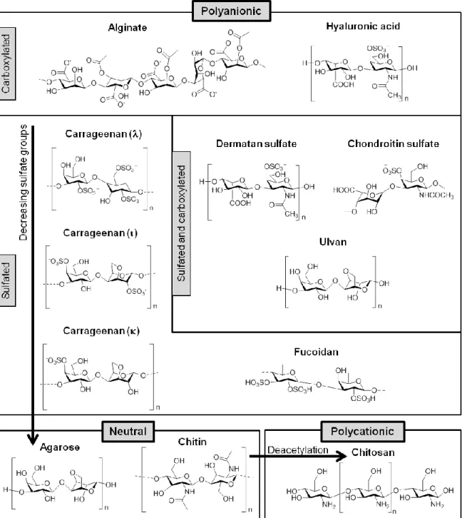
The vast marine source has proven to be valuable in different areas, such as pharmacy and cosmetic, biomedicine and also in food.32 One big class of interesting compounds is the polysaccharides from marine origin. These metabolites can be found in different biological origins, such as marine algae or animals and microorganisms, and consist of a large complex group of different macromolecules with diverse biological applications.9
Marine polysaccharides can be divided accordingly to their electrostatic characteristics (Figure 2).
Some authors have suggested that polysaccharide-based materials demonstrate the following advantages33:
6 - can be modified to form different materials using chemical and enzymatic
methods;
- are biodegradable and biocompatible and exhibit low immunogenicity; - can be useful in stimuli-responsive DDS;
- can be produced, complexed and conjugated with proteins and bioactives; - can be modified as gels;
- can give rise to interpenetrated polymeric networks; - the ionic polysaccharides are mucoadhesive.
Figure 2. Marine origin polysaccharides structures grouped according to their electrostatic nature.
7 1.3.1. Fucoidan
Fucoidan was isolated for the first time in 1913 and refers to a family of sulfated polysaccharides isolated from several brown algae and marine invertebrates (Figure 3).35 Depending on the source, fucoidan can present several structures, mainly composed by fucose and sulfate, but some structures can also have monosaccharides, uronic acids, acetyl groups and proteins.36
Figure 3. Chemical structure of fucoidan unit from Fucus vesiculosus.
Fucoidan has been studied regarding diverse biological activities, which are related to molecular size, type of sugar content, sulfation degree and molecular structure. Some of the evidenced properties are antitumor, antiviral, anti-inflammatory and a potent anticoagulant activity.32
Fucoidan already demonstrated potential anti-cancer activity in vivo in different types of cancers (Table 2)37. According to some studies, fucoidan acts through various mechanisms related with cell cycle arrest, immune system activation and apoptosis, and can be related to the induction of inflammation through immune system, oxidative stress and stem cell mobilization. An extensive study of the effects of fucoidan on cell cycle and apoptosis processes is reviewed by Atashrazm.38 This compound has a well known anticoagulant activity and some studies explain this effect partly due to its sulfate groups. Anti-viral activity is also reported, as well as anti-oxidant and anti-inflammatory activity.36, 39 From all exposed information, fucoidan shows promising characteristics that sustain further development of the substance as a future marine drug.
To overcome fucoidan low bioavailability, a lipid based delivery system was designed and the in vitro experiments showed that nanoparticles loaded with fucoidan induced apoptosis on osteosarcoma cell line more efficiently than free fucoidan. 40
Synergistic effect has also been reported when combining fucoidan with other chemotherapeutic agents, such as etoposide, cisplatin, tamoxifen or paclitaxel and lapatinib.41-43 Among the results, it can be predicted a potentiating effect of the chemotherapy drug and more relevant apoptotic effects.
Table 1 demonstrates that fucoidan from different sources present different mechanism of action on tumor growth, as it can inhibit angiogenesis effect in tumor
8 tissues, prevent metastasis, induce apoptosis and can delay tumor growth. Fucoidan is also suggested as a compound for the treatment of skin diseases.44-45
Table 2. Effect of fucoidan on tumor growth in tumor-bearing mice
Fucoidan source Route/ dose Tumor type Mechanism of
action Reference
Cladosiphon okamuranus
Oral
5 g/kg 26 colon cancer cells
Natural killer (NK) cell-mediated
46
Fucus
vesiculosus Intraperitoneal
5 mg/kg 4T1 breast cancer cells
Inhibition of angiogenesis and induction of apoptosis 47-48 Fucus evanescence 10 mg/kg
Lewis lung carcinoma
cells Unknown 49 From Ze Lang Nanjing Med. Tech Co. Intraperitoneal 200 mg/kg Bel-7402 hepatocellular carcinoma in nude mice
Inhibition of proliferation 50 Fucus vesiculosus Foot-pad injection 0.25 mg/mice
4T1-xenograft mice Prevention of metastasis 51 Sargassum plagiophyllum Oral 75 mg/kg Diethylnitrosamine-induced hepatocellular carcinoma Inhibition of carcinogen metabolism 52 Cladosiphon okamuranus Tokida Oral 100 mg/kg Sarcoma 180 (S-180)-xenograft Delayed tumor growth by nitric oxide produced by macrophages 53 Undaria pinnatifida Diet containing 1% Mekabu (34 mg/day)
A20 leukemia cells
Cytolytic activity by NK cell activation 54 Fucus vesiculosus Intravenous 5 mg/kg
Lewis lung carcinoma cells B16 melanoma cells Antiangiogenic effect 55 Ascophyllum nodosum 1 mg/mice
MOPC-315 plasma cell tumor
Prevention of angiogenesis in
tumor tissues
56
Adapted from Kwak37
Having in mind not only the use of fucoidan as a therapeutic compound, but turning the attention to their unique structural features, the idea of creating DDS from raw fucoidan was considered. As a DDS, fucoidan can be easily conjugated with chitosan by electrostatic interactions and this allows the creation of microspheres to treat burnt tissues, the design of hydrogels and the production of capsules for the controlled release of proteins and bioactive agents.34
9 1.3.2. Chitosan
Chitosan was first reported in 1884 and it is a linear polysaccharide which is obtained by the deacetylation of chitin, found mainly in the exoskeleton of arthropods and crustaceans (Figure 4).57
Figure 4. Structure of chitosan.
Chitosan is soluble in acidic conditions. One of the most interesting characteristics of this polymer is that it is pH sensitive. The presence of amine groups makes it positively charged in acidic environments and neutral in alkaline pH values due to a pKa value close to 6.58 This marine polysaccharide is one of the most abundant and it is widely used and studied for biomedical applications such as antimicrobial activity in wound infection, antitumor and anti-inflammatory activity.34
Considering its properties, chitosan is an excellent material to use in biological environments and to design drug delivery systems. This material is able to perform controlled drug release of cationic drugs, it has mucoadhesive properties due to the presence of cationic primary amino groups, allows the preparation of hydrogels, forms stable complexes with large polyanionic molecules such as DNA-based drugs and allows permeation enhancement because it interacts with cell membrane promoting a reorganization of tight junction-associated properties.59 As a DDS, chitosan can be used as a stable solid dosage forms in oral delivery; as hydrogels, nanoparticles and colloidal systems to apply by ophthalmic route; to give mucoadhesive properties in nasal route spray formulations and bucal drug delivery; confer polymer robustness to vaginal tablets; if it is purified, enables the use by parenteral route because it’s non toxic and also to combine antigens to produce vaccines.59
10 1.3.3. Fucoidan/Chitosan Nanoparticles
Taking advantages of both polysaccharides previously described, the interest to apply them as DDS has permitted the possibility to create a nanoparticle. The oppositely charged chitosan and fucoidan were explored, and their electrostatic interactions permitted the creation of pH sensitive nanoparticles.
Huang and co-workers proposed that fucoidan/chitosan nanoparticles could resist to gastrointestinal barriers and play an important role in oral drug delivery.60 The nanoparticles were successfully prepared and showed a sustained release of curcumin at acidic pH values but it rose significantly at physiological pH values. To release curcumin, the ionic linking of o-carboxymethyl chitosan/fucoidan nanoparticles was also tested, making the nanoparticles retain the curcumin at pH 2.5 but effectively release it at pH 7.4.61
These nanoparticles were also reported as effective carriers for the delivery of controlled release of stromal cell-derived factor 1 (SDF-1), very important to stem cells in tissue engineering applications.62
Another study with 99mTc-methylene diphosphonate showed that these nanoparticles were pH sensitive and manifested a preferential drug release at pH 7.4, making them suitable to be used as drug carriers in oral delivery.63
In the case of antibiotics, antioxidant nanoparticles of chitosan and fucoidan were also prepared to achieve pulmonary delivery. The antibiotic used was gentamicin, which was released with an initial burst effect followed by a slow drug release, providing good proof that this system might be effective in pulmonary drug delivery.64 This study was later explored for the biphasic release of gentamicin. It was possible to observe multiple antimicrobial effects from gentamitin and chitosan, the controlled release of the antibiotic provided a good solution to the adaptive resistance of antibiotics, which was also effective in reducing the risk of systemic toxicity by high gentamicin plasmatic levels. The aimed biphasic release was achieved, with 77% of encapsulated drug released in a first step to reach efficacy against bacteria and the following 22% slowly released to use a safe daily-dose of the drug.65 The possibility of using these nanoparticles in nerve tissue engineering was also reported, with the delivery of basic fibroblast growth factor (bFGF).66
The fucoidan/chitosan nanoparticles show great potential, and their preparation and characterization is being explored in the last years.67-68 Despite the fact that the preparation of these nanoparticles normally involved a bigger ratio of chitosan in the formulation, the current work was based in another approach. As it was enlightened before, fucoidan possesses several biological activities that might be very interesting to explore in the nanomedicine field (imaging agent, protein delivery, small drug delivery,
11 anti-coagulant, anti-inflammatory, gene delivery, regenerative medicine, therapeutic and diagnostic) as it is extensively reviewed by Chollet et al.69
1.4. Methotrexate
Methotrexate is an antimetabolite immunosuppressive drug widely used in cancer therapy, especially in leukemia, lymphoma and some solid tumors.70 It was first reported for use in cancer around 1940.71 Due to its potent anti-inflammatory activity against psoriasis72, it was also approved to this end in 1972. This compound also has immunomodulatory activity against inflammatory arthritis, so it has been used in rheumatoid arthritis as well.73
Methotrexate interferes on synthesis of deoxyribonucleic acid (DNA), ribonucleic acid (RNA) and proteins because it is a folic acid analogue with the ability to inhibit dihydrofolate reductase involved in the purine synthesis (Figure 5).74
Figure 5. Chemical structures of methotrexate (1) and folic acid (2). Adapted from Wong.75
As it is possible to confirm by the analysis of Figure 5, the structures of MTX and folic acid are very similar, featuring only the two different substitutions evidenced by the arrows.75
This drug has relatively good bioavailability and can be administered orally, intravenously or by subcutaneous injection. It is metabolized partially in the liver and in the intestine, and it is renal excreted. Nevertheless, methotrexate exhibits liver toxicity, causes gastrointestinal upset (gastritis, nausea, diarrhea and stomatitis) and fatigue in the next two days after drug administration.70, 76 Other less reported adverse effects include bone marrow suppression, hair loss and idiosyncratic interstitial pneumonitis (along with shortness of breath, fever, dry cough and hypoxemia). It is important also to point out that, being an immunosuppressive drug, patients should be aware of the higher risk of infection.70 Despite all these side effects, methotrexate is well tolerated by patients.
12 Methotrexate is currently being studied to treat uveitis77 and its potential use in other nanotherapeutic delivery approaches against solid resistant tumors is being evaluated and reviewed.75, 78 A recent technique tries to use methotrexate as a dual action drug, because it can target and bind to folate receptor and, at the same time, inhibit cell proliferation.21
Considering all the adverse side effects and the main characteristics of this compound, the use of DDS to improve the therapeutic effect of MTX were described, based not only in the route of administration but also the intended therapy. Some of the reported DDS used were the microspheres for prolonged release and reduced side effects, the use of liposomes to improve the pharmacokinetic of this low permeability drug, the design of nanoparticles to specifically release MTX in tumor environment due to enhanced permeability and retention (EPR) effect and also the exploitation of MTX characteristics to obtain a prodrug or drug conjugate which will only be effective after a specific activation mechanism, thus avoiding off target release.76
The enhancement of the therapeutic potential of methotrexate in psoriasis and rheumatoid arthritis has been reported, considering de use of DDS to aim the evasion from undesired side effects.79-80
1.5. Aim and Strategy
The marine biodiversity can be a source of new and promising compounds which bioactivity and polymeric properties can be used to create stable drug delivery systems. Considering that chitosan and fucoidan present inherent biological activities and can also be explored as DDS, these marine polysaccharides were investigated in this work.
Human organisms can be very challenging in terms of specific physiological conditions, as it is the case of the wide range of pH values in different parts of the body. This feature represents an external trigger of interest in DDS, as pH sensitive nanoparticles might deliver the active compound in the target region of the human body.
Methotrexate is an antimetabolite immunosuppressive drug widely used in cancer therapy, and inflammatory diseases (e.g. psoriasis, rheumatoid arthritis). 72 To overcome the associated side effects, methotrexate could be loaded in drug delivery systems.
Considering all the aforementioned considerations, chitosan and fucoidan were explored as pH sensitive nanoparticles for drug delivery. Two objectives were defined for this research work: 1) to design and characterize pH sensitive fucoidan/chitosan nanoparticles; 2) to assess the applicability of methotrexate-loaded pH sensitive fucoidan/ chitosan nanoparticles.
13 2. MATERIALS AND METHODS
2.1. Materials
Fucoidan (from Fucus Vesiculosus), chitosan, dimethyl sulfoxide and sodium chloride were supplied from Sigma-Aldrich (St Louis, MO, USA). Methotrexate was a gift from Excella (Feucht, Germany). Acetic glacial acid was obtained from VWR International LLC (Radnor, PA, USA). Double-deionized water was provided by an ultra pure water system (Arium Pro, Sartorius AG, Göttingen, Germany). The reagents were weighted in a digital analytical balance Kern ACJ/ACS 80-4 (Kern & Sohn; Balingen, Germany). pH measurements were achieved using a Crison pH meter GLP 22 with a Crison 52-02 tip (Crison; Barcelona, Spain). In cell culture, L929 fibroblast cell line and HaCaT human keratinocyte cell line were purchased from Cell Lines Service (CLS, Eppelheim, Germany). Dulbecco’s Modified Eagle’s Medium (DMEM), fetal bovine serum (FBS), penicillin-streptomycin (10000 U/mL) mixture and trypsin-EDTA 0.25% (v/v) were acquired from Gibco® (Invitrogen Corporation, UK),
2.2. Methods
2.2.1. Preparation of Fucoidan/Chitosan Nanoparticles
Fucoidan/ Chitosan (FC) nanoparticles were obtained by polyelectrolyte self-assembly method by ultrasonication at room temperature acording to a previously described method.63 The preparation process involves the mixing of the two polysaccharides under pulsed ultrasonication (pulse-on 3 seconds and pulse-off 7 seconds, totaling 30 seconds) using a probe sonicator (VCX130, Sonics and Material Vibra-CellTM with a CV-18 probe; 115 Newtown CT, USA
)
to promote the self-assembly of these compounds and produce the nanoparticles. Prior to this step, chitosan was dissolved in 1% (v/v) acetic acid solution to obtain a chitosan-acetic acid solution of 1 mg mL-1, using ultrasound bath to enhance the solubility. Fucoidan was dissolved in double-deionized water to obtain solutions of 1, 3 and 5 mg mL-1. With these solutions, different ratios of the fucoidan/chitosan nanoparticles were prepared, namely 1:1, 3:1 and 5:1. After the self assembly, the formulations were filtered using an 800 nm Minisart® Syringe Filter (Sartorius Stedim Biotech GmbH, Göttingen, Germany) to remove aggregates.14 2.2.2. Characterization of Fucoidan/ Chitosan Nanoparticles
2.2.2.1. Particle Size and Polydispersity Index
In Dynamic Light Scattering (DLS) the speed at which the particles are diffusing in their solvent due to Brownian motion is measured, giving information about the particles size and their polydispersity index (PDI). The speed of Brownian motion is defined by the translational diffusion coefficient. Since the particles are in suspension, the value of size is called hydrodynamic diameter, and it depends on the particles’ core and surface.81 The mean size and PDI of the formulations were determined using a Particle Size Analyzer (Brookhaven Instruments Corporation; Software: Particle Sizing v.5 Brookhaven Instruments; Holtsville, NY, USA). For each assay, 6 runs of 2 min were performed at 20ºC.
2.2.2.2. Zeta Potential
The hydrodynamic diameter in a DLS measurement shows the diffusion of the particle in a fluid. When the particle moves, the ions at the surface also move, showing the zeta potential value. The nanoparticles with surface potential values more positive than 30 mV and more negative than -30 mV are considered stable, and will not tend to aggregate.82 Zeta potential of the nanoformulations was determined using an electrode and Zeta Potential Analyzer (ZetaPALS, Brookhaven Instruments Corporation, Software: PALS Zeta Potential Analyzer v.5, Brookhaven Instruments; Holtsville, NY, USA) operating at a scattering angle of 90º at room temperature. For each assay, 6 runs of 10 cycles were performed.
2.2.2.3. Entrapment Efficiency and Loading Capacity
The amount of methotrexate in each formulation was determined in order to quantify the percentage of entrapped drug within the nanoparticles, when compared with the total amount added to produce them. To do so, 50 μL of each formulation was dissolved in 2 mL of DMSO to destroy the nanoparticles by vortex, and quantify the amount of MTX by UV-Vis spectrophotometer (Jasco V-660 Spectrophotometer, Software: Spectra Manager v.2, Jasco Corporation, USA).
15
The Drug Loading capacity was calculated accordingly to the following equation:
2.2.2.4. Fourier Transform Infrared Spectroscopy
Infrared spectroscopy is based on the vibrational motion of molecules and it is very useful as a chemical analysis technique.83 The vibrations in each molecule are so complex and unique that it is possible to assure that two different molecules do not produce the same spectra. A good strategy to be used as identification method is to compare an unknown spectrum with the spectrum of a compound that has already been identified. The infrared spectrum of a molecule is obtained measuring the energy in each vibration transmitted by the sample. The obtained spectrum is a plot of transmittance versus wavenumber (cm-1).84
The spectra of Fucoidan, Chitosan and Fucoidan/ Chitosan nanoparticles were obtained by placing an amount of the powder of each compound, after freeze-drying process, in a FTIR equipment (Perkin Elmer FT-IR Spectrometer Frontier) equipped with an attenuated total reflectance (ATR) device and zinc selenite crystals. The samples were transferred directly into the ATR compartment, and the result was obtained by combining the 16 scans. The spectra were recorded between 4000 and 600 cm−1 with a resolution of 4 cm−1.
2.2.2.5. Transmission Electron Microscopy
To assess the morphology of the formulations, the Transmission Electron Microscopy (TEM) technique was used. TEM uses an electron beam to irradiate a thin sample and produce an image. This technique uses electron as radiation source, allowing the ability to see samples of angstrom size, with good details of its’ surface.85
In each measurement, 20 L of each formulation were placed in the cooper grid for 1 minute. The excess was removed and then uranyl acetate was added to the sample as a contrast agent. The grid was exposed at the accelerating voltage of 60 kV. The images were obtained in a JEM-1400 Transmission Electron Microscope (TEM Jeol JEM-JEM-1400; JEOL, Ltd., Tokyo, Japan).
16 2.2.2.6. Cryo-Scanning Electron Microscopy
The nanoparticles were also visualized by cryo-Scanning Electron Microscopy (Cryo-SEM) using a JEOL JSM-6301F instrument (Tokyo, Japan) with an Oxford Instruments INCA Energy 350 energy source (Abingdon, UK) and a Gatan Alto 2500 cryo transfer system (Pleasanton, CA, USA). Each formulation was dropped on a grid, instantaneously frozen by nitrogen slush and transferred under vacuum to the cold stage of the preparation chamber. The samples were fractured at the surface, sublimated (-90ºC for 180 seconds), coated with a gold-palladium film, and then moved under vacuum into the SEM chamber at -150ºC.
2.2.2.7. pH Responsiveness
In order to evaluate the nanoparticles’ pH responsiveness, each formulation was submitted to different pH conditions and the information of the size, PDI and zeta-potential was collected. To do so, each formulation (initially at pH=3) was characterized. To change the pH environment, NaOH 1 M solution or HCl 1 M solution was added dropwise into the formulation to obtain the different physiological conditions (pH 2.5, 6.0, 7.0 and 7.4).
2.2.2.8. Lyophilization
The process of lyophilization consists in the dehydration of a sample. By this procedure, the water in the sample is frozen and removed by sublimation. This technique involves several steps including the pre-treatment, freezing, primary drying and secondary drying.86
In the current work, the nanoparticles were pre-treated with a chosen cryoprotectant (Aerosil, Mannose and Trealose) at different percentages (1 and 2% (w/v)) and then frozen overnight at -80ºC (Deep Freezer, GFL®, Germany). After this procedure, the nanoparticles were lyophilized using a freeze drier (LyoQuest -85 plus v.407, Telstar) for 72 hours at -80ºC.
17 2.2.2.9. Stability Studies
To assess the storage stability of the formulations through time, regular measurements of size, PDI, zeta potential and drug content were performed over a period of 16 weeks using the characterization techniques previously described. Formulations were stored at 4ºC and at room temperature to compare the results.
2.2.2.10. In vitro Drug Release
Since the oral route was considered, the in vitro release of methotrexate was performed in conditions to mimetize the gastrointestinal environment. The MTX loaded formulations (containing 250 μg of MTX as starting amount in all assays) were placed in a cellulose bag (Cellu.Sept®T2, Membrane Filtration Products, Inc., Seguin, Texas, USA) accordingly to the dialysis bag method. After this step, the membranes were placed in a beaker containing 80 mL of the intended media, and submitted to heat and stirring using a magnetic stirring plate (IKA-Werke, Staufen, Germany).
For gastrointestinal conditions, FaSSGF (Fasted State Simulated Gastric Fluid – pH 1.6), FaSSIF (Fasted State Simulated Intestinal Fluid – pH 6.5) and PBS (Phosphate Buffered Saline – pH 7.4) were the tested media at 37ºC and constant magnetic stirring. First, the membranes containing the formulations were placed in gastric media for 2 hours. At regular intervals of 30 minutes, a sample of the media was collected and replaced by the same amount of the corresponding buffer to maintain sink conditions. After 2 hours of assay, the membranes were placed in intestinal media for 4 hours. As previously reported, at regular intervals of 30 minutes, a sample of the media was collected and then replaced by the same amount of the corresponding buffer to maintain sink conditions. After 4 hours of assay, the membranes were placed in physiological media for 20 hours.
For topical conditions, PBS (Phosphate Buffered Saline – pH 5.5) was used to simulate skin environment at 32ºC under constant magnetic stirring. The formulations were placed in the dialysis bags and placed in 80 mL of buffer for 4 hours. At regular intervals of 30 minutes, a sample of the media was collected and replaced by the same amount of the corresponding buffer.
All collected samples were analyzed using a UVis spectrophotometer (Jasco V-660 Spectrophotometer, Software: Spectra Manager v.2, Jasco Corporation, USA). Before this procedure, the calibration curves in each medium (FaSSGF, FaSSIF, PBS 7.4 and
18 PBS 5.5) were obtained for the maximum absorbance of methotrexate to calculate the percentage of cumulative drug release through time.
2.2.2.11. In vitro Skin Permeation Using Franz Diffusion Cell
The skin permeation of methotrexate and formulations was evaluated using a Franz cell assembly (9 mm unjacketed Franz Diffusion Cell with 5 mL receptor, o-ring joint, clear glass, clamp and stir-bar; PermeGear, Inc., USA). Pig ear skin was used as a barrier. A volume of 0.5 mL of the formulations and free drug solution with a known amount of drug were placed in the apical medium, the basolateral medium was phosphate buffer at pH 7.4 and the diffusion area between cells was 0.785 cm2. The stirring rate of basolateral medium was 400 rpm and the temperature was 37ºC. At defined intervals of 30 minutes, 1 mL of the basolateral medium was collected and replaced with the same amount of fresh buffer. The cumulative amount of MTX was determined by HPLC rather than UV-Vis spectrophotometry to overcome interferences from skin components released during the experiment. The HPLC system included MD-2015 multi-wavelength detector (Jasco, Easton, MD, USA) programmed for peak detection at 303 nm, a high-pressure pump (PU-2089), an autosampler (AS-2057) and a controller (LC-Net II/ADC) mastered by ChromNAVsoftware. A Chromolith 4.6-mm reversed-phase monolithiccolumn (Merck, Darmstadt, Germany) connected to a guard column of the same material was used as a stationary phase. The MTX quantification was performed according to method previously described. In particular, standard MTX solutions were prepared at 0.05, 0.25, 0.5, 1, 2, 5, 10, 15 and 25 μg/mL in mobile phase. Samples were analysed after addition of 10% (v/v) of acetonitrile for defined times of 30 minutes for a period of 6h. The permeability results were expressed in percentage of release and apparent permeability (Papp). Papp was calculated using the following equation:
where C0 is the initial concentration in the apical compartment (g mL-1), A is the surface area of the Franz diffusion cell (cm2), Dt is the time during which the experiment occurred (seconds) and DQ is the amount of compound detected in the basolateral side (g).
19 2.2.3. Cellular Studies
2.2.3.1. Cell Culture
L929 fibroblasts cells were cultured at 37ºC in a 5% CO2 atmosphere (Unitherm CO2 Incubator 3503 Uniequip; Planegg, Germany) in Dubelco’s Modified Eagle Medium (DMEM) supplemented with 10% of Fetal Bovine Serum (FBS) and 1% Penicillin-Streptomycin. For every two to three days the medium was replaced by supplemented fresh DMEM. Cells reaching 90% of confluence were physically detached from the cell culture flask using a scrapper (NuncTM Cell Scrappers, Thermofisher Scientific; Waltham, MA USA).
HaCaT keratinocytes cells were cultured at 37ºC in a 5% CO2 atmosphere (Unitherm CO2 Incubator 3503 Uniequip; Planegg, Germany) in Dubelco’s Modified Eagle Medium (DMEM) supplemented with 10% of Fetal Bovine Serum (FBS), 1% Penicillin-Streptomycin and 1% Amphotericin B (250 μg mL-1). For every two to three days the medium was replaced by supplemented fresh DMEM. Cells reaching 90% of confluence were detached from the cell culture flask by chemical detachment using 0.25% (w/v) trypsin-EDTA.
In both cell lines after detachment, the cells were centrifuged using the HeraeusTM MultifugeTM X1R centrifuge (Thermo Fisher Scientific; Waltham, MA USA) at 300 xg for 5 minutes and re-suspended in fresh culture medium. Viable cells were counted using a Neubauer Chamber (Improved Neubauer Bright-Line, Boeco; Germany) in a Motic® AE2000 Binocular Inverted Microscope (Motic Electric Group Co., Ltd; Xiamen, Fujian, China).
2.2.3.2. Cell Viability Assay
To determine the effect of the formulations in cell viability, the 3-[4,5-dimethylthiazol-2-yl]-2,5-diphenyltetrazolium bromide (MTT) assay was performed. The collected cells were seeded in a 96-well plate at a density of 5 104 cells/well in 100 μL of supplemented DMEM. After cell adhesion, the medium was removed, different concentrations of the formulations were diluted in DMEM and added to the plated cells. To compare the results, a positive control of culture medium was added. The cells with the formulations were incubated for a determined period at 37ºC in 5% CO2 atmosphere. After this step, the volume of formulations was removed from each well and replaced by 100 μL
20 of MTT solution (0.5 mg mL-1 in DMEM), followed by incubation for 2 hours at 37ºC in 5% CO2. Then, the MTT solution was discarded and 100 μL of DMSO were added to each well to solubilize the crystals. The absorbance was measured using a microplate reader at 570 nm and 630 nm for background subtraction.
The percentage of cell viability was compared to control wells by the ratio of corrected absorbance measured for the tested conditions and the untreated cells.
2.3. Statistical Analysis
Statistical analysis was performed using GraphPad Prism Software (Version 6.01 for Windows; GraphPad Software Inc, San Diego, CA, USA).
2.4. Design of chemical structures
The chemical structures in this work were designed using ChemDraw Professional 16.0.
21 3. RESULTS AND DISCUSSIONS
Design and characterization of pH sensitive fucoidan/chitosan nanoparticles The purpose of this work was to produce a pH-sensitive drug delivery system based on marine polysaccharides to further deliver methotrexate. As so, the first step was to optimize the preparation of these drug delivery systems with the correct combination of the chosen polysaccharides, fucoidan and chitosan, to produce the pH sensitive nanoparticles.
3.1. Preparation of Fucoidan/Chitosan Nanoparticles
The preparation of fucoidan/chitosan nanoparticles involved the mixing of the two compounds, prepared individually and combined at three defined ratios. Fucoidan is water soluble, allowing the preparation of three aqueous solutions of different concentrations in double deionized water: 1, 3 and 5 mg mL-1. Chitosan is not soluble in water or alkaline solvents. This polysaccharide was dissolved in a 1% (v/v) acetic acid solution, to produce a chitosan-acetic acid solution of 1 mg mL-1. The prepared solutions were then added and mixed accordingly to Table 3.
Table 3. Ratios of Fucoidan/Chitosan (F/C) nanoparticles
Ratio F/C Fucoidan (F) Chitosan (C) [F] mg mL-1 Volume (mL) Weight (mg) [C] mg mL-1 Volume (mL) Weight (mg) 1F/1C 1 1.5 1.5 1 1.5 1.5 3F/1C 3 1.5 4.5 1 1.5 1.5 5F/1C 5 1.5 7.5 1 1.5 1.5
After the addition of the two marine polysaccharides the nanoparticles were produced by a simple polyelectrolyte self-assembly method using an ultrasonication probe, illustrated in Figure 6. The pulsed sonication is used to allow the best contact between the two polysaccharides and to promote electrostatic interactions when the pulse is off-cycle, so when the sonication is on, the two polysaccharides are mixed in the formulation.
22
Figure 6. Scheme of fucoidan/chitosan nanoparticles’ preparation.
3.1.1. Determination of size, polydispersity and surface potential of the nanoparticles
In order to obtain information about the size, polydispersity (PDI) and zeta potential of the prepared formulations, the DLS technique was applied. The collected information is summarized in Table 4.
Table 4. Characterization of nanoparticles
Formulation Size (nm) PDI ζ - potential (mV) 1F/1C 427 ± 26 0.12 ± 0.04 +61 ± 2 3F/1C 305 ± 10 0.14 ± 0.02 -39 ± 2 5F/1C 355 ± 9 0.18 ± 0.03 -44 ± 2
Data expressed as mean ± standard deviation (n >3)
The 1F/1C nanoparticles presented an average size of 427 ± 26 nm, which makes them possible to apply to oral and topical delivery.87-88 The polydispersity index of 0.12 ± 0.04 is below 0.2, which indicates that the nanoparticle population is homogeneous. In terms of surface potential analysis, the obtained value was +61 ± 2 mV. It is currently admitted that zeta potentials of |30| mV are required for electrostatic stabilization.89 Thus the 1F/1C nanoformulations should be stable and with no or low tendency to aggregate.
The 3F/1C nanoparticles displayed a size of 305 ± 10 nm and a polydispersity index of 0.14 ± 0.02. This formulation has particle sizes that are also suitable for oral and topical drug delivery and the polydispersity index indicates a narrow size distributions.
23 Analyzing the zeta potential, the obtained value was -39 ± 2 mV, indicating that the nanoparticles are also stable. This negative value of zeta potential is related with the increasing ratio of fucoidan in the nanoparticle matrix, since this is the negatively charged polysaccharide.
The measured size of 5F/1C nanoparticles was 355 ± 9 nm. In terms of polydispersity, the 0.18 ± 0.03 also indicates a short range of sizes in this formulation. For this formulation the zeta potential was -44 ± 2 mV, confirming the previously stated, with the increasing ratio of fucoidan in the nanoparticle, the zeta potential tends to be more negative.
In sum, regarding the results for particle size, it is possible to report the designed 1F/1C; 3F/1C and 5F/1C nanoparticles ranged from 300 to 450 nm, which is suitable for oral and topical drug administration.87, 90 The values obtained for polydispersity index are representative of formulations with a narrow range of particle sizes, all below 0.2. Regarding the information about zeta potential, all formulations tend to be stable, since their absolute values are equal to or higher than |30| mV.
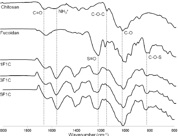
3.1.2. Fourier Transform Infrared Spectroscopy Studies
In order to provide evidences about the interaction of the two polysaccharides upon the nanoparticles’ production, infrared spectra of the reference compounds (chitosan and fucoidan) were obtained, as well as for the prepared ratios (1F/1C, 3F/1C and 5F/1C) of nanoparticles. The infrared spectra are presented on Figure 7.
Chitosan has shown the protonated amino group bending vibrations at 1560 cm-1 and the secondary amide carbonyl group stretching at 1650 cm-1. It was also visible the asymmetric C-O-C stretching at 1150 cm-1 and C-O vibration at 1026 cm-1.62
Fucoidan exhibited S=O asymmetric stretching between 1160 and 1260 cm-1 and C-O-S stretching of the sulfate groups at 845 cm-1.66 In fact, sulfate groups are very evident in this kind of compounds.
Fucoidan/Chitosan nanoparticles showed both characteristic peaks of fucoidan and chitosan spectrum. Although, it is noticeable the shift of the C=O group to a lower wavenumber which might be caused by a different environment around this group.64 The wavenumbers of the characteristic peaks were maintained in the nanoparticles, indicating that no covalent interactions were established between the two polysaccharides. With this analysis it is possible to assume that these nanoparticles were formed relying on the electrostatic interactions between the positively charged chitosan and the negatively charged fucoidan, in accordance with the production method.64