Mestrado Integrado em Medicina Veterinária Ciências Veterinárias
Contribution to the study of Feline Pemphigus Foliaceus
Daniela de Fátima Rua Matos Talhada
Supervisor name Pascal Prèlaud
Co-supervisor name
Adelina Maria Gaspar Gama Quaresma
UNIVERSIDADE DE TRÁS-OS-MONTES E ALTO-DOURO VILA REAL, 2011
iii Abstract
Pemphigus foliaceus is the most common auto-immune skin disease in cats. No age, breed or sex predisposition are reported.
A retrospective study of eleven cats diagnosed for pemphigus foliaceus over a period of three years in “ADVETIA” clinic was performed. Age at onset ranged from less than 1 year to 10 years, median 5 years. Clinical signs of the disease included crusts (n=11), paronychia (n=7), erosions (n=4) and alopecia (n=4). Lesions were most common on claw folds (n=7), outer pinnae (n=7) and/or inner pinnae (n=3), nasal planum (n=3) and muzzle (n=3). Cytologic evaluation revealed acantholytic keratinocytes in 7 cats. The histological examination of 4 biopsy specimens revealed acantholytic keratinocytes and neutrophilic crusts (n=2). Active acantholysis was not observed in 1 case. All cases were followed for 1 to 49 months. Most cats (n=9) achieved total remission with immunosuppressive treatment. Only one cat died due to systemic signs development during therapy.
iv Resumo
O pênfigo foliáceo é a doença de pele autoimune mais comum no gato, não estando descrita predisposição etária, genética ou sexual.
Neste trabalho, foi realizado um estudo retrospectivo de onze casos diagnosticados de pênfigo foliáceo na espécie felina, durante um período de três anos na clínica ADVETIA. O início das lesões variou de menos de 1 ano a 10 anos de idade, mediana de 5 anos. Os sinais clínicos observados incluíram crostas (n=11), paroníquia (n=7), erosões (n=4) e alopécia (n=4). As lesões foram mais frequentes nas pregas ungueais (n=7), pavilhão auricular externo (n=7) e/ou pavilhão auricular interno (n=3), plano nasal (n=3) e nariz (n=3). A avaliação citológica em 7 casos demonstrou a presença de queratinócitos acantolíticos. No exame histológico de 4 biópsias observaram-se queratinócitos acantolíticos e crostas neutrofílicas (n=2). Num caso não foi observado acantólise activa. Todos os casos foram seguidos por 1 a 49 meses. A maioria dos casos (n=9) atingiram remissão completa com tratamento imunosupressor. Apenas um gato foi eutanasiado, devido ao desenvolvimento de sinais sistémicos durante a terapia.
v Index 1. Literature review 1.1. Introduction... 1 1.2. Epidemiology ... 2 1.3. Pathogenesis ... 2 1.3.1. Autoantibodies involved ... 3 1.3.2. Target antigen(s) ... 3
1.3.3. Eosinophilic infiltrate and Apoptosis ... 4
1.4. Triggering factors ... 4
1.4.1. Ultraviolet light ... 5
1.4.2. Other skin disorders or other diseases ... 5
1.4.3. Drugs ... 5
1.4.4. Other factors ... 6
1.5. Clinical signs and lesions distribution ... 6
1.6. Differential diagnosis ... 9 1.7. Diagnosis ... 10 1.7.1. Cytology ... 10 1.7.2. Histopathology ... 11 1.7.3. Immunopathology ... 15 1.8. Treatment ... 15 1.8.1. Common therapeutics ... 16 1.8.1.1. GCs (Glucocorticoids) ... 16 1.8.1.2. Chlorambucil... 18
1.8.2. Current alternative therapeutics ... 18
1.8.2.1. CsA (Ciclosporin) and tacrolimus ... 18
1.8.3. Additional alternative therapy ... 19
1.8.3.1. Cyclophosphamide... 19
1.8.3.2. Chrysotherapy ... 19
1.8.3.3. IVIg (Human Intravenous Immunoglobulin) therapy ... 20
1.8.4. Other immunosuppressive drugs not used in feline patients ... 20
1.8.4.1. Azathioprine... 20
1.8.4.2. Niacinamide and tetracycline or doxycicline ... 21
1.8.4.3. Mycophenolate mofetil ... 21
1.8.4.4. Dapsone ... 21
1.9. Monitoring ... 22
vi
2. Objectives ... 25
3. Retrospective study 3.1. Material and Methods ... 26
3.2. Results ... 28 3.2.1. Incidence... 28 3.2.2. History... 28 3.2.3. Clinical signs ... 29 3.2.4. Diagnostic findings ... 31 3.2.4.1. Cytology ... 31 3.2.4.2. Histopathology ... 32 3.2.4.3. Other tests ... 33
3.2.5. Treatment and clinical follow-up ... 33
4. Discussion ... 35
5. Conclusion...40
6. Bibliography ... 41
vii List of tables
Table 1. Autoimmune (Primary) and Immune-mediated (Secondary) Skin Diseases………….. 1
Table 2. GC therapy for feline PF………. 17
Table 3. Immunosuppressive drugs recommended for feline PF……… 20
Table 4. Monitoring of drug therapy in feline PF……… 22
Table 5. Principles of treating PF in cats……… 23
Table 6. Dermatological lesions in cats with PF at diagnosis... 29
Table 7. Locations of dermatological lesions in cats with PF at diagnosis... 30
viii List of images
Figure 1. Location of Pemphigus lesions within epidermis in cats………. 2 Figure 2. Alopecia, erosions and crusts developed on the face of a 1.5-year-old Siamese
cat during a flare of PF……….. 7
Figure 3. Yellowish crusts are present on the convex or concave aspects of the pinnae of
two cats with PF……….. 7
Figure 4. An adult Birmain cat with PF lesions on the muzzle……… 7 Figure 5. In feline PF, crusts can be seen around footpads and claws or on pads paws….. 8 Figure 6. Same cat as in figure 4. Footpad crusts, characteristic of feline PF………. 8 Figure 7. Alopecia, erythema and erosions on the dorsum of the paw along with paronychia and crusting al the claw folds……… 8 Figure 8. Palmer surface of the paw in a cat with PF. Thick crusts are seen at the margins of the pads as well as in the interdigital spaces along with scaling of the surface of the
pads……….. 8
Figure 9. Same cat as figure 4 has also yellowish crusts at the claw folds. Another feline
PF case with claw involvement………. 9
Figure 10. Same cat as figure 4. Legs are an uncommon area involved in feline PF………. 9 Figure 11. Cytology from under crusts around claw folds, from the cat of Figure 3, stained with Diff-Quick. A first evaluation with 10x objective shows acantholytic cells with numerous neutrophils. With 40x objective, isolated acantholytic keratinocytes surrounded by non-degenerated neutrophils were seen and rare eosinophils were present too………… 11 Figure 12. Feline epidermis. An active area of acantholysis located within the stratum spinosum is forming beneath an older recornified pustule (x10) (a). Acantholytic cells can be seen “springing” up from the stratum spinosum (x20) (b). Staining with H&E………. 13 Figure 13. Feline dermis. A diffuse dermal infiltrate consisting predominately of mast cells and neutrophils are seen in the superficial dermis. Staining with H&E……….. 13 Figure 14. Sample from an auricular lesion of a cat with PF. Histological examination reveals a fluid-filled vesiculo-pustule cointaining few neutrophils and acantholytic cells. Staining with H&E……… 14 Figure 15. Feline PF. Large subcorneal pustule with neutrophils and acantholytic keratynocytes. Recornification is a characteristic feature of PF. Staining with H&E………… 14 Figure 16. Canine PF. Direct IF reveals intercellular epidermal IgG in the stratum spinosum and granulosum………. 15 Figure 17. Canine PF. Indirect IF demonstrates that only rare sera from dogs with PF recognize Dsg1. These results indicate that Dsg1 is a minor antigen in canine PF…………. 15 Figure 18. Histogram of age at the onset of lesions………. 27
ix
Figure 19. Seasonal onset for 10 cats with PF………. 28 Figure 20. Squeme of distribution pattern and the regional location of lesions in ten cats with PF……….. 30 Figure 21. Treatment outcomes for the 11 cats included in the study………... 35
x Abbreviations
ANA – Anti-nuclear Antibodies AKs – Apoptotic Keratinocytes CBC – Complete Blood Count CsA – Ciclosporin A
DM – Diabetes Mellitus
DNA – Desoxyribonucleic acid Dsc – Desmocollin
Dsg – Desmoglein
ELISA – Enzime Linked Immunosorbent Assay FAD – Flea Allergic Dermatitis
FeLV – Feline Leukemia Virus FIV – Feline Immunodeficiency Virus Gc(s) – Glucocorticoid(s)
H&E – Haematoxylin and Eosin
HVBV – Hospital Veterinário do Baixo Vouga IBD – Intestinal Bowel Disease
IF – Immunofluorescence IgG – Immunoglobulin G IM – Intramuscular IV – Intravenous
IVIg – Human Intravenous Immunoglobulin Kg - kilogram
LE – Lupus Erythematosus mg – milligram
ml – milliliters
PAS – Periodic Acid Schiff PD - Polydipsia PE – Pemphigus Erythematosus PF – Pemphigus Foliaceus PO – Per os PU - Polyuria PV – Pemphigus Vulgaris TPMT - Thiopurine methyltransferase ® - Registered Trademark ™ - Unregistered Trademark ºC – Degrees Celsius
xi Acknowledgments
I would like to leave my deepest appreciation to Doctor Pascal Prèlaud, who treated me so kindly and allowed me to achieve so much. I hope that in this thesis I have given something back to you. This work would not have been possible without your knowledge, encouragement and patience. I would also like to express appreciation to all ADVETIA team. Thank you all.
To my “Mon Cherry” Aurore Laprais, my first friend in Belgium, for her friendship and support.
I wish to express my gratitude to Doctor Adelina Quaresma, for her support, patience, friendship and advice. Your help was crucial for this work.
My gratitude is also to all HVBV team, without any exception. To Dr. Artur Alves, Dr. Sónia Miranda, Dr. Pedro Olivério, Dr. Pedro Oliveira, Dr. Hugo Vilhena, Dr. Inês Fonseca, Dr. Mafalda Sardinha, Dr. Sílvia Rios and Dr. José Campos. To the nurses Nuno Ferreira, Daniela Ribau and Ana Macedo and to the auxiliaries Daniel, Katia and Liliana. Thank you for receiving me and teaching me your knowledge.
To my recently friend, Anita, she knows how to make me smile and how to go on.
To all my university professors, who taught me to appreciate the value of education and the utility of knowledge. To my enthusiastic and entertaining veterinary colleagues, Andreia, Tânia, Teresa, Giselle, Catarina, Joana and Daniela Fragateiro, who enriched my life and continuously remind me of how fortunate we are to share the world with animals.
To my uncles Nuno and Tina and to my friends from France, for their hospitality and for all the good times that we spend together. I will never forget that.
To my family, my dad, my mom, and my brothers Catarina and Tiago, for their encouragement and enduring love. A special kiss to my cousin Sónia Matos, who offered her time to read and correct my English.
To my pets, Jonas, Cleo and Pequenote, that inspire me all the time.
To the many pets I have known and loved, because they have enriched my life immeasurably.
1 1. Literature review
1.1. Introduction
The immune-mediated skin disorders include a large group of inflammatory diseases that have in common the central participation of the immune system in their development. They are classified into primary or autoimmune, and secondary or immune-mediated (Table 1) (Torres, 2004).
Table1. Autoimmune (Primary) and Immune-mediated (Secondary) Skin Diseases (Torres, 2004)
Autoimmune Immune-mediated
Pemphigus complex
Subepidermal bullous diseases Bullous pemphigoid
Epidermolysis bullosa acquisita Linear IgA bullous dermatosis Mucous membrane pemphigoid Cutaneous lupus erythematosus Discoid lupus erythematosus
Vesicular cutaneous lupus erythematosus Exfoliative cutaneous lupus erythematosus
Cutaneous adverse drug reaction Erythema multiforme
Toxic epidermal necrolysis Vasculitis
Auricular chondritis Alopecia areata
Sterile nodular panniculitis
The pemphigus complex is a group of uncommon autoimmune diseases and described in humans, dogs, cats, horses and goats (Olivry and Chan, 2001; Bystryn and Rudolph, 2005; Olivry, 2006). In cats, the pemphigus group has been described from three decades (Thoday, 1981; Ackerman, 1985). It is characterized by the formation of autoantibodies against an autoantigen, creating vesicles in the skin. The clinical manifestations differences between pemphigus forms are based on which particular antigens are targeted by autoantibodies and in the distribution of these antigens in the different regions of the body (Torres, 2004; Waschake, 2008; Gershwin, 2010).
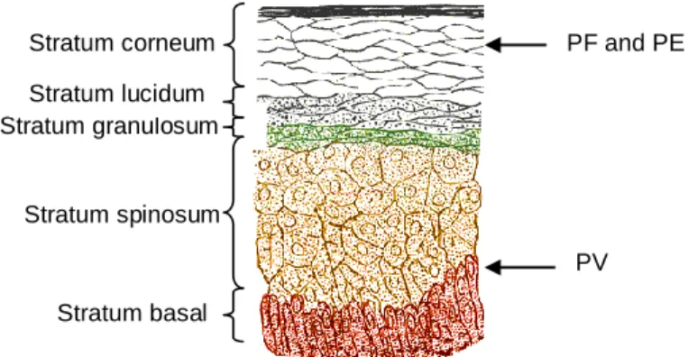
In cats, three different forms of pemphigus are distinguished: Pemphigus foliaceus (PF), Pemphigus erythematosus (PE), and Pemphigus vulgaris (PV). PF and PE are specific forms of superficial pemphigus, whereas PV is a deep form clinically distinct (Figure 1) (Scott et al., 2001; Paterson 2006; Tater and Olivry, 2010). PF is the most common form of pemphigus seen in cats, as well as the most common autoimmune dermatosis (Scott et al., 2001). PE may be a benign form of PF, with the skin lesions limited to the face, or a crossover between pemphigus and Lupus Erythematosus (LE) (Torres, 2004; Hnilika, 2011). The incidence of feline PF has been calculated as five per 1000 patients, per 10 years (Preziosi et al., 2003).
2 1.2. Epidemiology
A breed predisposition for PF has not been reported definitively in the feline species even though domestic short-haired cats were found to be most commonly affected with this disease. Many other breeds, including Orientals and long-haired and short-haired varieties were represented (Preziosi et al., 2003). PF has been reported in Siamese, Himalayan, Persian, Maine Coon, Somali, Ragamuffin, Scottish Fold and American Blue breeds (Angus, 2005).
No sex predisposition for development of PF in cats has been noted. Feline PF is most common in young adult cats, however the age of onset is highly variable, ranging from less than 1 year to greater than 17 years, with a median age of onset of 5 years (Preziosi et al., 2003).
1.3. Pathogenesis
The exact pathomechanism of PF lesions is not known. PF affects the epidermis, the outermost superficial skin layer and is a specific type of superficial pemphigus. About 85% of the epidermal cells are composed of tightly adherent keratinocytes. Four types of adhesion structures hold keratinocytes together: desmosomes, responsible for cell-to-cell adhesion, hemidesmosomes that bind the basilar epidermal keratinocytes to the basement membrane, adherens junctions and focal adhesions (Scott et al., 2001). Desmosomes occur predominantly in epithelial cells and their molecular components play an important role for cell-cell adhesion. The molecular basis of hiperadhesion is complex and incompletely understood, and more than one desmosomal component may be involved (Waschake, 2008; Garrod, 2010). Desmosomes contain two major transmembrane components of the cadherin-type: desmoglein (Dsg) and desmocollin (Dsc) (Amagai, 2009). The basis of pemphigus pathophisiology is that acquired autoantibodies attack these molecules in desmosomes and the resilience of epidermis is lost
Stratum corneum Stratum granulosum Stratum spinosum Stratum basal Stratum lucidum PF and PE PV
Figure 1. Location of Pemphigus lesions within epidermis in cats. PF and PE pustules form beneath or within the
stratum corneum. PV vesicles form above the basal cell layer. (Image adapted from http://en.wikipedia.org/wiki/File:Skinlayers.png)
3
resulting in superficial blister formation (Scott et al., 2001; Fassihi et al., 2006; Amagai, 2009; Tater and Olivry, 2010). This loss of cohesion between epidermal cells is known as acantholysis. Free epidermal cells in the vesicles or bullae are called acantholytic keratinocytes and not acanthocytes, which are erythrocytes with membrane projections (Scott et al., 2001). Indeed, in a canine PF case, autoantibodies titles were related with disease activity, suggesting that PF is an antibody-mediated autoimmune disorder (Nishifuji et al., 2005).
Unfortunately, studies to characterize autoantibodies and antigens in feline species with PF are scarce and pathogenesis of cats is extrapolated from other species.
1.3.1. Autoantibodies involved
In dogs with PF, recent reports were made to determine whether antikeratinocyte IgG (Immunoglobulin G) antibodies are pathogenic (Olivry et al., 2008). It was detected high titles of antikeratinocyte antibodies of IgG4 subclass in 80% of the serum of PF-affected dogs, but only rarely in the serum of control dogs. Antikeratinocytes antibodies of IgG1 subclass were found in both affected and non-affected dogs and there was no statistical difference between groups. The other subclasses, IgG2 and IgG3, were uncovered at low titles. This study supports the importance of IgG4 in the pathogenesis of PF, as their titles decrease during treatment-induced reduction of disease severity and can induce subgranular blisters when transferred passively in neonatal mice. In human PF, patients produce IgG1 and IgG4 anti-Dsg1. However, IgG1 isolated from PF sera failed to induce lesions when injected in neonatal mice (Hacker et al., 2002). All these data support the idea that IgG4 is common between species with PF and cats probably have a similar pathogenesis.
1.3.2. Target antigen(s)
In human PF, autoantibodies recognize more than one antigen, but Dsg1 is known as the principal autoantigen targeted by autoantibodies (Amagai, 1999; Cotell et al., 2000; Karlhofer et al., 2003; Fassihi et al., 2006; Culton et al., 2008), and the same process has been proposed for domestic animals (Scott et al., 2001). Limited immunoblotting studies suggested that the main autoantigen in canine PF might also be Dsg1 (Iwasaki et al., 1997). However, subsequent studies did not suggest Dsg1 to be the major autoantigen in dogs with PF (Olivry et
al., 2006; Olivry and Linder, 2009; Yabuzoe et al., 2009) suggesting that other desmosomal
proteins may be involved, such as desmoplakin. Microscopy techniques show that the binding site of autoantibodies is the extracellular region of desmosomes (Yabuzoe et al., 2009). Very recent immunomapping studies are strongly suggestive that Dsc1 may be a major target autoantigen in canine PF (Bizikova et al., 2011a; Bizikova et al., 2011b), although a previous study suggests that extracellular domains of Dsc1 are not involved in canine PF (Aoki-Ota et al.,
4
2004). Therefore, the results are not conclusive, and more research needs to be performed in order to identify major autoantigen(s) for this disease in dogs and cats. Once identified, it should be considered the suitability of immunological tests (e.g. ELISA (Enzime Linked Immunosorbent Assay) and immunoblotting tests) employing recombinant antigen as an aid for the diagnosis, disease monitoring and the development of targeted and specific immunotherapies (Stanley et
al., 2009).
Since canine PF appears to be clinically, histologically and immunologically heterogeneous, with rare PF sera autoantibodies targeting Dsg1, it has been proposed that PF may not be a single disease but a general term for all superficial pemphigus diseases, since all superficial pemphigus conditions are very similar (Olivry, 2006). Deep pemphigus conditions are clinical and immunological distinct from superficial pemphigus, and the term Pemphigus cannot be used alone as a diagnostic because it refers to a heterogeneous group of deep and superficial pemphigus.
1.3.3. Eosinophilic infiltrate and Apoptosis
To date, only one study investigates the importance of eosinophilic infiltrates in the pathogenesis of PF in dogs. There were no statistically significant differences in the occurrence of acantholytic cells or active acantholysis in dogs with or without an eosinophilic infiltrate (Vaughan et al., 2010). In cats with PF, eosinophils are rarely seen (Preziosi et al., 2003) and its importance in pathogenesis seems irrelevant. Further studies are warranted to confirm if there is a relationship between eosinophilic infiltrate and autoimmune diseases in domestic animals.
In humans, apoptosis contributes to the mechanisms by which autoantibodies induce acantholysis. Theoretically, the blockade of the caspase pathway could prevent apoptosis and acantholysis. This observation may be a promising therapeutic tool that can help in the treatment of pemphigus flare-ups (Schmidt and Waschake, 2009; Bektas et al., 2010; Pacheco-Tovar et al., 2011). Apoptosis is also seen in some cats with PF (Vogel et al., 2009), but it is not known if the activation of the apoptotic pathway can be an early consequence of the binding of autoantibodies to keratinocytes in this species.
A recent study indicates that inflammatory mediators released from eosinophils may be involved in triggering apoptosis in epidermal keratinocytes (Griffin et al., 2010). Further studies are needed to confirm and elucidate the inflammatory mediators involved in this process.
1.4. Triggering factors
The mechanisms leading to immune dysregulation and autoimmunity are complex and not fully understood. In humans both endogenous and exogenous factors have been implicated
5
in the pathogenesis of pemphigus (Culton et al., 2008). In domestic species it is unclear if several factors can exacerbate pemphigus, but they should be avoided whenever it is possible.
1.4.1. Ultraviolet light
Ultraviolet exposure may be a potential environment trigger for PF. However, such suspicion is referred only in dogs (Olivry, 2006), and no seasonal or environmental risk factors have been identified in cats. However, Griffin (1991) proposed a seasonal evolution for some cats.
1.4.2. Other skin disorders or other diseases
It is not clear if previous skin diseases are implicated in the development of feline PF. In a retrospective study, two of eleven cats with previous dermatological problems (including allergic dermatitis, indolent ulcer, otitis, feline acne, pyoderma and unspecified pruritic skin disease) had a history of chronic allergic skin disease for several years prior to developing PF, suggesting that chronic skin inflammation may be a trigger factor (Preziosi et al., 2003). A 3 year-old cat diagnosed with PF was treated before for an eosinophilic granuloma complex for one year, with prednisolone, essential fatty acids, hipoallergenic diet and ectoparasites control, but it is unlikely that eosinophilic granuloma complex leads to PF lesions (Chapelin et al., 2004). Previous skin allergy was also reported in canine PF (Scott et al., 2001). In dogs, PF is reported in patients with leishmaniasis (Ginel et al., 1993), thymoma (Day, 1997) and Systemic Lupus Erithematosus (Foster et al., 2000). Concurrent Leishmaniosis and PF have been described in a cat that lived in both Spain and Switzerland. It is not clear if Leishmania species infection had a causative relation with PF. However, the concurrence of both diseases seems to be rare (Rüfenacht et al., 2005). PF can be present in these patients by coincidence or these systemic diseases may produce autoantibodies against desmosomes.
1.4.3. Drugs
It is reported that in humans, some drugs may influence the development of PF. Some of them may activate proteolytic enzymes in the skin that alter desmosomes and results in biochemical acantholysis. They may also stimulate the development of autoantibodies against desmosomes resulting in immunologic acantholysis. These drugs include thiols compounds, containing sulfur groups (e.g. penicillamine) or compounds containing amide group (e.g. enalapril); or nonthiol drugs that contain sulfur and may undergo metabolic changes to form active thiol drugs (e.g. penicillins, cephalosporins) (Brenner et al., 1998; Brenner and Goldberg, 2011).
6
Drug eruptions resembling PF lesions have been reported in dogs and cats with antibiotics like ampicillin (Mason et al., 1987), cimetidine (McEwan et al., 1987), amoxicillin, enrofloxacin, sulfonamides and metronidazole (Willemse, 2000; Vaughan et al., 2010), cephalexin, trimethoprim-sulfadiazine and oxacillin (White et al., 2002). The administration of itraconazol, lime sulfur dip, methimazole and ipodate may also be implicated as potential triggers (Preziosi et al., 2003; Gross et al., 2005). More recently, the administration of a topical spot-on product containing metaflumizone and amitraz (Promeris-Fort Dodge Animal Health) has been associated with PF in 22 dogs (Oberkirchner et al., 2011). It is difficult to establish a relationship between a drug and PF because patients have often been exposed to multiple drugs and some drugs may have a prolonged latency period between exposure and onset of the disease. If any drug is suspected as a trigger of feline PF, it should be discontinued and avoided in the future.
1.4.4. Other factors
Genetic factors (Tron et al., 2005), food containing certain organic compounds, such as thiols and phenols (Brenner et al., 1997; Fedeles et al., 2010), viral infections (Brenner et al., 2002; Sagi et al., 2008) and skin infections caused by Staphylococcus aureus (Amagai, 2009) have also been associated with some cases of human PF, but it is unknown if any of these factors, especially the diet, are triggers in domestic species.
1.5. Clinical signs and lesions distribution
The signs of an attack on keratinocyte adhesion structures are clinically evident. Pemphigus is characterized by the development of intraepidermal vesicles or bullae which are rapidly infiltrated by leucocytes and so form pustules (Broek, 1991). Pustule is defined as a small, circumscribed elevation of epidermis that is filled with pus. The color is usually yellow, but may be green or red (Scott et al., 2001). Unfortunately, the thinness of feline epidermis promotes rupture of pustules and primary lesions are very difficult to find. Affected cats often present thick, yellowish to honey-colored adherent crusts with associated scale, alopecia and erosions (Figures 2-6) (Preziosi et al., 2003; Sparkes and Caney, 2005; Paterson, 2006; Peterson and McKay, 2010). Nikolsky’s sign (elicited by applying pressure on a vesicle, or at the edge of an ulcer or erosion or even on normal skin) may be positive, when the outer layer of the skin is easily rubbed off or pushed away, indicating poor cellular cohesion (Scott et al., 2001). Other clinical signs may include pruritus in variable degrees, lethargy, pyrexia, mild to moderate anorexia, tachycardia, dehydration, limb edema and apparent skin pain (Preziosi et
al., 2003; Chapelin et al., 2004; Medleau and Hnilika, 2006; Foster et al., 2007; Peterson and
7
lymphadenopathy, otitis externa, cystitis, seborrhea sicca and depression (Broek, 1991; Preziosi et al., 2003). The disease commonly has a waxing/weaning course. There may be hours to days when numerous new pustules form, followed by days to weeks of crusting during which few new lesions are found (Scott et al., 2001). Secondary bacterial infection can occur together with PF (Broek, 1991; Foster et al., 2007).
The initial distribution of feline PF lesions is often localized and mild, but most of them generalize to dorsum and ventrum (Preziosi et al., 2003). The most common affected area is the head (almost 80%), with lesions in the pinnae, face/head, nose, chin or periocular area (Figures 2-4) (Preziosi et al., 2003; Chapelin et al., 2004; Matousek, 2004; Sparkes and Caney, 2005; Friberg, 2006; Foster et al., 2007; Peterson and McKay, 2010).
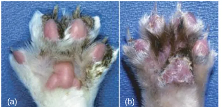
Paws are the next most likely area of involvement, with feet (Preziosi et al., 2003; Chapelin et al., 2004; Peterson and McKay, 2010) or claw folds lesions, these with sterile paronychia with purulent exudate (Figures 5-9) (Preziosi et al., 2003; Matousek, 2004; Crow, 2009; Peterson and McKay, 2010). In some cases, purulent claw fold discharge may be the only
(a) (b)
Figure 3. Yellowish crusts are present on the convex (a)
or concave (b) aspects of the pinnae of two cats with PF (Olivry, 2006)
Figure 2. Alopecia, erosions and crusts
developed on the face of a 1.5-year-old Siamese cat during a flare of PF (Olivry, 2006)
Figure 4. An adult Burmese cat with PF
lesions on the muzzle (photo courtesy of Dr Pascal Prélaud)
8
sign (Torres, 2004). Often, PF lesions are confined to the face and paw pads. Nail changes seem to be rare, and onychomadesis was reported by Guaguere et al. (2000). Interdigital skin, pads and junction of pad with haired skin are also common (Angus, 2005). Hyperkeratosis is an uncommon finding (Paterson, 2006) but in some cats may be the only sign (Broek, 1991).
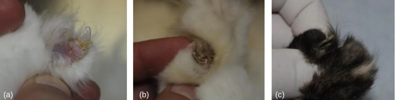
Other areas reported for initial involvement include the dorsum in 10%, area around the nipples, legs (Figure 10) or tail (Preziosi et al., 2003; Moriello, 2005). PF does not affect mucous membranes (Broek, 1991) although the mucocutaneous junctions and oral cavity has been reported as an unusual location for PF in cats (Gross et al., 2005; Rees, 2011).
Figure 6. Same cat as in figure 4. Footpad
crusts, characteristic of feline PF (photo courtesy of Dr Pascal Prélaud).
.
(a) (b)
Figure 5. In feline PF, crusts can be seen around
footpads and claws (a) or on pads paws (b) (Olivry, 2006).
Figure 7. Alopecia, erythema and erosions on
the dorsum of the paw along with paronychia and crusting at the claw folds (Preziosi et al., 2003).
Figure 8. Palmer surface of the paw in a cat with
PF. Thick crusts are seen at the margins of the pads as well as in the interdigital spaces along with scaling of the surface of the pads (Preziosi
9 1.6. Differential diagnosis
Differential diagnosis for feline PF, based in clinical findings (pustular or papular dermatosis) include bacterial folliculitis, notoedric mange, otodectic mange, cheyletiellosis, leishmaniasis, other autoimmune skin diseases (discoid and systemic lupus, PE), dermatophytosis (Trichophyton mentagrophytes), demodicosis, cutaneous epitheliotropic lymphoma, cutaneous adverse drug reactions, zinc responsive dermatosis, actinic dermatosis, dermatomyositis, eosinophilic pustulosis, superficial necrolytic migratory erythema and mosquito bite hypersensibility (Gross et al., 2005; Moriello, 2005; Medleau and Hnilika, 2006; Paterson, 2006; Peters et al., 2007; Nuttal et al., 2009).
When the lesions are localized in the face, feline miliary dermatitis, allergy (especially atopy), food intolerance and cowpox infection should be ruled out (Jackson and Foster, 2006; Paterson, 2006).
Differential for paronychial disease include Staphylococcal infection of the nail beds, yeast infection (Malassezia spp.) and neoplasia (metastatic bronchogenic carcinoma and squamous cell carcinoma of the lungs) (Paterson, 2006).
The frequent bilateral symmetry and the polycyclic pattern of lesions are important differentiating features (Gross et al., 2005).
Figure 10. Same cat as figure 4. Legs are an
uncommon area involved in feline PF (photo courtesy of Dr Pascal Prélaud).
Figure 9. Same cat as figure 4 has also yellowish crusts at the claw folds (a) and (b). Another feline PF case
with claw involvement (c) (photos courtesy of Dr Pascal Prélaud).
10 1.7. Diagnosis
The most important diagnostic aspects are the history, physical examination, direct smears, histopathological findings and in rare cases direct Immunofluorescence (IF) (Scott et
al., 2001; Tater and Olivry, 2010). Antinuclear antibody (ANA) test is not necessary for the
diagnosis (Tater and Olivry, 2010). A history of acute onset of crusted lesions in cats is suggestive of PF (Peterson and McKay, 2010). Footpads hyperkeratosis may be very suggestive of feline PF (Broek, 1991).
Other diagnostic tests to rule out differentials include fungal cultures, bacterial culture, skin scrapings, hair plucks and impression smears (Moriello, 2005; Paterson, 2006; Crow, 2009). Due to the potential of serious secondary effects, the diagnosis of PF should be made before the administration of immunosuppressive drugs.
Complete Blood Count (CBC) and biochemical profile abnormalities detected in the remaining cats are mild and nonspecific (Preziosi et al., 2003; Peterson and Mckay, 2010), so they are not relevant for the diagnosis. Many cases can have moderate to marked leukocytosis and neutrophilia, mild nonregenerative anaemia, mild hypoalbuminemia and mild elevations in globulins. A hemogram and biochemical exams cannot diagnose PF, but they are helpful to detect any systemic disease, that may appear with immunosuppressive therapy.
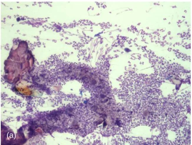
1.7.1. Cytology
The diagnosis of PF in cats begins with impression smears, usually under newly formed crusts, since intact pustules are transient and rare in cats. If a pustule is present, the procedure is rupture with a fine needle and put the contents carefully on a slide, to prevent cellular damage (Tzanck preparation) (Forsythe, 2007; Foster et al., 2007; Rees, 2011). However, when these are rare, they should be preserved for histopathology. After drying and staining with Diff-Quick™ or Rapistain, cytologic preparations should be evaluated with 10x, 40x and 100x (oil immersion) objectives (Paterson, 2006; Forsythe, 2007). The structures that are seen in normal skin include squamous (angular, anuclear keratinocytes) and occasional nucleated keratinocytes. Free melanin granules can be seen eventually and should not be mistaken for bacteria. Melanin granules may also be found in keratinocytes.
In PF disease, pustules contain neutrophils and are sterile. Cytology is useful to find acantholytic keratinocytes, which are nucleated epithelial cells with rounded shape that have lost their intercellular adhesions. Their cytoplasm is normally stained or can be hypereosinophilic (Scott et al., 2001). Acantholytic keratinocytes admixed with nondegenerated neutrophils are very suggestive of PF (Figure 11) (Broek, 1991; Preziosi et al., 2003; Paterson, 2006; Forsythe, 2007; Crow, 2009; Peterson and McKay, 2010; Tater and Olivry, 2010), but acantholysis may be absent (Chapelin et al., 2004). However, they are not pathognomonic
11
since they may be seen in other pustular and inflammatory dermatoses, such as superficial pyoderma and dermatophytosis (Torres, 2004; Forsythe, 2007; Nuttal et al., 2009). Acantholytic keratinocytes exhibit either microscopic characteristics of normal differentiated spinous or granular layer epithelial cells, or they present signs of apoptosis with eosinophilic cytoplasm, condensed chromatin or karyorrhexis. Occasionally, neutrophils can be seen in close apposition to detached keratinocytes (Olivry, 2006). Eosinophils can be present, but it is not common (Forsythe, 2007).
Cytologic study is helpful to discover bacteria, yeasts and fungi. When secondary infection is present, bacteria can be seen and neutrophils tend to be more degenerated, due to bacterial toxins exposition. An appropriate antibiotic can be administrated during 2 to 3 weeks, and then repeat cytology (Paterson, 2006; Forsythe, 2007; Tater and Olivry, 2010). If the lesions are resolved only with antimicrobial treatment, the condition is not pemphigus, but probably a bacterial pyoderma.
(a) (b)
(c)
Figure 11. Cytology from under crusts around claw
folds, from the cat of Figure 3, stained with Diff-Quick. A first evaluation with 10x objective shows acantholytic cells with numerous neutrophils (a). With 40x objective, isolated acantholytic keratinocytes (red arrows) surrounded by non-degenerated neutrophils were seen (b) and rare eosinophils were present too (black arrows) (c). Bacterial infection was not observed (photos courtesy of Dr Pascal Prélaud)
12 1.7.2. Histopathology
Histological examination of lesional skin is usually diagnostic of PF (Yager and Wilcock, 1988; Broek, 1991; Preziosi et al., 2003; Chapelin et al., 2004; Peterson and McKay, 2010; Hnilika, 2011). Anti-inflammatory agents can dramatically affect the histological appearance of PF. Cats that receive some form of corticosteroid at the time of biopsy had a significant reduction of samples that contain adequate diagnostic criteria (Preziosi et al., 2003). The administration of such agent should optimally be stopped for 2 to 3 weeks before biopsy. Secondary bacterial pyoderma often obliterate the histopathological features of PF and it is imperative to eliminate these secondary infections with appropriate antibiotic therapy before biopsies are performed, to increase the chances of a clear diagnosis from histological examination (Scott et al., 2001; Crow, 2009; Tater and Olivry, 2010). Ideally, samples should include an intact pustule and adjacent normal skin. The pustule should be centered in the biopsy specimen. However, most of times this is not possible and newly developed crusts are the second choice (Broek, 1991; Scott et al., 2001; Paterson, 2006; Forsythe, 2007). The crust often contains diagnostic elements and should be included as part of the sample (Torres, 2004). Other possibility is to hospitalize the patient and check every 2 to 4 hours if primary lesions develop (Paterson, 2006). The clinician should take multiple samples and obtain specimens from a variety of lesions (Scott et al., 2001).
Technique
Sedation and local anesthesia is usually adequate before the excision. The local anesthetic should not contain vasoconstrictor properties, once vasoconstriction affects the histopathological pattern. Lidocaine 1% to 2% injected subcutaneously can be used for this purpose. Do not surgically prepare the skin, because it removes diagnostic lesions. A 4-6mm biopsy punch is placed perpendicular to the skin surface and the incision is made with unidirectional downward and rotational movement, to minimize shearing artifacts. In some body areas, like ear pinnae or around the nipples, a scalpel blade is more appropriate to do the biopsy. The samples must be performed very carefully, should be fixed in 10% neutral buffered formalin and sent to a laboratory, with a complete history, physical examination findings, a list of differential diagnoses and previous treatment (Broek, 1991; Torres, 2004; Moriello, 2005; Paterson, 2006; Forsythe, 2007; Foster and Foil, 2007).
Histological examination
Haematoxylin and eosin (H&E) stain is the most widely used routinely for skin biopsies. Due to acanthosis, the epidermis of most cases it is hyperplastic in various degrees, and may or may not present hypergranulosis (Preziosi et al., 2003). The stratum corneum exhibits
13
orthokeratotic hyperkeratosis, with focal parakeratosis in some cases (Preziosi et al., 2003; Gross et al., 2005).
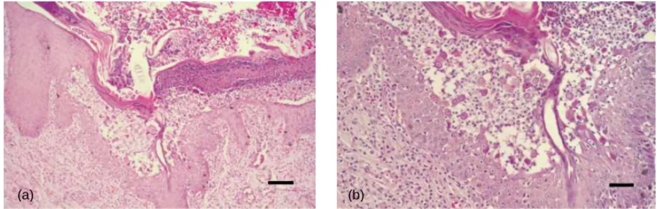
The dermis has perivascular to interstitial infiltrate in most cases and it is accompanied by edema, vascular ectasia and congestion (Gross et al., 2005). Neutrophils predominate but almost all samples have mast cells in the dermis and in some samples, the mast cells can be the prominent cell type in the dermis (Gross et al., 2005; Peterson and McKay, 2010). The mast cells may be present in epidermis too, but it is uncommon. As an explanation for this findings, it was suggested a possible association with allergic dermatoses. Eosinophils are usually present, in the dermis or marginating dermal blood vessels, but they are not the predominant cell type (Figure 13) (Gross et al., 2005). Apparently, there is no significant difference in the type or intensity of dermal infiltrate in the biopsies of cats that had a prior diagnosis of allergic dermatitis or acute inflammatory dermatitis (Preziosi et al., 2003).
Brightly, eosinophilic, contracted Apoptotic Keratinocytes (AKs) may also be present, surrounded by neutrophils and sometimes a few lymphocytes or histiocytes (Gross et al., 2005). AKs undergo a series of morphologic changes: the cells shrink, become denser and more eosinophilic and lose its normal contacts. Nuclear changes include pyknosis, margination of
Figure 13. Feline dermis. A diffuse dermal
infiltrate consisting predominately of mast cells and neutrophils are seen in the superficial dermis. Staining with H&E (Preziosi et al., 2003).
Figure 12. Feline epidermis. An active area of acantholysis located within the stratum spinosum is forming
beneath an older recornified pustule (x10) (a). Acantholytic cells can be seen “springing” up from the stratum spinosum (x20) (b). Staining with H&E (Preziosi et al., 2003).
14
chromatin and karyorrhexis (Scott et al., 2001). In a recent retrospective study, only in 24% of skin biopsies from cats with PF were found AKs. Besides, AKs are found also in other feline inflammatory dermatoses, thus other histopathological findings are needed for accurate diagnosis (Vogel et al., 2009).
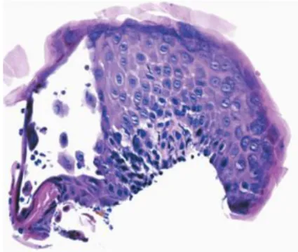
Intact pustules may be found, but it is more common the presence of overlying degenerating pustules, especially in samples of paw pads. Recornification or newly reformed stratum corneum at the base of neutrophlic pustules is very suggestive of PF (Figure 15) (Preziosi et al., 2003). Caution is warranted because re-epithelialization may cause subepidermal location. Re-epithelialization is usually recognized as a single layer of elongated basal epidermal cells at the base of the vesicle or pustule (Scott et al., 2001). Acantholysis also may occur at the level of stratum spinosum or granulosum (Figure 12), and some samples may present both subcorneal and intracorneal pustules. Rafts of acantholytic cells clinging or adhered to the overlying stratum corneum are characteristic (Preziosi et al., 2003; Gross et al., 2005). The pustules content are predominantly neutrophils and acantholytic cells and some samples may present eosinophils. Neutrophils may encircle and cling to individual acantholytic cells (Figure 14 and 15). Pustules may span various follicular hairs, depending on the density of hair and size of the pustule (Preziosi et al., 2003; Chapelin et al., 2004; Gross et al., 2005; Foster and Foil, 2007).
Some samples should be stained with Periodic Acid Shiff (PAS), to evaluate the presence of pustular dermatophytosis.
Figure 14. Sample from an auricular lesion of
a cat with PF. Histological examination reveals a fluid-filled vesiculo-pustule cointaining few neutrophils and acantholytic cells. Staining with H&E (Olivry, 2006).
Figure 15. Feline PF. Large subcorneal pustule with
neutrophils and acantholytic keratynocytes. Recornification is a characteristic feature of PF. Staining with H&E (Olivry, 2006).
15 1.7.3. Immunopathology
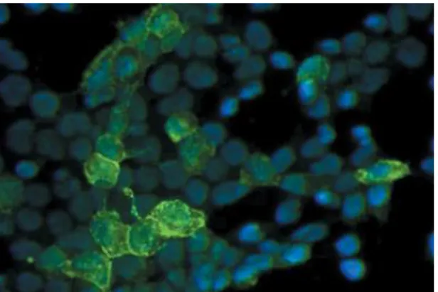
Biopsy specimens can be stained with anti-IgG fluorescein to demonstrate the deposition of antibody (Gershwin, 2010), which may be helpful for diagnosis (Figures 16 and 17). Detection of pemphigus antibody by IF or immunohistochemical testing are not routinely recommended, due to the costs, technical problems and poor diagnostic sensitivity and specificity (Scott et al., 2001). These techniques are more available for research and to distinguish the forms of subepidermal vesiculobullous diseases (Torres, 2004).
In a recent review, three criteria for the diagnosis of PF in animals with a skin disease were suggested by Olivry (2006): 1- clinical examination: transient pustules that rapidly evolve to erosions and crusts, predominantly on the face and feet; 2- histopathology: superficial epidermal or follicular pustules, with non-degenerated neutrophils and acantholytic keratinocytes; 3- differential diagnoses: rule out other acantholytic neutrophilic pustular diseases, such as bacterial skin infections and pustular dermatophytosis.
1.8. Treatment
To date, this disease is chronic and not curable. PF is managed with drugs that suppress the immune system, affecting autoreactive B cells and decreasing autoantibody production, and therefore blister formation. There are many treatments available, however it is not known which is the most effective or safest treatment option, or which is the best combination for PF, both in humans (Martin et al., 2009) and in domestic species (Rosenkrantz, 2009). The therapy’s goal is suppress disease, maintaining the quality of life and minimizing
Figure 16. Canine PF. Direct IF reveals
intercellular epidermal IgG in the stratum spinosum and granulosum (Olivry, 2006).
Figure 17. Canine PF. Indirect IF demonstrates that
only rare sera from dogs with PF recognize Dsg1. These results indicate that Dsg1 is a minor antigen in canine PF (Olivry et al., 2006).
16
drug side effects. If secondary infection is concurrent with PF, an antimicrobial therapy combination should be considered.
Treatment options should be chosen based on the severity of clinical presentation, after careful evaluation of the benefits and side effects, in the context of the individual’s other medical conditions, like Feline Leukemia Virus (FeLV) and Feline Immunodeficiency Virus (FIV), initial response and response to treatment. Owner compliance and financial resources are important too (Dunn, 1998). It is very important that owners are fully informed of the prognosis and medication side effects before starting treatment (Tater and Olivry, 2010).
A division in three treatment categories of treatment is suggested by Rosenkrantz (2004): common therapeutics, current alternative therapeutics and additional alternative therapeutics.
1.8.1. Common therapeutics
Actually, the most common immunosuppressive agents used in domestic animals consist in the powerful drugs of the steroid group and cytotoxic drugs.
1.8.1.1. GCs (Glucocorticoids)
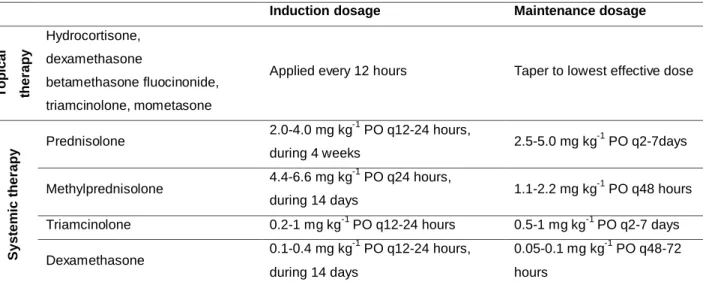
Localized forms of PF can be treated with topical GCs, initially with a potent topical GC and then, if adequate response is seen, it is recommended to switch to a less potent topical GC. These cats may not need a systemic therapy later if topical preparation induces clinical remission of signs and no further development of new lesions (Rosenkrantz, 2004; Paterson, 2006). Suitable topical GCs for initial therapy are represented in Table 2. Persistent use of more potent topical GC can create skin atrophy, alopecia and localized pyoderma. If no positive response is seen within 14 days, the introduction of systemic therapy is recommended (Rosenkrantz, 2004; Paterson, 2006). Topical therapy can be used in conjunction with systemic therapy on more persistent focal areas that remain active despite systemic treatment (Rosenkrantz, 2004; Rosenkrantz, 2009).
Systemic GCs are actually the most common form of therapy used in PF lesions management, because of their rapid onset. The oral GCs of choice have been prednisolone or prednisone (Willemse, 2000; Scott et al., 2001; Chandler et al., 2004; Merchant, 2007; Peterson and McKay, 2010). However oral prednisolone is a better choice than prednisone for feline patients, because of the lower bioavailability of prednisone compared with other GCs (Graham-Mize and Rosser, 2004). The majority of feline cases respond well and it is maintained in clinical remission with prednisolone monotherapy (Preziosi et al., 2003; Rosenkrantz, 2004). Some authors prefer methylprednisolone, due to the reduced mineralocorticoid effects and better response in some cases (Chapelin et al., 2004; Rosenkrantz, 2009). Cats are few sensitive to
17
the immunosuppressive effects of GCs and often require high doses for remission (Thacker, 2010) and if no improvement in clinical signs is evident within one or two weeks, the dose may be increased (Tater and Olivry, 2010). Oral triamcinolone (Preziosi et al., 2003) or oral dexamethasone are alternative GCs, 6 to 10 times more potent than prednisolone or prednisone (Rosenkrantz, 2009; Rees, 2011). If response is seen within 10 to 14 days, the dosage is reduced gradually on a weekly basis over 30 to 40 days and then lowered to an alternate day basis to the lowest effective dosage of 1 mg kg-1 q 48 hours or less (Rosenkrantz, 2009). The patient should be revaluated every time that the dosage is adjusted. Induction and maintenance doses are detailed in Table 2. If the PF recurs when the GC is tapered, another immunosuppressive drug should be added as adjunctive therapy.
Nowadays, injectable GCs such as methylprednisolone acetate are not recommended for PF treatment (Tater and Olivry, 2010).
The most common GCs side effects include poor dull scaly hair coats, muscle atrophy, polyuria and polydipsia (PU/PD), polyphagia, weight gain, behavioral changes, panting and increased risk for infections. Bladder infections and skin infections, especially dermatophytosis,
Malassezia infections and demodicosis, may develop due to chronic steroid usage (Paterson,
2006; Rosenkrantz, 2009). When secondary pyoderma is present, antibiotics according to bacteriogram or culture should be used (Peterson and McKay, 2010). Other skin changes include atrophic skin, calcinosis cutis, atrophic scars, comedones and miliary follicular cysts. Gastrointestinal ulcerations, diarrhea, pancreatitis, Diabetes mellitus (DM), adrenal gland suppression, iatrogenic hyperadrenocorticism and hipotiroidism can also occur (Rosenkrantz, 2009; Thacker, 2010).
Table 2. GC therapy for feline PF
Induction dosage Maintenance dosage
T o p ic a l th e ra p y Hydrocortisone, dexamethasone betamethasone fluocinonide, triamcinolone, mometasone
Applied every 12 hours Taper to lowest effective dose
S y s te m ic t h e ra p y Prednisolone 2.0-4.0 mg kg-1 PO q12-24 hours, during 4 weeks 2.5-5.0 mg kg -1 PO q2-7days Methylprednisolone 4.4-6.6 mg kg -1 PO q24 hours, during 14 days 1.1-2.2 mg kg -1 PO q48 hours Triamcinolone 0.2-1 mg kg-1 PO q12-24 hours 0.5-1 mg kg-1 PO q2-7 days Dexamethasone 0.1-0.4 mg kg -1 PO q12-24 hours, during 14 days 0.05-0.1 mg kg-1 PO q48-72 hours
References: (Chandler et al., 2004; Rosenkrantz, 2004; Medleau and Hnilika, 2006; Paterson, 2006; Foster and Foil, 2007; Crow, 2009; Rosenkrantz, 2009; Hnilika, 2011; Rees, 2011).
18 1.8.1.2. Chlorambucil
Chlorambucil is an alkylating agent that affects the Deoxyribonucleic acid (DNA) synthesis. In cats whose PF lesions fail to respond to GCs, chlorambucil is the most commonly cytotoxic drug used and can be used in combination with GCs or as sole therapy. When combined with prednisolone, these drugs are given on alternating days to prevent gastrointestinal irritation. The onset of action is slow and results appear within 3 to 6 weeks. Therapy has to be given 4 to 8 weeks and then one should be able to stop chlorambucil and use GCs alone (Rosenkrantz, 2009; Tater and Olivry, 2010). Initial and maintenance dosages are detailed in Table 3.
Chlorambucil associated adverse effects include myelosuppression, hepatotoxicity in addition to vomiting, diarrhea and anorexia with weight loss. When bone marrow suppression occurs, the drug should be withdrawn until parameters return to normal (Chandler et al., 2004; Paterson, 2006; Rosenkrantz, 2009; Irwin et al., 2011).
1.8.2. Current alternative therapeutics
Current immunosuppressive drugs can be introduced when common therapeutics fail or have undesirable secondary effects (Rosenkrantz, 2009). Combination therapy can also be used as an initial treatment strategy (Paterson, 2006).
1.8.2.1. CsA (Ciclosporin) and tacrolimus
CsA is a potent inhibitor of cell-mediated immunity, and a less potent inhibitor of humoral immunity. CsA properties include immunosuppressive effects, anti-inflammatory effects, antiproliferative effects, inhibits antigen presentation and it is antiparasitic and lacrimomimetic (Robson, 2003b). CsA has been used successfully in veterinary medicine to treat several dermatologic diseases (Robson and Burton, 2003). Topical CsA and tacrolimus is not recommended in cats because of the potential toxicity and/or lack of efficacy and the cat’s tendency to ingest topical medication (Paterson 2006; Foster et al., 2007). The efficacy of transdermal formulation of CsA (Atopica – Novartis, Animal health) in cats was recently studied, due to the difficulties of oral administration in this species. The absorption via transdermal was inconstant therefore oral administration remains the most recommended (Miller et al., 2011). Initial and maintenance dosages are detailed in Table 3.
In dogs and cats, CsA is often used in conjunction with GCs. In human patients with pemphigus, apparently the combination treatment with GCs and CsA offers no advantage over treatment with GC alone (Ioannides et al., 2000).
Irwin et al. (2011) compared oral CsA with chlorambucil in management of feline PF. It’s interesting that there was no significant difference in remission times or disease response
19
between CsA and chlorambucil, and the cats managed with CsA required significantly less GCs for remission induction and for maintenance therapy. It seems that CsA provides comparable efficacy for management of feline PF.
Few clinically relevant drug interactions with CsA have been reported in veterinary medicine. A very useful drug interaction is with ketoconazol, which inhibits CsA metabolism, resulting in CsA dose reduction and costs saving to the client (Robson, 2003a; Rosenkrantz, 2009; Katayama et al., 2010). Few adverse effects of CsA have been reported in cats. Diarrhea is the most frequently reported adverse reaction (Robson, 2003a; Rosenkrantz, 2004). The administration of gastric protectants, starting with lower dosages or more frequently dividing dosages may prevent gastrointestinal disturbances (Rosenkrantz, 2009). Gingival hyperplasia, hypertrichosis, pyoderma, self-limiting diarrhea, vomiting, nephrotoxicity and intermittent soft stools were reported too (Foster et al., 2007; Irwin et al., 2011).
1.8.3. Additional alternative therapy 1.8.3.1. Cyclophosphamide
This drug is an alkylating agent and it is more toxic than chlorambucil. Induction dosage is 1.5-2.5 mg kg-1 q 48 hours, and reduction of dosage and frequency is recommended once remission is achieved (Table 3) (Rosenkrantz, 2009).
Cyclophosphamide has no advantages over chlorambucil in cats in the treatment of pemphigus (Paterson, 2006). The use of cyclophosphamide has been associated with bone marrow suppression, hemorrhagic cystitis with hematuria and dysuria, nausea, vomiting and anorexia, infertility and teratogenic effects. It is potentially carcinogenic. Wound healing disturbance, alopecia and loss of whiskers can occur (Rosenkrantz, 2009).
1.8.3.2. Chrysotherapy
Chrysotherapy can be used alone or with systemic GCs. Remission can be successfully obtained with this protocol (Kofod, 1993; Rosenkrantz, 2009). Gold salts are available as oral (auranofin) formulations. Injectable forms (aurothiomalate or aurothioglucose) are quite effective in feline PF but are no longer available (Rosenkrantz, 2009). About 25% of cats that have not results to GC treatment respond to chrysotherapy (Willemse, 2000).
Side effects with gold therapy include myelosuppression, thrombocytopenia, oral ulcers, glomerulonephropathy, hepatotoxicity, stomatitis, sterile abscesses and cutaneous drug eruptions (Chandler et al., 2004; Foster et al., 2007).
20
1.8.3.3. IVIg (Human Intravenous Immunoglobulin) therapy
IVIg is a high-purified IgG preparation from human plasma. IVIg therapy may be useful in patients to whom conventional therapies have failed not only in humans (Ahmed and Sami, 2002) but also in animals (Rosenkrantz, 2009). This form of therapy is not completely evaluated in domestic species due to limited number of cases treated with IVIg, but appears to be safe. The animal’s response to IVIg is inconstant, showing 50% of efficacy. IVIg should be started at 0.01 ml kg-1 minute intravenously and gradually increased every 30-60 minutes to a maintenance fluid rate not exceeding 0.08 ml kg-1 minute. IVIg is administered over 6 to 12 hours. Anaphylaxis may be seen after the first treatment and concurrent GC therapy is recommended (Rosenkrantz, 2009).
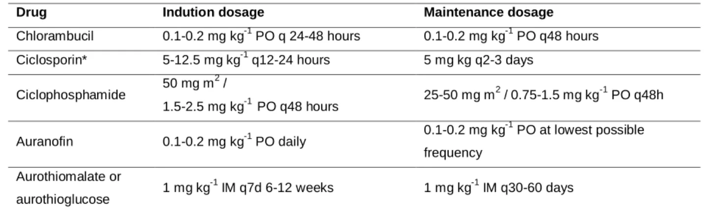
Table 3. Alternative Immunosuppressive drugs recommended for feline PF
Drug Indution dosage Maintenance dosage
Chlorambucil 0.1-0.2 mg kg-1 PO q 24-48 hours 0.1-0.2 mg kg-1 PO q48 hours Ciclosporin* 5-12.5 mg kg-1 q12-24 hours 5 mg kg q2-3 days
Ciclophosphamide 50 mg m 2 / 1.5-2.5 mg kg-1 PO q48 hours 25-50 mg m 2 / 0.75-1.5 mg kg-1 PO q48h Auranofin 0.1-0.2 mg kg-1 PO daily 0.1-0.2 mg kg -1 PO at lowest possible frequency Aurothiomalate or aurothioglucose 1 mg kg -1 IM q7d 6-12 weeks 1 mg kg-1 IM q30-60 days
References: (Chandler et al., 2004; Rosenkrantz, 2004; Medleau and Hnilika, 2006; Paterson, 2006; Foster et al., 2007; Crow, 2009; Rosenkrantz, 2009; Hnilika, 2011; Rees, 2011).
*The ciclosporin dose for feline PF has not been established
1.8.4. Other immunosuppressive drugs not used in feline patients 1.8.4.1. Azathioprine
Azathioprine is a cytotoxic drug to T cells. Despite this drug was successful in PF remission in cats (Caciolo et al., 1984 cit. by Broek, 1991), it is contraindicated due to profound myelosuppression and potential fatal reactions in cats. This may be related to the low levels of activity in cats of Thiopurine methyltransferase (TPMT), the enzyme responsible for the metabolism of azathioprine, and therefore cats are more susceptible to azathioprine adverse effects (Foster et al., 2000; White et al., 2000). Other secondary effects include hepatotoxicosis, vomiting, diarrhea, panniculitis, drug eruption, alopecia and increased susceptibility to opportunistic infections (Chandler et al., 2004; Rosenkrantz, 2009).
21
1.8.4.2. Niacinamide and tetracycline or doxycicline
These drugs can be used in dogs and cats for PF treatment, however are not typically used in feline patients, due to the difficulty of oral administration.
1.8.4.3. Mycophenolate mofetil
This drug has been used limitedly in dogs (Rosenkrantz, 2009), but in cats it is unknown its efficacy for the treatment of autoimmune diseases.
1.8.4.4. Dapsone
Dapsone is avoided in cats, because this species commonly develop hemolytic anemia and neurotoxicity (Paterson, 2006).
There is a variation in dosage plan and combination of drugs used, and the response to treatment can vary between animals, which makes the choice of treatment schedule complex. It is important that clinicians do not underestimate the difficulty in managing this disease. Relapsing or refractory cases are very common and it is important to differentiate immunosuppression-induced pyoderma, demodicosis or dermatophytosis from an actual disease flare, otherwise lesions may worsen from immunosuppression (Paterson, 2006; Tater and Olivry, 2010; Hnilika, 2011).
Further studies are needed to determine the optimal treatment protocol and especially to assess the optimal GC dose and the role of adjuvant immunosuppressive drugs, to improve benefit results and decrease adverse effects.
A very recent experimental study in mice suggested that superficial expression of Dsg-2 activates multiple growth and survival pathways and can limit epidermal blister formation mediated by PF autoantibodies (Brennan et al., 2010). Thus, a drug that increases Dsg2 levels on the skin offers a potential therapeutic treatment.
Recently, rituximab has been used in refractory and severe human PF (Schmidt et al., 2009; Kasperkiewicz et al., 2010; Lipozenčić and Marinović, 2011). Rituximab is a monoclonal antibody that reduces the number of B lymphocytes and therefore autoantibodies production. However, efficacy in remission of PF lesions was not proved due to lack of patients and adjuvant therapy with other immunosuppressive drugs. In future, this drug may be studied and adapted for pemphigus treatment in animals.
22 1.9. Monitoring
Often cats require lifelong therapy to maintain remission, and under any immunosuppressive treatment, the patient should be monitored with physical examination, CBCs and chemistry profiles to monitor medication side effects, urinalysis and urine bacterial cultures to monitor occult urinary tract infections, especially during induction periods (Paterson, 2006; Rosenkrantz, 2009). Re-evaluation is recommended before and after each change in medication type or dose to help monitor clinical signs (Tater and Olivry, 2010). Monitoring of drug therapy is detailed in Table 4.
Table 4. Monitoring of drug therapy in feline PF (Adapted from Paterson, 2006; Rosenkrantz, 2009)
Drugs Monitoring
GCs Twice yearly CBC, chemistry profiles, urinalysis, and urine cultures
Chlorambucil Routine hematology (including platelet count), liver function tests every 2-4 weeks for 2 months, then 3-4 times yearly
CsA CBC, Chemistry profiles and urinalysis every 3-4 months
Cyclophosphamide CBC, urinalysis and chemistry profiles every 2 weeks for the first 8-12 weeks
Gold salts Routine hematology (including platelet counts), biochemistry and urinalysis every 2-3 weeks for 4 months, then 2-4 times yearly.
Cutaneous side effects of immunosuppression, such as skin infection, demodicosis or dermatophytosis may mimic PF flares. It is important to rule out any possibility of these conditions instead of assuming that a new skin lesion is a PF flare, otherwise, lesions may worsen with immunosuppressive therapy, and refractory pemphigus may be erroneously diagnosed (Tater and Olivry, 2010).
The principles of treating PF are summarized in Table 5.
1.10. Prognosis
In most cases, the prognosis is fair to good, but some of them can have an unfavourable clinical evolution (Kofod, 1993; Chapelin et al., 2004). Some animals have significant improvement after discontinued immunosuppressive therapy, but most of them require lifelong therapy to maintain remission. Rare cases evolve unfavourable, provoking death or were euthanized due to disease or complications of therapy (Preziosi et al., 2003).
23
Chronic blistering can result in pain, dehydration, secondary infections and rarely, death (Preziosi et al., 2003; Chapelin et al., 2004). In several conditions, pemphigus may have a significant impact of the quality of life. Psychological impact on owners may be profound, due to chronicity of the disease, lifelong therapy, long-term costs of recheck examinations and test to monitor PF patients receiving therapy and side effects from treatment. The successful treatment of PF depends to a large extent on the client, who administers most prescribed therapies. It is important to make the owner aware of potential problems, the likely course of disease as well as the need for follow-up and therapeutic modifications. The owner’s compliance is a very important factor to obtain good results (Scott et al., 2001).
Table 5. Principles of treating PF in dogs and cats (Tater and Olivry, 2010)
Control any concurrent secondary bacterial infection. Consider antibiotic selection based on bacterial culture and antimicrobial sensivity results, especially with deep pyoderma.
Select an immunosuppressive therapy after reviewing indications, dosages, administration regimens, and adverse effects. Immunosuppressive therapy should only be used in dogs and cats with a confirmed diagnosis.
Recheck patients at regular intervals to monitor for recurrence of pemphigus lesions. Perform laboratory tes ts (CBC, serum chemistry profiles, urinalysis, and urine bacterial culture) to monitor for adverse effects.
If lesions decrease in extent and severity, the dose or frequency or both, of immunosuppressive therapy should be decreased.
If new cutaneous lesions occur during the treatment, first rule out bacterial skin infections, demodicosis, or dermatophytosis.
If new cutaneous lesions are determined to be due to a flare-up of PF, adjust the dose or frequency, or both, of the medication. If systemic GCs are being used, adding another medication may enable GCs to be decreased and then discontinued in the future.
Cases of PF that cannot be maintained in remission or develop adverse effects should be referred to a veterinary dermatologist.
24 Objectives
The purpose of this retrospective study is to increase the knowledge in feline PF, by describing the epidemiological, clinical features, cytological and histopathological findings of 11 cases of cats diagnosed for PF. The therapeutic options employed and their outcomes are also characterized. The results obtained are discussed, compared with the literature and conclusions about these cases are exposed.