367
CASE REPORTSarcoidosis simulating metastasis in patient
with Merkel cell carcinoma
Sarcoidose simulando metástase em paciente com carcinoma de células de Merkel
Nathália Bordeira Chagas1; Lucimar Retto da Silva de Avó2; Victor Hugo Maion3; Michel Antonio Kiyota Moutinho4; Débora Gusmão Melo5; Carla Maria Ramos Germano6
ABSTRACT
Merkel cell carcinoma (MCC) is a rare and aggressive skin neuroendocrine tumor whose etiology remains little known. The immune system dysfunction is considered one of the possible causes, shared with other diseases, like sarcoidosis. This article presents the case of a male patient, in the seventh decade of life, which had a prior MCC diagnosis and was subsequently diagnosed with sarcoidosis, which radiologically simulated lung and mediastinum metastasis. The association between MCC and sarcoidosis is infrequent and rarely described in literature, found in only two case reports.
Key words: Merkel cell carcinoma; sarcoidosis; immune system.
J Bras Patol Med Lab, v. 50, n. 5, p. 367-370 outubro 2014
INTRODUCTION
Merkel cell carcinoma (MCC) is a rare and highly aggressive skin cancer. Considered a neuroendocrine tumor derived from mechanoreceptor cells located in the basal layer of the epidermis, it mainly affects areas chronically exposed to the sun(4).
The annual incidence rates of diagnosed MCC cases are 0.23 and 0.01 per 100 thousand individuals, in Caucasians and Afro-descendants, respectively. It occurs predominantly in older people, with mean age of 69 years at diagnosis, affecting more men (61%) than women (39%)(4). Besides age, other risk factors are associated
to its little known etiology: exposure to ultraviolet radiation, Merkel cell polyomavirus infection, and immunosuppression(4, 5).
In the literature, immune system dysfunction is pointed as an important factor that may contribute to the occurrence of MCC(5)
and, in a less evident manner, sarcoidosis(2) – a systemic
non-infectious granulomatous disease of uncertain etiology(9).
This paper describes the case of a MCC patient who, during the investigation of metastatic disease, presented radiologic imaging
suggestive of pulmonary and mediastinal metastasis. Through the histological study, the possibility of metastasis was excluded, and the diagnosis of sarcoidosis was achieved. The association between MCC and sarcoidosis is not common, and it is rarely described in the literature (only two case reports).
CASE REPORT
The patient was a 70-year old white male, engineer, retired professor, and non-smoker. At the beginning of his career, he worked as an engineer at a metallurgy company for a month and, later, at a car factory for three years. He had noticed a nodule in the gluteal region two years before, which displayed faster growth nine months ago. The patient had a dermatologist check the lesion, and was referred to a general surgeon to excise it.
The pathology report described a lesion with reddish irregular plain surface, measuring 0.5 × 0.5 cm, 0.3 cm apart from the nearest surgical resection margin. Microscopic examination revealed no epidermal alterations. In the deep dermis and
First submission on 25/10/13; last submission on 13/02/14; accepted for publication on 17/07/14; published on 20/10/14
1. Graduating student of Medicine at Universidade Federal de São Carlos (UFSCar). 2. Pathologist; assistant professor at the Medicine Department of UFSCar. 3. Oncological surgeon.
4. Pathologist.
5. Clinical geneticist; assistant professor at the Medicine Department of UFSCar. 6. Pediatric endocrinologist; assistant professor at the Medicine Department of UFSCar.
368
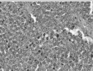
hypodermis, a multinodular malignant neoplasm was identiied, formed by small cell-blocks, of scant cytoplasm, round nuclei with uniformly distributed dense chromatin and inconspicuous nucleoli. Mitotic igures and apoptotic corpuscles were frequently observed. Lateral and deep surgical margins coincided with the neoplasm (Figure 1).
After diagnosis, the patient was referred to an oncologic surgeon to widen the margins. At clinical assessment, he did not present any respiratory symptom, as well as fever, weight loss, fatigue or night sweats. His physical examination was described as normal. At the previously operated site, there was a newly-formed scar with no signs of relapse. The widening of surgical margins was performed, and a thoracic computed tomography (CT) with contrast enhancement was required for staging. The CT revealed mediastinal adenomegaly, multiple pulmonary nodules and micronodules, suggestive of metastatic neoplasia (Figure 3).
Based on these indings, sentinel lymph node biopsy, right inguinal lymph node dissection and segmental left lung resection through videothoracoscopy were proposed. The histological examination of surgical specimens showed granulomatous inlammation, in the different analyzed tissues, besides a chronic nonspeciic inlammatory process in the scar and in tissue from the widened surgical margins (Figure 4). The investigations
of alcohol-acid-resistant bacilli (AARB) (using Ziehl-Neelsen
staining) and fungi (using methenamine silver staining)
were negative, both at lymph nodes and pulmonary segment, reinforcing the diagnostic hypothesis of sarcoidosis.
After the diagnosis of sarcoidosis, the patient was examined by a pneumologist, who performed a pulmonary function test. This exam revealed a mild chronic obstructive process, and the regular use of an inhaled glucocorticoid was prescribed. The patient is currently on regular oncologic follow-up, with no evidence of active neoplastic disease.
Sarcoidosis simulating metastasis in patient with Merkel cell carcinoma
FIGURE 1 – Photomicrograph of MCC, formed by cells with round homogeneous nuclei, scant cytoplasm, and numerous mitotic figures. HE, 400×
MCC: Merkel cell carcinoma; HE: hematoxylin and eosin.
Immunohistochemical staining was positive for cytokeratin 20, chromogranin A, Ki67 and synaptophysin; and negative for thyroid transcription factor (TTF-1)/lung, CD20 and CD3, conirming the diagnosis of MCC (Figure 2).
FIGURE 2 – Immunohistochemical staining of biomarkers used in the diagnosis of MCC: A) positive for cytokeratin 20 (clone KS20.8); B) positive for synaptophysin (clone SY38); C) positive for chromogranin A (clone DAK-A3); D) negative for T-cell receptor-CD3 (epsilon chain) (polyclonal)
MCC: Merkel cell carcinoma.
FIGURE 3 – Thoracic computed tomography showing mediastinal adenomegaly, multiple pulmonary nodules and micronodules
369
DISCUSSION
MCC typically presents as a rapid growing (weeks or months) painless violaceous nodule. It usually arises on sun-exposed regions, like the head and neck (41%-50%), followed by the extremities (32%-38%) and the trunk (12%-14%)(1, 4).
Occurrence in the gluteal region, as observed in this patient, is uncommon, with few cases described in the literature.
Diagnosis is established by biopsy, and to differentiate MCC from other tumors, immunohistochemistry is performed(5).
This study highlights positivity for cytokeratin 20 (whose expression is detected in 89%-100% of MCC)(10) and negativity
for TTF-1 (which is speciic for small cell lung carcinoma)(6) as
important tools for the differential diagnosis of MCC.
Due to the aggressive behavior of this cancer, besides exeresis of the primary tumor, the sentinel lymph node biopsy has been adopted. It serves as a complementary diagnostic strategy for detecting metastasis(5), since lymph node involvement at
diagnosis is considered an important prognostic factor(11),
being present in 50%-70% of MCC patients. In the present case, sentinel lymph node biopsy revealed the presence of noncaseating epithelioid granulomas, with negative results for AARB and fungi. Histological examination of the resected lung segment presented the same aspect, reinforcing the diagnostic hypothesis of sarcoidosis.
Sarcoidosis is a systemic inlammatory non-infectious disease in which noncaseating epithelioid granulomas are the microscopic elementary lesions. The lung and the lymphatic system are the predominantly affected sites(9). The prevailing
etiological hypothesis, still not deinite, is that multiple non-identiied antigens, both infectious and environmental, can trigger an exaggerated immune reaction in genetically susceptible hosts. Its incidence is slightly higher in women (1.3%) than in men (1%), and higher in Blacks (2.4%) than in Caucasians (0.8%). It is generally sporadic, although a positive family history is found in 17% of the cases(9).
The disease spectrum is wide, and the clinical presentation may comprise asymptomatic patients, whose sarcoidosis is incidentally discovered through an abnormal chest radiograph (8%-60% of the cases), a situation compatible with our patient’s. Likewise there are symptomatic patients, whose manifestations may be: a) respiratory, like persistent dry cough (30%); b) extrathoracic, principally affecting peripheral lymph nodes, eyes and skin; c) erythema nodosum (3%-44%); or inally,
Nathália Bordeira Chagas; Lucimar Retto da Silva de Avó; Victor Hugo Maion; Michel Antonio Kiyota Moutinho; Débora Gusmão Melo; Carla Maria Ramos Germano
d) unspeciic constitutional symptoms such as fatigue (27%), weight loss (28%), fever (10%-17%) and night sweats(9).
The case reported in this paper generates two discussions: the value of histological examination in the differential diagnosis of metastatic disease suggested by imaging, and the association between sarcoidosis and MCC.
Differentiation between sarcoidosis and metastasis is probably not a simple process. Fluorodeoxyglucose positron-emission tomography (FDG-PET), widely used in the assessment of neoplastic diseases, is quite effective to differentiate malignant from inlammatory lesions(6). Kaira et al. (2007)(7)
report that the use of PET with L[3-18F]-alpha-methyltyrosine
(18F-FMT-PET) in combination with FDG-PET may distinguish
sarcoidosis from malignancy, and may be a useful tool in this differential diagnosis. It is worth emphasizing that these exams are of dificult access far from large diagnostic centers, and they were not available for use in the cited case.
In a literature review, we found one case, published in 2005, in which the association between sarcoidosis and MCC was described in an 84-year-old female with chronic sarcoidosis who had MCC diagnosed on her face nine years later(8).
A more recent case, described in 2011, reports a 57-year-old woman with MCC diagnosed in the left inguinal region, with an associated granulomatous reaction suggestive of mediastinal lymph node sarcoidosis(6). Other similar studies
were not found in the medical literature, what attests that the association between both diseases is rare and may represent just a coincidence.
The pathogenesis of sarcoidosis, as well as that of MCC, although poorly understood, seems to involve immune dysfunction, what may represent a causal factor for the coexistence of these diseases in a patient(8). Cohen and Kurzrock
(2007)(3), in a literature review, state that sarcoidosis may
develop in association with different neoplasms. A possible explanation lies in the administration of antineoplastic drugs that could both lead to the occurrence and activate a focus of latent sarcoidosis. This does not apply to the present case, since the patient did not undergo chemotherapy.
370
RESUMO
O carcinoma de células de Merkel (CCM) é um tumor neuroendócrino de pele, raro e agressivo, cuja etiologia permanece pouco compreendida. Disfunção do sistema imune é apontada como um possível fator causal, tanto para o CCM quanto para a sarcoidose. Este artigo descreve o caso de um paciente do sexo masculino, na sétima década de vida, com diagnóstico de CCM, posteriormente diagnosticado com sarcoidose, que, à radiologia, simulava metástase para pulmão e mediastino. A associação entre CCM e sarcoidose é rara e muito pouco descrita na literatura, encontrada em apenas dois relatos de casos.
Unitermos: carcinoma de célula Merkel; sarcoidose; sistema imune.
MAILING ADDRESS
Lucimar Retto da Silva de Avó
Alameda das Pitangueiras, 97; Parque Faber; CEP: 13561-359; São Carlos-SP, Brazil; e-mail: [email protected].
REFERENCES
1. ALMEIDA, M. W. R. et al. Carcinoma de células de Merkel em extremidade inferior. Rev Col Bras Cir, v. 39, n. 2, p. 165-7, 2012. 2. BZHALAVA, D. et al. Risk of second cancers after the diagnosis of Merkel
cell carcinoma in Scandinavia. Br J Cancer, v. 104, n. 1, p. 178-80, 2011.
3. COHEN, P. R.; KURZROCK, R. Sarcoidosis and malignancy. Clin Dermatol, v. 25, n. 3, p. 326-33, 2007.
4. DUPRAT, J. P. et al. A review of the epidemiology and treatment of Merkel cell carcinoma. Clinics, v. 66, n. 10, p. 1817-23, 2011.
5. GOMES, C. A. et al. A importância da linfocintilograia no tratamento do carcinoma de células Merkel. Rev Col Bras Cir, v. 38, n. 5, p. 361-5, 2011.
6. HIGASHI, Y. et al. Sarcoid reaction associated with Merkel cell carcinoma revealed by luorodeoxyglucose positron emission tomography: a case report. J Med Case Rep, v. 5, n. 282, 2011.
7. KAIRA, K. et al. Diagnostic usefulness of luorine-18-alfa-methyltyrosine
positron emission tomography in combination with 18F-luorodeoxyglucose in sarcoidosis patients. Chest, v. 131, n. 4, p. 1019-27, 2007.
8. McLOONE, N. M. et al. Merkel cell carcinoma in a patient with chronic sarcoidosis. Clin Exp Dermatol, v. 30, n. 5, p. 580-2, 2005.
9. NUNES, H. et al. Sarcoidosis. Orphanet J Rare Dis, v. 2, n. 46, 2007. 10. PERMAN, M. J. et al. Giant Merkel cell carcinoma masquerading as a
benign cyst on the buttock of an African American man. Case Rep Oncol Med, v. 2011; 2011: 849767.
11. POULSEN, M. Merkel-cell carcinoma of the skin. Lancet Oncol, v. 5, n. 10, p. 593-9, 2004.