Cardiac Aging in Female Wistar Rat Models With Sedentary or Physicaly Active Lifestyle
62
0
0
Texto
(2)
(3) CARDIAC AGING IN FEMALE WISTAR RAT MODELS WITH SEDENTARY OR PHYSICALY ACTIVE LIFESTYLE. Dissertação apresentada com vista à obtenção do grau de mestre em Atividade Física e Saúde,. da. Faculdade. de. Desporto. da. Universidade do Porto ao abrigo do Decreto de Lei nº.74/2006 de 24 de Março, orientada pelo Professor Doutor José Alberto Ramos Duarte e co-orientada pelo Professor Doutor Hélder Fonseca. Morgana Hoffmann Porto, 2013. I.
(4) HOFFMANN, M. (2013). Cardiac aging in female Wistar rat models with sedentary or physicaly active lifestyle. Dissertação de Mestrado em Atividade Física e Saúde. Faculdade de Desporto da Universidade do Porto, Porto.. Key words: Exercise; Cardioprotection; Voluntary running, Cardiovascular function; Sedentarism.. II.
(5) Funding Sources This present work was conducted in the Research Center for Physical Activity, Health and Leisure (CIAFEL), which is a research unit housed in the Faculty of Sports, University of Porto, Portugal. The experimental studies were supported by Fundacão para a Ciência e Tecnologia. (FCT). grants. PTDC/DES/104567/2008. OE/SAU/UI0617/2011.. III. and. PEst-.
(6) IV.
(7) To family, my base and my support. Thank you for everything. With love…. V.
(8) VI.
(9) Acknowledgements. I would like to thank God for allowing me to achieve this objective.. The decision to take a master degree in another country, in the beginning was not easy because, if I really wanted to, I would have to chose the certain by the uncertain, make new friends, know a different culture and face the challenges of these two years of working in an area that was unfamiliar to me and far from family and friends. Fortunately, I was lucky to be surrounded by the people that helped me to overcome through all the obstacles along this way, and it is for these people that I would like be thankful.. First, I would like to thank my supervisor, Prof. José Duarte, for receiving me in the biochemistry laboratory, sharing all his knowledge throughout this time. I am very grateful to all your help and support to perform this work. Thank you for the unique opportunity and all your supervision.. To my co-supervisor, Prof. Hélder Fonseca, I run out of words to express how grateful I am for all the tireless time you dedicated to this work and the patience and also the disposition at any time. Your help and support was essential to complete this journey.. To Prof. Daniel Gonçalves, for all the time shared, confidence and support in this work and over these time. Thank you Dani, for helping me through all over this journey. To my dear “Família 18”, Diana, Toni e Re, thank you for the friendship and all the special moments you proportionate to me in these time. I´m very lucky for you letting me to be part of your life and I hope that this friendship last forever! Thank you, you are one of the most important part of it.. To my friend Wagner, that since the first classes of the master has being such a big friend. Be sure that all the moments listening to me, making me VII.
(10) laughing, encouraging me, will not be forgotten. “Gracias tchê!” And also to my special friends that I did here in Portugal, specially César, Cris, Teresa, Ana e Nórton, thanks for all the great moments, conversations, laughters, “indiadas”, “mates na Ribeira”, and of course, the parties and to the best histories of this years. I wish that this friendship is for life! Thank you for making my days better, and together with “Família 18”, unforgettable.. To my longtime dear friends, that even being on the other side of the ocean, always were with me, specially Nabi, Maia, Mogui, Quel, Feni e Ví. Thanks for all the messages, emails, good vibes and the words on the difficult times that I needed for. Thank you “chinocas”, for being with me at anytime and anywhere.. To my colleges on the laboratory, Ana Carvalho, Gonçalo, Ricardo, Eduardo and Dani Girardi, thanks for the time spent, the company, conversations and for the knowledge shared in the laboratory. Also a special thank to Dona Celeste, for all the technical support and all the friendship through this time. From your “bruxinha”. To my “Cycling” practitioners of the gym, especially, Alcina, Cris, Helder e Marlene, for making my work be a pleasure, my weeks full of energy and to be my motivation in the classes. Thank you to be part of this experience.. To all the people that work on the faculty, especially Lurdes, for all the help even before arriving in Porto and for always being this kind person with Brazilians. Finally I wish to thank… … minha família amada, pai, mãe e mano, por serem meu porto seguro, minha inspiração, meu orgulho e minha base. Obrigada pelo amor incondicional, pelos valores, carinho, força, apoio, suporte e por terem entendido minha ausência física durante estes dois anos. Vocês estiveram sempre comigo nessa longa caminhada. Eu sempre vou ser grata a vocês. Amo vocês! VIII.
(11) This was the biggest challenge of my life and that just could be possible with all the support of these people. This would not be so special without you all, my eternal gratitude!. IX.
(12) X.
(13) Index of Contents. Funding Sources. III. Acknowledgments. VII. Index of Contents. XI. Resumo. XIII. Abstract. XV. List of Abbreviations. XII. 1. GENERAL INTRODUCTION. 1. 2. STATE OF THE ART. 6. 3. EXPERIMENTAL STUDY: Regular physical exercise attenuates the. 14. cardiac degenerative changes related with age in female Wistar rats. 4. MAIN CONCLUSION. 41. XI.
(14) XII.
(15) Resumo As alterações cardíacas relacionadas com a idade ao longo da vida são progressivas e irreversíveis, comprometendo a habilidade cardíaca de tolerar estímulos. de. estresse.. A. diminuição. no. número. de. cardiomiócitos. acompanhado pela hipertrofia dos mesmos, assim como o aumento do conteúdo de colagênio em ambos os ventrículos, direito e esquerdo, promove um estado deletério na estrutura cardíaca e uma gradual incapacidade de uma eficaz resposta do coração quando submetido a estímulos de estresse. Além disto, a função sistólica e diastólica dos ventrículos também é prejudicada pelas alterações relacionadas com o envelhecimento. Por outro lado, o exercício físico regular e voluntário tem sido associado com a atenuação e prevenção destas alterações relacionadas com o processo de envelhecimento promovendo um efeito cardioprotetivo. Neste sentido, o nosso objetivo era determinar se a prática regular e voluntária de corrida através de roda livre é capaz de atenuar os danos estruturais e funcionais relacionadas com a idade em ratas fêmeas Wistar. Dezenove Wistas ratas com 5 meses de idade foram mantidas em condições laboratoriais padrão e divididas aleatoriamente em três grupos: 1- Jovens (Y, n=7; sacrificadas uma semana após aclimatação); 2idosas sedentárias (SO, n=6; movimentos limitados ao espaço da caixa por 9 meses) e 3- idosas activas (AO, n=6; alojadas em caixas com uma roda livre por 9 meses). Após o final dos seus protocolos, todos os animais foram preparados para avaliação hemodinâmica do ventrículo esquerdo (LV) e do ventrículo direito (RV) com cateteres de condução. Imediatamente após isto, os animais foram sacrificados e as amostras do ventrículo esquerdo e direito foram coletadas para avaliação histológica da área de secção transversal (CSA; H&E) e fibrose (Pricosirius red staining). Nossos resultados mostraram que o exercício físico regular e voluntário durante a vida melhorou as funções sistólica e diastólica como também o relaxamento rápido. A melhoria destas funções foi associada com a prevenção da hipertrofia dos ventrículos e com os reduzidos níveis de fibrose intersticial (P<0,05). A idade cronológica induz alterações graduais e deletérias no coração e que comprometem a função cardíaca. Estes danos estruturais e funcionais relacionados com a idade XIII.
(16) podem ser prevenidos e atrasados pela prática regular de exercício físico ao longo da vida.. Palavras chave: Exercício; Cardioproteção; Corrida voluntária; Função cardiovascular; Sedentarismo.. XIV.
(17) Abstract Lifelong age-related cardiac changes are gradual and irreversible, ultimately compromising its ability to tolerate stressful stimulus. The decrease on the number of cardiomyocytes followed by hypertrophy of the remaining, as well as the increase of fibrosis in both left and right ventricles, provides a systematic deleterious state on heart structure and a gradual incapacity of the heart to efficiently respond when submitted to stressful stimuli. Moreover, systolic and diastolic function is impaired by the changes of aging process. On the other hand, a voluntary physical exercise lifelong is associated with the attenuation and prevention of the changes related with aging process, providing a cardioprotective phenotype. In this sense, it was our objective to determine if long-term voluntary wheel running is able to attenuate the age-related impairment of cardiac function and structure in female Wistar. Nineteen female Wistar rats with 5 months of age were maintained in standard laboratory conditions and randomly divided into three groups: 1- Young (Y, n =7; sacrificed one week after acclimation); 2- Sedentary Old (SO, n =6; movements confined to the cage’s space for 9 months) and 3- Active Old (AO, n =6; housed in cages with a running wheel for 9 months). After the end of their respective protocols, all animals were prepared for hemodynamic evaluation of the left (LV) and right ventricle (RV) with conductance catheters. Immediately after that, the animals were sacrificed and LV and RV samples were collected for histological evaluation of cardiomyocytes cross-sectional area (CSA; H&E) and fibrosis (Pricosirius red staining). Our data showed that voluntary regular physical activity improved systolic and diastolic function and faster relaxation in the AO animals. This functional improvement was associated with the prevention ventricles hypertrophy and with the lower levels of interstitial fibrosis (P<0.05). Chronological age induces gradual and deleterious alterations in heart and compromises cardiac functionality. These structural and functional age-related impairments can be prevented and delayed by regular physical exercise lifelong.. XV.
(18) Key words: Exercise; Cardioprotection; Voluntary running, Cardiovascular function; Sedentarism.. XVI.
(19) XVII.
(20) List of Abbreviations. ROS: reactive oxygen species VO2max: aerobic capacity DNA: deoxyribonucleic acid ATP: adenosine triphosphate CP: creatine phosphate Ca2+: calcium CSA: cross-sectional area LV: left ventricle RV: right ventricle. XVIII.
(21) XIX.
(22) 1. GENERAL INTRODUCTION Aging is a process that is described to be irreversible and to commit all living species. Several factors are involved in this process and are responsible for the many biological changes associated with aging. These injurious stimulus decline the body´s systems capacity of adaptation, being associated with organic alterations and deleterious disorders [1]. In industrialized countries, elder people are the age group that has increased mostly during the last decades, with average life expectancies going from around 65 years in 90s to more than 68 years in 2010 in both sexes [2]. This is overall attributable to better public health policies and treatment options available to the populations [3-5]. The changes that are associated with the development of age also increase the age-related diseases and disturbances [1], leading, in this sense, to a gradual structural impairment, compromising the functionality of the systems. This inadaptability to sustain sudden conditions of stress is directly associated with metabolic syndromes, obesity, diabetes mellitus, decline on aerobic fitness (VO2max) and also hypertension, cardiovascular diseases, atherosclerosis, coronary artery diseases, among others, that are closely associated with the major causes of death [6]. Concerning this, the major causes of death in elderly are associated with the dysfunction of the heart and the structural commitments that decreases its capacity of adaptability [7-9]. In industrialized countries, cardiovascular diseases represent the leading cause of morbidity and mortality, and indeed, around 800.000 individuals remaining elder people would suffer some kind of heart dysfunction in the next years [6]. In addition, cardiovascular disease has been an increasingly concern in another younger age groups, as they are demonstrating alarming signs of cardiac disease at more premature ages, which highlights the need for intervention and prevention [10, 11]. Regarding the deleterious changes in the systems, the pressure that is exerted against the heart is enhanced, inducing it to work under overload conditions impairing its function. Cardiac dysfunction related with aging also leads to the impairment of the systolic and diastolic function in right (RV) and left ventricles (LV) which triggers a cascade of maladaptive responses. Initially, 1.
(23) the maximum heart rate decreases with the advancing age while the ejection fraction (EF) is maintained or slightly decreased in LV. Moreover, a prolonged contraction in aging heart is secondary to the early-diastolic filling decrease leading to a compensatory late-diastolic filling increase. Those events lead to a delayed relaxation time and consequently enhance LV systolic and diastolic dysfunction [12]. In RV, although both systolic and diastolic functions are impaired, the pulmonary circulation pressure exerted upon the RV is lower compared to the pressure imposed by the systemic circulation to the LV, which allows that RV adapts faster to preload alterations [13]. Nevertheless, the adaptation by the RV to prolonged and constant pressure overload is impaired, and it seems that this is due to a decline in contractibility and afterload increase [13]. However, there are few studies involving RV dysfunction and the mechanisms behind this phenomena still remains unclear [13, 14]. The impairment on the cardiac function is also induced by several disturbances of structural changes associated with aging. Namely, the myocytes. are. impaired,. inducing. various. others. alterations. in. heart. compromising its structure [15]. For some reason, there is a loss – and apparently irreversible – of cardiomyocytes [16] and a consequent adaptation of the viable ones to increase their dimensions [15]. Therefore, as a protective reaction, there is an abnormal synthesis of collagen by the fibroblasts increasing the interstitial collagen deposition. Such cellular damage leads to the increase of the weight of the senescence heart as well as to its hypertrophy and stiffness. Concomitantly, in the vascular level the thickness of the arterial walls are increased and also, associated to the increased fibrosis, the complacency of the vessels is impaired [15]. Nevertheless, the mechanisms that triggers these age-related changes still remain largely unclear despite recent important discoveries in this context [17]. Molecular and cellular damage accumulation seems to initiate a cascade of maladaptive mechanisms to “protect” the organism against impairment. Thus, the deoxyribonucleic acid (DNA) integrity is affected through those protective mechanisms (i.e., reactive oxygen species, chemicals, and also DNA response agents,) [17]. Meanwhile, the abnormal mRNA production through transcriptional factors, stem cells deregulation [18], telomerase length shortening. and impaired of its function [1] as well as 2.
(24) oxidative stress increase, seem to jeopardize the maintenance of homeostasis and accelerate the process of aging [17]. In this sense, some interventions as physical exercise, have been suggested to attenuate and prevent this unbalance and the disturbances associated with aging, since the maintenance of an active lifestyle is associated with the improvement of the organic systems function and structure [19-22], decreasing the morbidity and mortality risk factors enhancing the life expectancy. The decrease on the aerobic capacity related with the aging process can be also attenuated by physical exercise during life. These effects were recently confirmed by studies involving old men (75->80 years) that aged healthy (i.e. without any severe pathology or disease) with endurance training through their life as athletes and maintained physical active during life, and another group on the same conditions but with a sedentary lifestyle through their entire life. The remaining studies showed that in those men that maintained activity even after interrupting their career as athletes, the VO2max levels were great compared to those that were sedentary [23, 24]. Moreover, an active lifestyle is also associated with a cardioprotective phenotype, attenuating cardiac structural changes and the impairment on its function by age-related processes. In the rat model, regular and voluntary physical exercise through wheel running seems to prevent and also improve cardiac dysfunction maintaining or attenuating the impairment on cardiac structure age-related deleterious changes [20, 25]. Nevertheless the mechanisms involved to this protective response still remain unclear and have being vigorous debated [20]. Remarkably, a sedentary behaviour during life linked to aging process may accelerate these irreversible deleterious biological and cardiac changes, which, on the other hand, are prevented or improved by long-term physical exercise [26]. Regarding this, the purpose of this study is to verify if long-term physical exercise attenuates cardiac structural and functional age-related changes in an animal model.. 3.
(25) References 1. 2. 3. 4. 5. 6.. 7. 8. 9. 10. 11. 12. 13. 14. 15.. 16. 17. 18. 19. 20.. 21.. 22. 23.. Lakatta, E.G. and F.C. Yin, Myocardial aging: functional alterations and related cellular mechanisms. Am J Physiol, 1982. 242(6): p. H927-41. The state of US health, 1990-2010: burden of diseases, injuries, and risk factors. JAMA, 2013. 310(6): p. 591-608. Oeppen, J. and J.W. Vaupel, Demography. Broken limits to life expectancy. Science, 2002. 296(5570): p. 1029-31. Mackenbach, J.P., Political conditions and life expectancy in Europe, 1900-2008. Soc Sci Med, 2013. 82: p. 134-46. Karavidas, A., et al., Aging and the cardiovascular system. Hellenic J Cardiol, 2010. 51(5): p. 421-7. Lloyd-Jones, D., et al., Heart disease and stroke statistics—2009 update a report from the American Heart Association Statistics Committee and Stroke Statistics Subcommittee. Circulation, 2009. 119(3): p. 480-486. Jahangir, A., S. Sagar, and A. Terzic, Aging and cardioprotection. J Appl Physiol, 2007. 103(6): p. 2120-8. Gaziano, T.A., Cardiovascular disease in the developing world and its cost-effective management. Circulation, 2005. 112(23): p. 3547-53. Lakatta, E.G., Age-associated cardiovascular changes in health: impact on cardiovascular disease in older persons. Heart Fail Rev, 2002. 7(1): p. 29-49. Bleakley, C., et al., Lifetime risk of cardiovascular disease: the next generation in risk prediction. Can J Cardiol, 2013. 29(2): p. 147-50. Lloyd-Jones, D.M., et al., Prediction of lifetime risk for cardiovascular disease by risk factor burden at 50 years of age. Circulation, 2006. 113(6): p. 791-8. Ferrari, A.U., A. Radaelli, and M. Centola, Invited review: aging and the cardiovascular system. J Appl Physiol, 2003. 95(6): p. 2591-7. Bogaard, H.J., The right ventricle under pressure. Piao, L., G. Marsboom, and S.L. Archer, Mitochondrial metabolic adaptation in right ventricular hypertrophy and failure. J Mol Med (Berl), 2010. 88(10): p. 1011-20. Goldspink, D.F., Ageing and activity: their effects on the functional reserve capacities of the heart and vascular smooth and skeletal muscles. Ergonomics, 2005. 48(11-14): p. 1334-51. Olivetti, G., et al., Cardiomyopathy of the aging human heart. Myocyte loss and reactive cellular hypertrophy. Circ Res, 1991. 68(6): p. 1560-8. Lopez-Otin, C., et al., The hallmarks of aging. Cell, 2013. 153(6): p. 1194-217. Anversa, P., et al., Myocardial aging--a stem cell problem. Basic Res Cardiol, 2005. 100(6): p. 482-93. Rosa, E.F., et al., Habitual exercise program protects murine intestinal, skeletal, and cardiac muscles against aging. J Appl Physiol, 2005. 99(4): p. 1569-75. Budiono, B.P., et al., Voluntary running in mice beneficially modulates myocardial ischemic tolerance, signaling kinases, and gene expression patterns. Am J Physiol Regul Integr Comp Physiol, 2012. 302(9): p. R1091-100. de Moura, L.P., et al., Moderate physical activity from childhood contributes to metabolic health and reduces hepatic fat accumulation in adult rats. Lipids Health Dis, 2013. 12: p. 29. Geffken, D.F., et al., Association between physical activity and markers of inflammation in a healthy elderly population. Am J Epidemiol, 2001. 153(3): p. 242-50. Trappe, S., et al., New records in aerobic power among octogenarian lifelong endurance athletes. J Appl Physiol, 2013. 114(1): p. 3-10.. 4.
(26) 24.. 25. 26.. Pimentel, A.E., et al., Greater rate of decline in maximal aerobic capacity with age in endurance-trained than in sedentary men. Journal of Applied Physiology, 2003. 94(6): p. 2406-2413. Bronikowski, A.M., et al., Lifelong voluntary exercise in the mouse prevents age-related alterations in gene expression in the heart. Physiol Genomics, 2003. 12(2): p. 129-38. Leon, A.S., Interaction of Aging and Exercise on the Cardiovascular System of Healthy Adults. American Journal of Lifestyle Medicine, 2012. 6(5): p. 368-375.. 5.
(27) STATE OF THE ART. 6.
(28) 2. STATE OF THE ART 2.1 The process of aging Aging is a process that is liable and irreversible to all species. Several changes occurring with time, either social, environmental, biological, or behavioural, commit all living organisms and are responsible for several chronic transformations that occur during life. In this sense the aging process leads to several alterations that are deleterious to the organism which increase the susceptibility to develop several pathologies and diseases and enhance exponentially the risk of death [1, 2]. During the last century, the expectancy of life without any major diseases (i.e. a healthy aging) has increased significantly in industrialized countries for both sexes (≥68 years) [3-5], which consequently has lead to governmental concern and to the implementation of health measures of care to ensure adequate treatment of the progressively aged population [5]. Elderly individuals are the population group that has mostly increased in the last decades in industrialized countries, with projections showing an ascending trend for the increase in life expectancy [3, 4, 6]. This has motivated the creation of several lines of investigation in order to elucidate the mechanisms that are involved in the aging process. Namely, it is now known that aging is related with several physiological disturbances that decrease the individual’s autonomy and well-being [7]. These are caused by the progressive impairment of the organic systems function and structure, leading to a progressive installation of disease such as metabolic syndrome, cardiovascular disease, hypertension, atherosclerosis, coronary artery diseases, severe aerobic capacity (VO2max) decline, obesity, diabetes mellitus type 2, among others [8, 9]. The concern to elucidate what are the mechanisms that trigger the cascade of aging-related disturbances in the organism that lead to the physiological impairment of the body systems [10], has prompted intense research in this field over the last years. Recent research has described some of the mechanisms that initiate, or that seem to be directly involved in the aging process [11]. One of the molecular and cellular events that contribute to this process is the increased accumulation of genetic damage during life and the 7.
(29) several aging diseases that are anticipated due to this phenomenon. DNA integrity can be compromised by exogenous and endogenous insults with a physical, biological, and chemical nature, which initiate a cascade of protective processes to repair the DNA damage. Further, similar errors can occur in the mitochondrial DNA, and these will be transferred to the daughter cells of a mitotic line, which will result in a vicious cycle of accumulation of aberrant mitochondria, with modified proteins, that lead to further mitochondrial dysfunction [10, 11]. In fact, results from animal studies, have demonstrated that the impairment of the capacity to repair DNA is associated to the speed of the process of aging. This accumulation of damage seems to be the main culprit of ageing as it triggers progressive cellular and, ultimately, organic dysfunction [11]. Further, alterations at the transcriptional level seem to play an important role in aging. Abnormal production and maturation of mRNA has been observed in tissue samples from old individuals in comparison to young, which are thought to influence lifespan by affecting components of longevity networks or by interfering with cellular behaviour [11].. 2.2 Cardiovascular aging Aging, has been described as a biological complex process that affects human body throughout the life, leading to a gradual decline on several organic systems, namely of the cardiac structure and function. With age there is a decline in the hearts capacity of adaptation to stressful conditions which is a consequence of deleterious physiological changes such as the loss of functionality of macromolecules such as proteins and lipids, and oxidative system disturbances which compromise the organ homeostasis [1, 2]. Organic alterations caused by aging, seems to affect directly the heart as the organ responsible to sustain the circulatory system function of body and the disturbances related to oxygen consumption by the several age-related changes [12]. These deleterious changes progressively lead to cardiac dysfunction which consequently increases risk factors for coronary and cardiovascular disease, hypertension, atherosclerosis, among others [13-16]. It is estimated that cardiovascular disease is the leading cause of death in United 8.
(30) States and that approximately 800.000 people remaining the same country would have some coronary attack in the next few years [8]. Unfortunately, those cardiac insults are also targeting younger population, concerning health care in industrialized countries [17, 18]. Regarding cardiac age-related changes, during life the capacity of the heart to sustain stressful conditions and cardiac overload suffers a progressive decline, culminating in cardiac dysfunction and failure [7, 19]. Such disturbances and imbalances seem to impair both left (LV) and right ventricle (RV) [20-22].. 2.3 Cardiac structural changes Throughout the life, the cardiac structure undergoes several changes that, in a progressive manner, jeopardizing the capacity of the heart to deal with different daily overloading challenges. Likewise, vasculature system also undergoes alterations, with an increase in arterial thickness and stiffness being a hallmark, which has serious repercussions to the heart [1, 16, 19]. As a consequence, arterial´s wall capacity to distend in systolic phase and to retract in diastolic phase are impaired, leading to a progressive increase in systolic pressure [10]. Also contributing to the increase in systolic pressure, there is a decrease on the sensibility of the baroreceptors, induced by the stiffening of both ascending aorta and carotid sinus, which impairs the functionality of central nervous system to normalize pressure levels and to maintain intravascular pressure homeostasis [10]. In response to this pressure overload, the heart develops an hypertrophic maladaptive phenotype that is characterized by the progressive loss of cardiomyocytes by apoptosis/necrosis [7, 16, 23] and the consequent hypertrophy of the surviving ones, as well as by extracellular matrix remodelling, namely with the expansion of cardiac fibrosis [23, 24]. These changes will ultimately affect some cardiac proprieties, mainly diastolic proprieties, by increasing myocardial stiffness, end-diastolic pressure and eventually, compromising myocardial filling [25].. 9.
(31) 2.4 Cardiac functional changes Cardiac function suffers several changes with aging, affecting more diastolic than systolic function. Concerning systolic function, in baseline it is relatively well preserved. However, during exercise, ejection fraction (EF) may be impaired on sedentary aged individuals [20]. Peak cardiac output is impaired by around 20-30% compared to healthy younger individuals during maximal effort [22]. This seems to reflect heart rate alterations rather than stroke volume changes. In fact, maximum HR is jeopardized by advancing age and tends to decline [10] due to impaired β-adrenergic responsiveness as well as disturbances in the signalling cascade of Ca2+ handling [10, 22]. Alteration in Ca2+ handling compromises cardiac contractibility, and therefore can lead to systolic dysfunction [22, 26]. Regarding to diastolic function, both human and animal studies show that the senescent heart suffers significant alterations on relaxation proprieties [4]. It becomes slower, affecting the early diastolic filling [27]. In order to compensate that, more filling occurs in the late diastolic phase, in part due to a more vigorous atrial contraction, but at the expense of an increase in both atrial and ventricular filling pressures [27, 28]. This compensatory mechanism helps to maintain LV filling at the cost of atrial hypertrophy and increased risk for atrial fibrillation [29].. 2.5 Lifelong physical activity Despite all the changes that commit the organic systems in the body, physical exercise seems to have the ability to attenuate and prevent some of the age-associated cardiac alteration. Some findings have demonstrated that physical exercise can improve the organic system’s function, attenuating the deleterious changes and disturbances that accompany the aging process, and decrease risk factors associated with increased mortality in both humans and animals [2, 30-32]. Studies that analyzed older individuals (75->80) that exercised during their entire lives, showed an improvement on maximal oxygen uptake levels (VO2max) compared to older individuals that had a lifelong sedentary behaviour [33, 34]. This parameter (VO2max) was shown to be independently linked to the mortality risk factors [33]. In this sense, inactivity 10.
(32) has been associated with several metabolic disorders and dysfunctions, increasing the risk to heart diseases [30]. In the recent years, several studies have been made in order to comprehend the mechanisms underlying the cardioprotective effect of life-long physical activity. The cardiac dysfunction and structural impairment, as well as in other systems, is attenuated in mammals that maintain long-term physical activity behaviour. Voluntary running-wheel activity was shown to confer a protective phenotype from cardiac dysfunction and to prevent molecular and cellular damage that has a direct influence on cardiac aging [30, 35]. In addition, moderate levels of physical activity during life also attenuate the extent of damage to genes, attenuating the consequent cascade of deleterious events [2, 35]. Regular physical activity is also associated with increased anti-oxidant defences, which was associated to the prevention of myocardial ischemiainduced damage [23, 30]. It was also showed to be an effective way to avoid cardiomyocyte loss and to reduce the levels of fibrosis [36]. Overall, the available scientific evidence supports the notion that regular physical exercise can modulate cardiac aging [35, 37, 38]. However, one question that remains under debate is to know to what extent are the cardiac structural and functional changes that occur with aging a consequence of aging per se, or a consequence of a sedentary behavior, that globally characterizes aging.. 11.
(33) References 1. 2. 3. 4. 5. 6. 7. 8.. 9. 10.. 11. 12.. 13. 14. 15. 16. 17. 18. 19. 20.. 21.. 22.. Lakatta, E.G. and F.C. Yin, Myocardial aging: functional alterations and related cellular mechanisms. Am J Physiol, 1982. 242(6): p. H927-41. Rosa, E.F., et al., Habitual exercise program protects murine intestinal, skeletal, and cardiac muscles against aging. J Appl Physiol, 2005. 99(4): p. 1569-75. Oeppen, J. and J.W. Vaupel, Demography. Broken limits to life expectancy. Science, 2002. 296(5570): p. 1029-31. Karavidas, A., et al., Aging and the cardiovascular system. Hellenic J Cardiol, 2010. 51(5): p. 421-7. The state of US health, 1990-2010: burden of diseases, injuries, and risk factors. JAMA, 2013. 310(6): p. 591-608. Mackenbach, J.P., Political conditions and life expectancy in Europe, 1900-2008. Soc Sci Med, 2013. 82: p. 134-46. Terman, A. and U.T. Brunk, Autophagy in cardiac myocyte homeostasis, aging, and pathology. Cardiovasc Res, 2005. 68(3): p. 355-65. Lloyd-Jones, D., et al., Heart disease and stroke statistics—2009 update a report from the American Heart Association Statistics Committee and Stroke Statistics Subcommittee. Circulation, 2009. 119(3): p. 480-486. Fonseca, H., et al., Lifelong sedentary behaviour and femur structure. Int J Sports Med, 2011. 32(5): p. 344-52. Goldspink, D.F., Ageing and activity: their effects on the functional reserve capacities of the heart and vascular smooth and skeletal muscles. Ergonomics, 2005. 48(11-14): p. 1334-51. Lopez-Otin, C., et al., The hallmarks of aging. Cell, 2013. 153(6): p. 1194-217. Cury, D.P., et al., Morphometric, quantitative, and three-dimensional analysis of the heart muscle fibers of old rats: transmission electron microscopy and high-resolution scanning electron microscopy methods. Microsc Res Tech, 2013. 76(2): p. 184-95. Jahangir, A., S. Sagar, and A. Terzic, Aging and cardioprotection. J Appl Physiol, 2007. 103(6): p. 2120-8. Ballard, V.L. and J.M. Edelberg, Stem cells and the regeneration of the aging cardiovascular system. Circ Res, 2007. 100(8): p. 1116-27. Gaziano, T.A., Cardiovascular disease in the developing world and its cost-effective management. Circulation, 2005. 112(23): p. 3547-53. Lakatta, E.G., Age-associated cardiovascular changes in health: impact on cardiovascular disease in older persons. Heart Fail Rev, 2002. 7(1): p. 29-49. Bleakley, C., et al., Lifetime risk of cardiovascular disease: the next generation in risk prediction. Can J Cardiol, 2013. 29(2): p. 147-50. Lloyd-Jones, D.M., et al., Prediction of lifetime risk for cardiovascular disease by risk factor burden at 50 years of age. Circulation, 2006. 113(6): p. 791-8. Anversa, P., et al., Myocardial aging--a stem cell problem. Basic Res Cardiol, 2005. 100(6): p. 482-93. Lakatta, E.G. and D. Levy, Arterial and cardiac aging: major shareholders in cardiovascular disease enterprises: Part II: the aging heart in health: links to heart disease. Circulation, 2003. 107(2): p. 346-54. Lakatta, E.G., Arterial and cardiac aging: major shareholders in cardiovascular disease enterprises: Part III: cellular and molecular clues to heart and arterial aging. Circulation, 2003. 107(3): p. 490-7. Ferrari, A.U., A. Radaelli, and M. Centola, Invited review: aging and the cardiovascular system. J Appl Physiol, 2003. 95(6): p. 2591-7.. 12.
(34) 23. 24. 25.. 26. 27.. 28.. 29. 30.. 31.. 32. 33. 34.. 35. 36. 37. 38.. Nicoletti, A. and J.B. Michel, Cardiac fibrosis and inflammation: interaction with hemodynamic and hormonal factors. Cardiovasc Res, 1999. 41(3): p. 532-43. Orlandi, A., et al., Role of ageing and coronary atherosclerosis in the development of cardiac fibrosis in the rabbit. Cardiovasc Res, 2004. 64(3): p. 544-52. Hessel, M.H., et al., Characterization of right ventricular function after monocrotalineinduced pulmonary hypertension in the intact rat. Am J Physiol Heart Circ Physiol, 2006. 291(5): p. H2424-30. Fleg, J.L., et al., Impact of age on the cardiovascular response to dynamic upright exercise in healthy men and women. J Appl Physiol, 1995. 78(3): p. 890-900. Oh, J.K., S.-J. Park, and S.F. Nagueh, Established and Novel Clinical Applications of Diastolic Function Assessment by Echocardiography. Circulation: Cardiovascular Imaging, 2011. 4(4): p. 444-455. Miyatake, K., et al., Augmentation of atrial contribution to left ventricular inflow with aging as assessed by intracardiac Doppler flowmetry. Am J Cardiol, 1984. 53(4): p. 5869. Dai, D.F., et al., Cardiac aging: from molecular mechanisms to significance in human health and disease. Antioxid Redox Signal, 2012. 16(12): p. 1492-526. Budiono, B.P., et al., Voluntary running in mice beneficially modulates myocardial ischemic tolerance, signaling kinases, and gene expression patterns. Am J Physiol Regul Integr Comp Physiol, 2012. 302(9): p. R1091-100. de Moura, L.P., et al., Moderate physical activity from childhood contributes to metabolic health and reduces hepatic fat accumulation in adult rats. Lipids Health Dis, 2013. 12: p. 29. Geffken, D.F., et al., Association between physical activity and markers of inflammation in a healthy elderly population. Am J Epidemiol, 2001. 153(3): p. 242-50. Trappe, S., et al., New records in aerobic power among octogenarian lifelong endurance athletes. J Appl Physiol, 2013. 114(1): p. 3-10. Pimentel, A.E., et al., Greater rate of decline in maximal aerobic capacity with age in endurance-trained than in sedentary men. Journal of Applied Physiology, 2003. 94(6): p. 2406-2413. Bronikowski, A.M., et al., Lifelong voluntary exercise in the mouse prevents age-related alterations in gene expression in the heart. Physiol Genomics, 2003. 12(2): p. 129-38. Leon, A.S., Interaction of Aging and Exercise on the Cardiovascular System of Healthy Adults. American Journal of Lifestyle Medicine, 2012. 6(5): p. 368-375. Ford, E.S., Does exercise reduce inflammation? Physical activity and C-reactive protein among U.S. adults. Epidemiology, 2002. 13(5): p. 561-8. Navarro, A., et al., Beneficial effects of moderate exercise on mice aging: survival, behavior, oxidative stress, and mitochondrial electron transfer. Am J Physiol Regul Integr Comp Physiol, 2004. 286(3): p. R505-11.. 13.
(35) EXPERIMENTAL STUDY. 14.
(36) Authors Morgana Hoffmann1 Daniel Moreira-Gonçalves1-2 Christine Schmidt1 Antônio Bolovini1 Gonçalo Castro1 Daniel Girardi1 Tiago Henriques-Coelho2 Adelino Leite-Moreira2 Hélder Fonseca1 José Alberto Duarte1 REGULAR PHYSICAL EXERCISE ATTENUATES THE CARDIAC DEGENERATIVE CHANGES RELATED WITH AGE IN FEMALE WISTAR RATS. Affiliation 1. CIAFEL, Faculty of Sport, University of Porto. 2. Department of Physiology and Cardiovascular Surgery, Faculty of Medicine,. University of Porto, Porto, Portugal. Corresponding author address Hélder Fonseca Adress: Faculdade de Desporto da Universidade do Porto Rua Dr. Plácido Costa, 91 4200-450 Porto, PORTUGAL Email: helder.rmf@gmail.com Telephone: +351 225 074 700 Fax: +351 225 500 689 Keywords: Exercise; Cardioprotection; Voluntary running, Cardiovascular function; Sedentarism. 15.
(37) Abstract Introduction: Conceptually, lifelong age-related cardiac changes are progressive and irreversible, ultimately compromising its ability to tolerate stressful stimulus. The loss of cardiomyocytes accompanied by hypertrophy of the remaining, as well as the increase of collagen content in both left and right ventricles, provides a systematic damage state on heart structure and a gradual incapacity of the heart to efficiently respond when submitted to any demand. On its turn, voluntary and regular physical exercise is supposed to prevent or delay these organic alterations and damages, by mitigating the loss of contractility and the progressive augment of collagen and fibrosis. In this sense, it was our objective to determine if long-term voluntary wheel running is able to attenuate the age-related impairment of cardiac function and structure in female Wistar rats. Material and Methods: Female Wistar rats (n =19) with 5 months of age were maintained in standard laboratory conditions and randomly divided into three groups: 1- Young (Y, n =7; sacrificed one week after acclimation); 2Sedentary Old (SO; n =6; movements confined to the cage’s space for 9 months) and 3- Active Old (AO; n =6; housed in cages with a running wheel for 9 months). After ending their respective protocols, all rats were prepared for hemodynamic evaluation of the left (LV) and right ventricle (RV) with conductance catheters. After that, animals were sacrificed and LV and RV samples were collected for histological evaluation of cardiomyocytes crosssectional area (CSA; H&E) and fibrosis (Pricosirius red staining). Results: Regular physical activity improved systolic (higher dP/dtmax) and diastolic function (higher dP/dtmin) and faster relaxation (evaluated by Tau) in the AO. This functional improvement was associated with lower levels of interstitial fibrosis (P<0.05). Conclusion: Chronological age induces gradual and deleterious alterations in heart and compromises cardiac functionality. These structural and functional age-related impairments can be mitigated by regular voluntary exercise, which seems to provide a cardioprotective phenotype.. 16.
(38) INTRODUCTION Aging can be broadly defined as a gradual functional decline and an increased probability of mortality over time [1]. It is characterized by a gradual deterioration of the body’s cells, tissues and organs, leading to a reduction in the efficiency of the several physiological systems [2, 3]. Age-induced changes include a decrease in reparative and regenerative capacity [4], which can jeopardize the cellular homeostasis and the organism’s capacity to adapt itself to the demands or insults that are imposed to it on a regular basis. During the past century, several theories have been proposed to explain the mechanisms leading to biological ageing. These mechanisms have been thoroughly revised elsewhere [1, 4] and are related, for instance, with alterations in mitochondrial function, cellular senescence, genomic instability, telomere shortening and accumulation of oxidative stress damage. One of the notable features of ageing is the progressive deterioration of the cardiovascular performance. This is reflected by a significant decline in maximal oxygen uptake (VO2max) per decade of life [5, 6], a feature that is mostly determined by cardiac output [7], as well as by the increased prevalence of cardiovascular diseases such as myocardial infarction, heart failure or atrial fibrillation, in elder individuals [8, 9]. The onset of decreased cardiac performance and the increased susceptibility to cardiac disease are the outcome of several cardiac structural and functional changes that occur with ageing [8]. For instance, it has been shown that advanced age is associated with abnormalities in diastolic filling [10], left ventricular wall hypertrophy [11] and in beta-adrenergic signaling [12], which may undermine cardiac output. Cardiac wall hypertrophy can globally compromise myocardial contractility and relaxation, as well as coronary arteries blood flow reserve [7] and the decrease in β-adrenergic signaling is associated with a decrease in maximal heart rate [13]. Aging is also associated with changes in the myocardium extracellular matrix turnover leading to the accumulation of collagen and ultimately to cardiac fibrosis [14] which further impairs relaxation and diastolic filling. Another feature of cardiac aging is cardiomyocyte death and compensatory hypertrophy of the remaining cardiomyocytes, which also influences both systolic and diastolic function [15-18]. 17.
(39) Some evidence suggests that structural and functional cardiac changes typically associated with the aging process can be modulated by physical exercise. For instance, there is evidence showing that decreases in VO 2max with age are less steeper in men undergoing vigorous endurance training [12] and that elder man that were lifelong physically active have an almost 2-fold higher VO2max than untrained individuals of the same age [6]. Moreover, physical exercise was shown to decrease cardiovascular mortality [19] by inducing several favorable myocardial adaptations [20]. Therefore, regular physical exercise seems to be able to protect the heart from several age-associated disorders [21-23]. This observation has leads us to pose the question of to what extent are the cardiac structural and functional changes that occur with aging a consequence of ageing per se, or a consequence of the lack of physical activity. Our purpose is then to investigate if cardiac features normally associated with aging are in fact a physiological destiny or if, in turn, they are a consequence of gradually increased sedentary lifestyle.. 18.
(40) Materials and methods. Overview of the experimental study To investigate the cardiac structural and functional changes associated with normal aging and with sedentary behavior, we performed the following experimental study. We used female Wistar rat models that were caged in sedentary conditions or in physically active conditions (through the presence or not of a running wheel inside the cage) during 9-months, from the age of 5 months until the age of 14 months. At the end of the experimental procedure all animals were subjected to hemodynamic evaluation, sacrificed and the heart prepared for histological evaluation.. Study design The University of Porto ethics committee approved all procedures involving animal care. Following one week of quarantine after arrival, 19 nulliparous female Wistar rats with 5 months of age (Charles River Laboratories, Barcelona), were either immediately sacrificed (sedentary young group, Y; n=7) or housed in cages with (active old group, AO; n=6) or without a running wheel (sedentary old group, SO; n=6) for 9 months. AO animals had free access to running wheel allowing them to perform voluntary daily physical activity. Running wheels were provided only to AO group and were equipped with a monitor to register the distance traveled by the animals, which was recorded daily. Animals from SO and AO groups were maintained in an inverted 12h light versus dark cycle with a controlled environmental temperature (20oC). The animals body weight and food consumption were recorded weekly. Food consumption was determined as the difference in chow in the food rack at the beginning and at the end of the week. Water and chow were provided ad libitum.. Hemodynamic evaluation At the day of sacrifice animals were anaesthetized with a mixture of 4% of sevoflurane with oxygen enriched air, and submitted to tracheal intubation for mechanical ventilation (60 cpm, tidal volume set at 1mL/100g; Harvard Small 19.
(41) Animal Ventilator, Model 683). The heart was exposed by sternotomy and the pericardium was removed. During the procedure, the animal was kept over a heating pad to maintain body temperature at 37 oC. To compensate the perioperative fluid losses, pre-warmed Ringers Lactate solution was infused through the right jugular vein (1mL-1.100g-1.1h-1) during the experiment. The ascending vena cava was also dissected and a suture was placed around it to allow constriction during the experimental protocol. Two 1.9F microtip pressurevolume catheters (FTS-1912B-8018, Scisense) were then inserted on the right (RV) and left ventricle (LV) along the ventricular axis, by apical puncture. An MVP-300 conductance system, connected through an interface cable (PCU2000 MPVS, FC-MR-4, Scisense) and a PowerLab 16/30 converter (AD Instruments) were linked to the catheters and to a personal computer for data collection. The animal was then allowed to stabilize for 15 minutes before collection of the hemodynamic parameters. Hemodynamic parameters were collected in baseline conditions, or during isovolumetric cycles induced by abrupt narrowing of the ascending aorta (afterload increases), or during the occlusion of the inferior vena cava (preload decreases). All the hemodynamic data were collected during the suspension of the animal’s breathing by momentarily turning off the ventilator. All data were recorded at a sampling rate of 1000 Hz and analyzed with PVAN3.5 software (Millar Instruments). Parallel conductance values were obtained by the injection of proximately 100 L of 10% NaCl into the right atrium. Calibration from the relative volume units (RVU) conductance signal to absolute values (L) was undertaken using a previously validated method for comparison of known values in Perspex wells [24]. The hemodynamic parameters assayed were heart rate (HR), peak systolic pressure (Pmax), end-systolic pressure (ESP), end-diastolic pressure (EDP), peak rate of pressure rise (dP/dtmax), peak rate of pressure decay (dP/dtmin), time-constant Tau (); index of ventricular relaxation time, (i.e. the greater the value the slower the relaxation is), end-systolic volume (ESV), enddiastolic volume (EDV), stroke volume (SV), ejection fraction (EF), cardiac output (CO), stroke work (SW) and arterial elastance (Ea). To determine cardiac hemodynamic function under pre-load reduction conditions recordings of maximal elastance (Emax), preload-recrutable stroke work (PRSW), end-. 20.
(42) diastolic pressure–volume relation (EDPVR), and peak rate of pressure rise end-diastolic volume (dP/dtmax-EDV) were performed from pressure–volume loops during the transient occlusion of the inferior vena cava by gently pulling a silk suture previously placed around it. Animal’s sacrifice and tissue harvesting Following the hemodynamic evaluation, animals were sacrificed by sectioning the aortic artery. The heart was then carefully dissected and weighted with a precision balance (resolution 0.01mg; Kern 870). Right and left ventricle (left ventricle + septum) were separated, weighted individually and incubated in a solution containing 4% paraformaldehyde, 2.5% sucrose (Sigma) and 1% gluteraldehyde (TAAB) in PBS (pH 7.2) at 4ºC for fixation. The left tibia was also removed and its length determined with a digital caliper to allow normalization of the heart weight to the animal body size [25].. Histomorphometry Heart tissue was processed for light microscopy according to the standard procedures of our laboratory. Immediately after sacrifice, tissue samples from both ventricles were fixed for 24 hours by immersion on the fixative solution, rinsed in cold PBS, dehydrated in graded ethanol solutions, cleared in xylene and included in paraffin blocks. Five μm sections from each heart tissue sample were then cut with a rotary microtome (Leica 2125 rotary microtome; Leica Microsystems Inc.) and collected on glass slides. Sections were then stained with hematoxylin and eosin to determine cross sectional area (CSA) and general qualitative evaluation of the cardiac structure or by the Picrosirius Red Staining technique to quantity the amount of fibrosis on the cardiac tissue. All sections were analyzed with a light microscope and images were recorded with a coupled digital camera (Axio Imager A1, Carl Zeiss; Germany). For the assessment of cardiomyocytes cross sectional area, 2 to 3 random fields with 40x magnification were recorded per tissue section, and a total of 60 cardiac muscle fibers were analyzed in those fields, per ventricle, with Image J software (NIH, Bethesda, MD). For the quantification of the degree of fibroses, 10 random fields with 40x magnification were recorded from each tissue section, per ventricle, stained with Picrosirius Red. The images were then 21.
(43) analyzed with Image-Pro Plus 6.0 software (Media Cybernetics, Inc.) by determining the percentage of bright red (collagen) and yellow (cardiac muscle) on each image.. Statistical analysis All statistical analysis were performed using GraphPad Prism (6.0 version) software. To analyze the normality of the data distribution, Kolmogrov-Smirnov test was performed. The Kruskal-Wallis, followed by Dunns test were performed to compare means whenever the data lacked normality (cardiomyocyte CSA, degree of fibrosis of LV and hemodynamic of LV-Preload Maximal Power analyzes). Whenever the necessary assumptions were met, comparisons between groups were performed using one-way ANOVA with Tukey´s post-hoc test. Differences were considered significant when p<0.05.. 22.
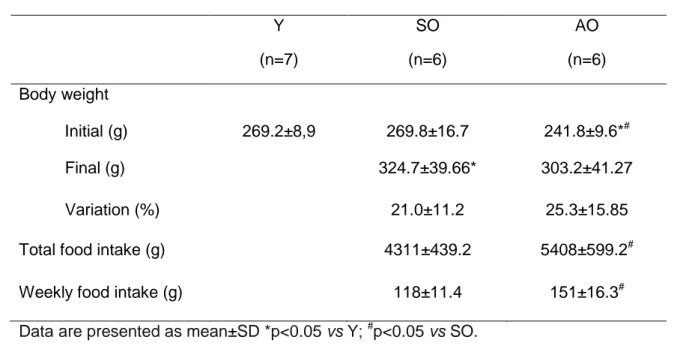
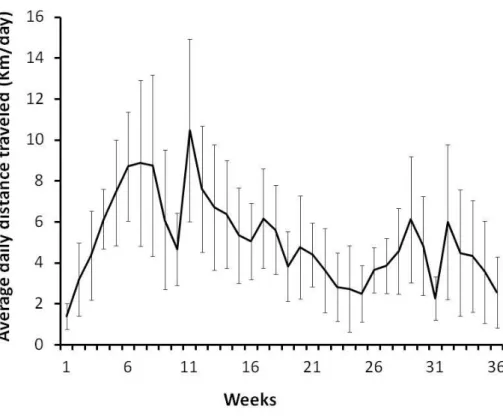
(44) RESULTS. Physical activity The evolution of the distance traveled by the active animals throughout the study period is shown in Figure 1. Daily average distance traveled by the AO animals in the first 8 to 10 weeks progressively increased reaching a peak of about 10Km/day. The distance traveled in the wheel gradually decreased thereafter to about 5 Km/day. Nevertheless, the running pattern of the animals was variable throughout the 9 months of the study period as well as the differences in distance traveled between animals, as it is observable from the high standard deviations.. Body weight and food intake Body weight and food intake throughout the study period are detailed in Table 1. At the beginning of the experiment body weight was slightly lower in AO animals (241.8±9.6g) compared to Y (269.2±8.9) and SO (269.8±16.7), while there were no differences between SO and Y. During the subsequent 9 months of the experimental protocol, body weight increased about 21% and 25% in SO and AO animals, respectively. Total food intake during the 36-weeks of the experimental period was also 25% higher in AO animals compared to SO (4311±439.2g vs 5408±599.2g; p<0.05).. Cardiac morphometry Morphometric analysis shows that heart weight was significantly higher in AO compared to Y animals (p<0.01). Despite the mean heart weight was also higher in AO animals compared to SO, the differences did not reached statistical significance (p=0.058). Differences in heart weight between groups were maintained after normalization to the left tibia length. In addition, there was also a significantly higher RV weight in AO compared to Y animals (p<0.02), but no differences between AO and SO (p=0.368). Nevertheless, these differences were lost after normalization to the tibia length. Regarding the LV morphometry, both raw and normalized values show that LV weight was higher in AO 23.
(45) compared to Y (p<0.01). There was also a tendency for mean LV weight to be higher in AO compared to SO animals, but differences did not reached statistical significance both for raw (p=0.07) and normalized values (p=0.063).. Cardiac histomorphometry The CSA of LV’s cardiomyocytes was significantly higher in AO (316.5±112.1 m2) compared to SO (257.6±112.2 m2; p<0.05) and Y (267.9±107.6 m2; p<0.05) animals. No significant difference in LV’s cardiomyocyte CSA were identified between SO and Y animals (Figure 2). In the RV, the average CSA of cardiomyocyte was significantly higher in Y (232.3±103.9 m2) animals compared to both AO (199.9±100.5 m2; p<0.05) and SO (181.0±91.9 m2; p<0.05) and higher in AO animals compared to SO (p<0.05). In both ventricles, total collagen content was significantly lower in cardiac tissue sections from AO animals (LV=4.71±2.66%, RV=4.24±1.16%) compared to SO animals (LV=7.31±1.67%, RV=7.6±1.64%; p<0.05). SO animals also had a significantly higher total collagen content in both ventricles compared to Y animals (LV=4.81±1.41%, RV=4.60±1.16%; p<0.05). Regarding the collagen to muscle ratio in the LV, AO animals (0.09±0.06; p<0.001) had a significantly lower value compared to SO animals (0.13±0.04; p<0.01). SO animals also had a significantly higher collagen to muscle ratio compared to Y group (0.09±0.03; p<0.03) in the LV. In the RV animals from the AO (0.08±0.03) group also had a significantly higher collagen to muscle ratio comparing to SO (0.13±0.04; p<0.05) and Y (0.09±0.03; p<0.05). Moreover, the mean collagen to muscle ratio in SO animals was also significantly higher compared to Y animals (p<0.1).. Hemodynamic outcomes Left ventricular function Data from the baseline hemodynamic evaluation of LV is detailed in table 3. According to our analysis, SV (81.56±35.67 vs. 146.8±39.24 uL P< 0.05), CO (34625±6149 vs. 62784±17515 uL, P<0.05) and SW (8164±3990 vs. 13556±3786 uL, P<0.05), were significantly affected in SO when compared to Y animals, suggesting a worse systolic function. Moreover, arterial elastance was 24.
(46) significantly elevated in SO group, supporting the notion that arterial stiffness is increased (1.91±1.03 mmHg/L; P<0.05 vs. Y). Concerning to AO group, voluntary physical activity was able to prevent the above-mentioned alterations and, in addition, provided a significant increase in contractility as suggested by the rise in dP/dtmax (9620±892.6 mmHg/sec P<0.05 vs. SO group). Regarding the diastolic function, it was impaired in SO group as the constant time Tau was significantly elevated in comparison to Y and to AO. In order to test the LV functionality under hemodynamic stress, temporary occlusion of the inferior vena cava (table 5) was performed. We found a significantly higher in Ees and EDPVR in animals from the SO group (Ees=1.90±1.6; EDPVR=0.0130±0.01 mmHg/L P<0.05 vs Y). No significant alterations were noted in any of the remaining parameters.. Right ventricular function Data from the baseline hemodynamic evaluation of the RV is presented in table 4. The only significant modification detected in SO group was the decrease in dP/dtmax in comparison to Y group (1502±186.4 vs 2154±151.1 mmHg/sec), suggesting that RV contractility was affected. Regarding to AO group, they also showed a significant decrease in dP/dtmax in relation to Y group, together with a significant reduction in Pmax. About diastolic parameters, only the AO group exhibited a significant decrease in EDP. RV functionality was also tested under hemodynamic stress induced by a sudden and acute occlusion of pulmonary artery. We detected an apparent inability of the RV from both AO and SO to deal with this insult since Pmax, dP/dtmax and dP/dtmin were significantly decreased in comparison to Y group. However, after normalization to the baseline values, no differences were detected in any of the parameters.. 25.
(47) DISCUSSION. This experimental study addressed the question of whether ageassociated alterations on cardiac structure and function would be mostly a consequence of a sedentary lifestyle or whether it could be largely prevented and attenuated by regular physical activity. Our experimental study shows that regular physical activity through voluntary wheel running prevents cardiac functional deterioration, cardiomyocyte atrophy and collagen deposition. These data corroborates the notion that part of the deleterious alterations that occur with aging are secondary to the lower levels of physical activity that usually follows with advancing age and not to aging per se. Aging has a progressive impact on the heart and arterial system, leading to impairment of cardiac function, with a special incidence on diastolic proprieties. Tau and dP/dtmax are two widely accepted invasive measures of the rate of relaxation [26]. We found that the time constant Tau was significantly increased in the LV of SO group, suggesting disturbances in relaxation, an alteration that is conforming to previous studies in rats and humans [27]. This alteration has direct consequences to cardiac function since it can compromise the early diastolic filling of LV [28]. In response, more filling occurs in the late diastolic phase, in part due to a more vigorous atrial contraction, and rising both atrial and ventricular filling pressures. [28, 29] This compensatory mechanism helps to maintain LV filling at the expense of atrial hypertrophy and increased risk for atrial fibrillation [30]. A compromised filling would lead to a lower stroke volume, which we indeed observed in the SO group, that was not avoided even with the decrease in ESV. Exercised animals, by their turn, exhibited normalization of LV Tau and improved dP/dtmax, which is suggestive of increased activity of SERCA2a [31] and a more efficient transport of calcium to the sarcoplasmic reticulum [32]. Interestingly, the RV evidenced apparent contractile impairment in SO and AO since their dP/dtmax was significantly inferior than Y group. The RV adaptations to exercise are currently a topic of big interest. Elevated filling pressures are another important physiologic consequence of diastolic dysfunction that occurs with aging [26] but we did not detect any 26.
(48) alteration in EDP. However, we found a significant increase in EDPVR in the SO group, suggesting decreased ventricular compliance [8, 30, 33, 34]. Aging is intimately associated with decreased ventricular compliance [8, 30, 33, 34]. Extracellular matrix remodeling, manifested by increased fibrosis and alteration of the collagen properties, is thought to be the main responsible for this particular alteration that greatly affects diastolic function [35, 36] [34]. It also promotes arrhythmias and impairs the diffusion of oxygen to cardiomyocytes increasing the susceptibility for heart failure development [37-39]. Sedentary old animals had elevated collagen levels and we found it to be significantly correlated with EDPVR (R2=0.98; P<0.05). Thus, reduced compliance can also explain in part the lower EDV that we observed in SO. By its turn, physical activity was effective in preventing fibrosis and diastolic stiffness (normal EDPVR and EDP at baseline) in AO animals, therefore suggesting that longterm physical activity can preserve the intrinsic myocardial function [40]. Despite a reduction in the total number of cardiomyocytes, the aging heart exhibits a progressive increase in left ventricular mass due to hypertrophy of the surviving cardiomyocytes and accumulation of fibrotic tissue [41]. Agerelated arterial stiffening increases hemodynamic load, contributing to the development of cardiac hypertrophy [41]. Despite the finding of increased Ea in SO group suggesting decreased arterial compliance, we did not find significant hypertrophy (only a trend) at the organ or at the cellular level. This could be explained by the fact that our animals were only fourteen months old (±half of their life span). Thus it is possible that cardiac remodeling was already on process but only on its early steps. After peaking at the 8-9th week of protocol, the overall volume of voluntary physical activity performed by AO animals went to a gradual decline, corroborating previous studies [21]. Despite that reduction, the hemodynamic overload was sufficient to promote significant cardiac hypertrophy in both ventricles. Besides hyperplasia [42, 43], increased cardiac mass is a result of increased cross sectional area, in part due to increased synthesis and reorganization of sarcomeric proteins [44] that supports the generation of contractile force [21-23, 45, 46]. This is probably why the AO animals, in face of higher levels of afterload (basaline Pmax), showed significantly more stroke work than their sedentary counterparts. Indeed, there. 27.
(49) is a straight relationship between ventricular contractile force and ventricular stroke work [47]. Concerning to the RV, it evidenced apparent contractile impairment in SO and AO since their dP/dtmax was significantly inferior to Y group. The RV adaptations to exercise are currently a topic of big interest with some data suggesting maladaptive remodeling in consequence of long-term intensive exercise training [48]. However, and contrarily to SO, we did not detect any other feature of maladaptive remodeling, such as fibrosis, in the RV of AO. Moreover, we detected a significant decrease in Pmax of AO group, which we interpret as a possible decrease in pulmonary vascular resistances [49]. More studies are needed focusing specifically in the RV adaptations to exercise training.. 28.
(50) CONCLUSION In conclusion, our overall findings suggest that regular physical exercise lifelong attenuates cardiac structural deleterious changes related with aging through the prevention of LV and RV hypertrophy and fibrosis. Further, our data also showed that an active lifestyle can prevent or even improve a cardiac dysfunction.. ACKNOWLEDGEMENTS This present work was conducted in the Research Center for Physical Activity, Health and Leisure (CIAFEL), which is a research unit housed in the Faculty of Sports, University of Porto, Portugal. The experimental studies were supported by Fundacão para a Ciência e Tecnologia. (FCT). grants. PTDC/DES/104567/2008. OE/SAU/UI0617/2011.. 29. and. PEst-.
(51) References 1. 2. 3. 4. 5. 6. 7. 8.. 9. 10. 11. 12.. 13.. 14. 15. 16. 17. 18. 19. 20. 21. 22. 23.. Lopez-Otin, C., et al., The hallmarks of aging. Cell, 2013. 153(6): p. 1194-217. Rosa, E.F., et al., Habitual exercise program protects murine intestinal, skeletal, and cardiac muscles against aging. J Appl Physiol, 2005. 99(4): p. 1569-75. Figueiredo, P.A., et al., Aging impairs skeletal muscle mitochondrial bioenergetic function. J Gerontol A Biol Sci Med Sci, 2009. 64(1): p. 21-33. Lombard, D.B., et al., DNA repair, genome stability, and aging. Cell, 2005. 120(4): p. 497-512. Pollock, M.L., et al., Twenty-year follow-up of aerobic power and body composition of older track athletes. J Appl Physiol, 1997. 82(5): p. 1508-16. Trappe, S.W., et al., Aging among elite distance runners: a 22-yr longitudinal study. J Appl Physiol, 1996. 80(1): p. 285-90. Bassett, D.R., Jr. and E.T. Howley, Limiting factors for maximum oxygen uptake and determinants of endurance performance. Med Sci Sports Exerc, 2000. 32(1): p. 70-84. Lakatta, E.G. and D. Levy, Arterial and cardiac aging: major shareholders in cardiovascular disease enterprises: Part II: the aging heart in health: links to heart disease. Circulation, 2003. 107(2): p. 346-54. Juhaszova, M., et al., Protection in the aged heart: preventing the heart-break of old age? Cardiovasc Res, 2005. 66(2): p. 233-44. Bryg, R.J., G.A. Williams, and A.J. Labovitz, Effect of aging on left ventricular diastolic filling in normal subjects. Am J Cardiol, 1987. 59(9): p. 971-4. Gerstenblith, G., et al., Echocardiographic assessment of a normal adult aging population. Circulation, 1977. 56(2): p. 273-8. Davies, C.H., N. Ferrara, and S.E. Harding, Beta-adrenoceptor function changes with age of subject in myocytes from non-failing human ventricle. Cardiovasc Res, 1996. 31(1): p. 152-6. Rodeheffer, R.J., et al., Exercise cardiac output is maintained with advancing age in healthy human subjects: cardiac dilatation and increased stroke volume compensate for a diminished heart rate. Circulation, 1984. 69(2): p. 203-13. Nicoletti, A. and J.B. Michel, Cardiac fibrosis and inflammation: interaction with hemodynamic and hormonal factors. Cardiovasc Res, 1999. 41(3): p. 532-43. Anversa, P., et al., Myocardial aging--a stem cell problem. Basic Res Cardiol, 2005. 100(6): p. 482-93. Ashton, K.J., et al., Age-associated shifts in cardiac gene transcription and transcriptional responses to ischemic stress. Exp Gerontol, 2006. 41(2): p. 189-204. Jahangir, A., S. Sagar, and A. Terzic, Aging and cardioprotection. J Appl Physiol, 2007. 103(6): p. 2120-8. Ferrari, A.U., A. Radaelli, and M. Centola, Invited review: aging and the cardiovascular system. J Appl Physiol, 2003. 95(6): p. 2591-7. Miller, T.D., G.J. Balady, and G.F. Fletcher, Exercise and its role in the prevention and rehabilitation of cardiovascular disease. Ann Behav Med, 1997. 19(3): p. 220-9. Golbidi, S. and I. Laher, Molecular mechanisms in exercise-induced cardioprotection. Cardiol Res Pract, 2011. 2011: p. 972807. Bronikowski, A.M., et al., Lifelong voluntary exercise in the mouse prevents age-related alterations in gene expression in the heart. Physiol Genomics, 2003. 12(2): p. 129-38. Ford, E.S., Does exercise reduce inflammation? Physical activity and C-reactive protein among U.S. adults. Epidemiology, 2002. 13(5): p. 561-8. Navarro, A., et al., Beneficial effects of moderate exercise on mice aging: survival, behavior, oxidative stress, and mitochondrial electron transfer. Am J Physiol Regul Integr Comp Physiol, 2004. 286(3): p. R505-11. 30.
(52) 24.. 25. 26.. 27. 28.. 29.. 30. 31. 32.. 33. 34. 35. 36. 37. 38.. 39. 40.. 41. 42. 43.. 44.. Pacher, P., et al., Left ventricular pressure-volume relationship in a rat model of advanced aging-associated heart failure. Am J Physiol Heart Circ Physiol, 2004. 287(5): p. H2132-7. Yin, F.C., et al., Use of tibial length to quantify cardiac hypertrophy: application in the aging rat. Am J Physiol, 1982. 243(6): p. H941-7. Nagueh, S.F., et al., Recommendations for the Evaluation of Left Ventricular Diastolic Function by Echocardiography. European Journal of Echocardiography, 2009. 10(2): p. 165-193. Karavidas, A., et al., Aging and the cardiovascular system. Hellenic J Cardiol, 2010. 51(5): p. 421-7. Oh, J.K., S.-J. Park, and S.F. Nagueh, Established and Novel Clinical Applications of Diastolic Function Assessment by Echocardiography. Circulation: Cardiovascular Imaging, 2011. 4(4): p. 444-455. Miyatake, K., et al., Augmentation of atrial contribution to left ventricular inflow with aging as assessed by intracardiac Doppler flowmetry. Am J Cardiol, 1984. 53(4): p. 5869. Dai, D.F., et al., Cardiac aging: from molecular mechanisms to significance in human health and disease. Antioxid Redox Signal, 2012. 16(12): p. 1492-526. Demirel, H.A., et al., Short-term exercise improves myocardial tolerance to in vivo ischemia-reperfusion in the rat. J Appl Physiol, 2001. 91(5): p. 2205-12. Miyamoto, M.I., et al., Adenoviral gene transfer of SERCA2a improves left-ventricular function in aortic-banded rats in transition to heart failure. Proc Natl Acad Sci U S A, 2000. 97(2): p. 793-8. Sheydina, A., D.R. Riordon, and K.R. Boheler, Molecular mechanisms of cardiomyocyte aging. Clin Sci (Lond), 2011. 121(8): p. 315-29. Lakatta, E.G., et al., Human aging: changes in structure and function. J Am Coll Cardiol, 1987. 10(2 Suppl A): p. 42A-47A. Anversa, P., et al., Myocyte cell loss and myocyte hypertrophy in the aging rat heart. J Am Coll Cardiol, 1986. 8(6): p. 1441-8. Anversa, P., et al., Myocyte cell loss and myocyte cellular hyperplasia in the hypertrophied aging rat heart. Circ Res, 1990. 67(4): p. 871-85. Harvey, P.A. and L.A. Leinwand, The cell biology of disease: cellular mechanisms of cardiomyopathy. J Cell Biol, 2011. 194(3): p. 355-65. Bernardo, B.C., et al., Molecular distinction between physiological and pathological cardiac hypertrophy: experimental findings and therapeutic strategies. Pharmacology & therapeutics, 2010. 128(1): p. 191. Dhesi, P., et al., How does the heart (not) die? The role of autophagy in cardiomyocyte homeostasis and cell death. Heart failure reviews, 2010. 15(1): p. 15-21. Hessel, M.H.M., et al., Characterization of right ventricular function after monocrotaline-induced pulmonary hypertension in the intact rat. American Journal of Physiology - Heart and Circulatory Physiology, 2006. 291(5): p. H2424-H2430. Biernacka, A. and N.G. Frangogiannis, Aging and Cardiac Fibrosis. Aging Dis, 2011. 2(2): p. 158-173. Ellison, G.M., et al., Physiological cardiac remodelling in response to endurance exercise training: cellular and molecular mechanisms. Heart, 2011. Ellison, G.M., et al., Exercise-induced cardiac stem cell activation and ensuing myocyte hyperplasia contribute to left ventricular remodelling. Proceedings of The Physiological Society, 2008. 11: p. C17. Bernardo, B.C., et al., Molecular distinction between physiological and pathological cardiac hypertrophy: Experimental findings and therapeutic strategies. Pharmacology and Therapeutics, 2010. 128(1): p. 191-227. 31.
(53) 45.. 46.. 47.. 48. 49.. Budiono, B.P., et al., Voluntary running in mice beneficially modulates myocardial ischemic tolerance, signaling kinases, and gene expression patterns. Am J Physiol Regul Integr Comp Physiol, 2012. 302(9): p. R1091-100. Moreira-Goncalves, D., et al., Moderate exercise training provides left ventricular tolerance to acute pressure overload. Am J Physiol Heart Circ Physiol, 2011. 300(3): p. H1044-52. Cotten, M.d. and H.M. Maling, Relationships Among Stroke Work, Contractile Force and Fiber Length During Changes in Ventricular Function. American Journal of Physiology -- Legacy Content, 1957. 189(3): p. 580-586. Benito, B.a., et al., Cardiac Arrhythmogenic Remodeling in a Rat Model of Long-Term Intensive Exercise Training / Clinical Perspective. Circulation, 2011. 123(1): p. 13-22. Kovacs, G., et al., Pulmonary vascular resistances during exercise in normal subjects: a systematic review. European Respiratory Journal, 2012. 39(2): p. 319-328.. 32.
(54) Legend of figures. Figure 1. Voluntary motor activity recorded on the running wheel from the AO animals throughout the 9 months of the experimental procedure. Avarage daily distance traveled through the 36 weeks. Data are mean±standard deviation. Figure 2. a) Average cardiac myocyte cross-sectional area (CSA) of right ventricle (RV) and left ventricle (LV). A lower fiber CSA is evident in both ventricles of SO animals. b) Group comparisons of cardiac myocyte CSA of LV and RV. Bars are 50 μm. Y-Young; SOSedentery Old; AO-Active Old. *Significant difference upon Y (p<0.05 vs Y); #Significant difference upon SO (p<0.05 vs SO). Figure 3. a) Representative cardiac tissue sections stained with Picrosirius red, showing collagen stained red and skeletal muscle stained yellow; b) Group comparisons of the total collagen expression; c) Group comparisons of the total collagen to muscle ratio. Bars represent 50 μm. Y-Young; SO-Sedentery Old; AO-Active Old; *Significantly different compared to Y (p< 0.05 vs Y); #Significantly different compared to SO (p<0.05 vs SO). Figure 4. Hemodynamic evaluation of left (LV) and right (RV) ventricles in baseline conditions. a) LV function: dP/dtmax: peak pressure rise; dP/dtmin: peak pressure decay; Tau: time-constant. b) end-systolic volume; stroke volume; cardiac output c) RV function: dP/dtmax: peak pressure rise; d) dP/dtmin: peak pressure decay; Pmax: peak systolic pressure; Tau: time-constant; Y-Young; SO-Sedentery Old; AO-Active Old; *Significant differences upon Y (p< 0.05 vs Y); #Significant difference upon SO (p<0.05 vs SO).. 33.
(55) List of Tables. Table1: Animals body weight and food intake Y. SO. AO. (n=7). (n=6). (n=6). 269.2±8,9. 269.8±16.7. 241.8±9.6*#. 324.7±39.66*. 303.2±41.27. 21.0±11.2. 25.3±15.85. 4311±439.2. 5408±599.2#. 118±11.4. 151±16.3#. Body weight Initial (g) Final (g) Variation (%) Total food intake (g) Weekly food intake (g). Data are presented as mean±SD *p<0.05 vs Y; #p<0.05 vs SO.. Table 2: Heart morphometry Y. AO. SO. Heart weight (mg). 711.5±49.0. 920.1±105.1*. 810.8±66.4. Heart weigth/ Tibia length ratio (mg/mm). 18.47±1.35. 22.94±2.46*. 20.36±1.48. RV weight (mg). 128.0±9.1. 157.7±21.7*. 143.7±20.2. Ratio RV weigth/ Tibia length (mg/mm). 3.33±0.29. 3.93±0.54. 3.61±0.53. LV weight (mg). 499.9±30.2. 639.8±74.4*. 565.7±49.9. Ratio LV weigth /Tibia length (mg/mm). 12.97±0.69. 15.95±1.73*. 14.20±1.14. RV: right ventricle; LV: left ventricle. Data are presented as mean±SD *p<0.05 vs Y; # p<0.05 vs SO.. 34.
Imagem
Documentos relacionados
Esta trajetória me leva hoje a pensar a identidade feminina, a homossexualidade e o travestimento não só como experiências que apenas digam respeito, respectivamente, a
On the contrary, in the present study, vertebral heart size showed the lowest coefficient of variation in compa- rison to the cardiac ratios, such as cardiac height and/or width
As semelhanças entre Thomas Schimidt e Santiago Nazarian devem-se, sobretudo, à imagem que o segundo construiu e divulgou antes mesmo de se lançar como escritor.. Haja vista
Objetivo: Na presente investigação procurou-se avaliar a fiabilidade de um instrumento de avaliação de manobras com cordas, comparar os efeitos de várias
Portanto, este estudo evidencia que o processo de Classificação de Tronzo seguindo o índice de Kappa, apresenta como resultado final uma categorização pobre
Animals in con- trol group (IR) were subjected to 30 min ischemia followed by 60 min reperfusion by the Langendorff method and saline (NS) was administered intravenously
Quanto ao grupo SB, as participantes que possuíam a mastigação bilateral alternada permaneceram, apesar de a ativação muscular ter aumentado e ter sido possível
In fact, increased levels of HSP72 were reported in rat skeletal muscle in re- sponse to nandrolone administration for 8 weeks, but only by injecting higher doses (10 mg/kg body
