RESUMO.- [Mensurações radiográficas relacionadas às dimensões cardíacas em borregas Bergamácia.] No
exame radiográfico torácico, rotineiramente utilizado em casos em que a avaliação cardíaca é indicada, a análise quantitativa do coração é um recurso útil a ser usado em combinação com a avaliação subjetiva. Diversos estudos relacionados à análise objetiva do tamanho cardíaco têm sido realizados em várias espécies, incluindo ovinos; no entanto, há pouca informação sobre os parâmetros car
-díacos de ovinos jovens da raça Bergamácia. Dessa for
-Radiographic measurements related with the cardiac size
in young female Bergamasca sheep
1Viviam R. Babicsak2*, Lidiane S. Alves2, Miriam H. Tsunemi3 and Luiz C. Vulcano2
ABSTRACT.- Babicsak V.R., Alves L.S., Tsunemi M.H. & Vulcano L.C. 2017. Radiographic measurements related with the cardiac size in young female Bergamasca sheep. Pes-quisa Veterinária Brasileira 37(12):1526-1530.Departamento de Reprodução Animal e Ra
-diologia Veterinária, Faculdade de Medicina Veterinária e Zootecnia, Universidade Estadual Paulista, Campus de Botucatu, Rua Prof. Doutor Walter Mauricio Correa s/n, Botucatu, SP 18618-681, Brazil. E-mail: viviam.babicsak@gmail.com
In thoracic radiographic examination, routinely used in cases which cardiac evalua
-tion is indicated, quantitative assessment of the heart is a useful role to be used in com
-bination with subjective analysis. Numerous studies about objective assessment of the cardiac size have been performed in several species, including sheep; however, there is scarce information regarding cardiac parameters of young Bergamasca sheep. Therefore, the purpose of this study was to determine the average results and suggest the range of expected normal values for parameters related to the heart size of young female Berga
-masca sheep by radiographic evaluation. Fifteen healty 8 months-old female Berga-masca sheep (mean weight: 41.13±4.71kg) were submitted to right lateral recumbency thoracic radiography. The length of the fourth and third to fifth thoracic vertebrae, cardiac height and width, vertebral heart size, cardiophrenic contact, caudal vena cava height, aorta caliber and tracheal angle were measured and the mean results found were, respective
-ly: 2.46±0.11cm (95% CI 2.41-2.52), 7.53±0.30cm (95% CI 7.38-7.68), 13.83±0.57cm, (95% CI 13.54-14.12), 8.99±0.37cm (8.80-9.17), 8.99±0.27 vertebrae (95% 8.85-9.13), 4.55±0.70cm (95% CI 4.19-4.90), 1.88±0.19cm (95% CI 1.79-1.97), 2.05±0.11cm (95% CI 2.00-2.11) and 14.36±2.73° (95% CI 12.98-15.75). Cardiac height and width and the sum of these parameters were compared to the length of third to fifth thoracic vertebrae, resulting in the respective mean values: 1.84±0.08 (95% IC 1.80-1.88), 1.20±0.05 (1.17-1.22) and 3.04±0.11 (95% IC 2.98-3.09). Ratios of cardiophrenic contact to cardiac hei
-ght and caudal vena cava hei-ght to length of fourth thoracic vertebra were also evaluated and the mean values obtained were 0.33±0.05 (95% IC 0.30-0.35) and 0.76±0.08 (95% IC 0.72-0.81), respectively. Authors suggest that the values available in this study may be used as reference for normal heart size in young female Bergamasca sheep and as basis for further studies.
INDEX TERMS: Radiography, heart, sheep, cardiology, radiology, morphometry.
1 Received on October 11, 2016.
Accepted for publication on April 6, 2017.
2 Departamento de Reprodução Animal e Radiologia Veterinária, Fa
-culdade de Medicina Veterinária e Zootecnia (FMVZ), Universidade Es
-tadual Paulista (Unesp), Campus de Botucatu, Rua Prof. Doutor Walter Mauricio Correa s/n, Botucatu, SP 18618-681, Brazil. *Corresponding author: viviam.babicsak@gmail.com
3 Departamento de Bioestatística, Instituto de Biociências, Unesp, Cam
ma, o objetivo deste estudo foi determinar as médias e sugerir o intervalo de valores esperados para os parâme
-tros relacionados ao tamanho cardíaco de borregas Ber
-gamácia por meio da avaliação radiográfica. Quinze bor
-regas Bergamácia de 8 meses de idade (média de peso: 41,13±4,71kg) foram submetidas à radiografia torácica em decúbito lateral direito. O comprimento do quarto e da terceira a quinta vértebras torácicas, a altura e a largu
-ra cardíaca, o tamanho do co-ração em relação à unidade de vértebra torácica, o contato cardiofrênico, a altura da veia cava caudal, o calibre da aorta e o ângulo traqueal foram mensurados, sendo encontrados os seguintes valo
-res médios, -respectivamente: 2,46±0,11cm (95% IC 2,41-2,52), 7,53±0,30cm (95% IC 7,38-7,68), 13,83±0,57cm, (95% IC 13,54-14,12), 8,99±0,37cm (95% IC 8,80-9,17), 8,99±0,27 vértebras (95% IC 8,85-9,13), 4,55±0,70cm (95% IC 4,19-4,90), 1,88±0,19cm (95% IC 1,79-1,97), 2,05±0,11cm (95% IC 2,00-2,11) e 14,36±2,73° (95% IC 12,98-15,75). A altura e a largura cardíaca e a soma desses parâmetros foram comparados com o compri
-mento da terceira a quinta vértebras torácicas, resultan
-do nos respectivos valores médios: 1,84±0,08 (95% IC 1,80-1,88), 1,20±0,05 (1,17-1,22) e 3,04±0,11 (95% IC 2,98-3,09). Também foram avaliadas as relações entre o contato cardiofrênico e a altura cardíaca e entre a altu
-ra da veia cava caudal e o comprimento da quarta vérte
-bra torácica, sendo determinados os valores médios de 0,33±0,05 (95% IC 0,30-0,35) e 0,76±0,08 (95% IC 0,72-0,81), respectivamente. Os autores sugerem que os valo
-res disponíveis no p-resente estudo podem ser utilizados como referência na avaliação das dimensões cardíacas de borregas Bergamácia e como base para estudos futuros.
TERMOS DE INDEXAÇÃO: Radiografia, coração, borregas, ovinos, cardiologia, radiologia, morfometria.
INTRODUCTION
Although echocardiography plays a substantial role in as
-sessment of the heart, thoracic radiography contributes a lot in the evaluation of the cardiovascular system and it is often the first diagnostic imaging technique performed in cases of suspicion of cardiac diseases.
Radiographic abnormalities of the cardiac silhouet
-te, however, are only detected if normal parameters are known and, to this end, abundant studies related to the radiographic assessment of the cardiac size have been performed in several species, including cats (Lord & Zon
-tine 1977, Litster & Buchanan 2000a, Litster & Buchanan 2000b, Ghadiri et al. 2008), dogs (Lehmkuhl et al. 1997, Lamb et al. 2001, Bavegems et al. 2005, Hansson et al. 2005, Marin et al. 2007, Spasojević-Kosić et al. 2007, Kra
-etschmer et al. 2008), ferrets (Stepien et al. 1999), llamas (Mattoon et al. 2001), psittacines (Straub et al. 2002), cattle (Jilintai et al. 2006), monkeys (Harada et al. 2009), rabbits (Onuma et al. 2010) and alpacas (Nelson et al. 2011).
In young individuals, radiographic evaluation of the cardiac size is challenger, since heart normally appears to be larger in relation to thoracic size in these animals
in comparison to its aspect in maturity (Owens & Biery 1999). The relative variation in heart size in young indi
-viduals have motivated investigations related to the nor
-mal radiographic anatomy and, in consequence, studies have been conducted in young individuals of some spe
-cies, including cats (Gaschen et al. 1999), dogs (Sleeper & Buchanan 2001), cattle (Jilintai et al. 2006), monkeys (Harada et al. 2009), alpacas (Nelson et al. 2011) and go
-ats (Ukaha et al. 2013), in order to provide normal refe
-rences and, consequently, aid in the diagnosis of cardiac diseases.
In young sheep, there are only a few reports available in the literature related to the radiographic evaluation of the cardiac size, which were conducted in young Santa Ines (Souza et al. 2012) and neonatal Ile de France (Ulian 2015) individuals. Due to the paucity of information, the purpose of this study was to determine the mean results and sug
-gest the range of expected normal values for radiographic parameters related to the cardiac size in young female Ber
-gamasca sheep.
MATERIALS AND METHODS
Ethics approval was obtained from Ethics Committee on Ani -mal Experimentation of the School of Veterinary Medicine and Animal Science of São Paulo State University (Protocol number 107/2013), in accordance with the ethical principles adopted by the Brazilian College of Animal Experimentation.
Fifteen healthy 8 months-old female Bergamasca sheep from a herd maintained by the School of Veterinary Medicine and Ani -mal Science of São Paulo State University were radiographed in this study. All animals were considered clinically normal and none presented any alteration during pulmonary and cardiac ausculta -tion. The average weight of the animals of this study was 41.13 kg and the standard deviation was 4.71 kg.
Right lateral recumbency radiographs were obtained in a digi -tal equipment (Pioneer DR 50kw System, GE Healthcare, Chalfont St Giles, UK) in full inspiration using a focal-film distance of 100 cm and exposure factors of 4.0mA, 200mAs and 80-90kVp. In all individuals, the entire thorax (defined by inclusion of the entire lung field, the thoracic spine and sternum) could be included in only one image. The animals were not sedated or anesthetized for this study.
Radiographic images were evaluated using a software pro -gram for medical imaging analysis (Clear Canvas, Clear Canvas Inc, Toronto, Canada) by two independent experienced radiolo -gists. All measurements were performed in triplicate by the two observers and then the mean of all values obtained for each varia -ble was calculated.
the thoracic vertebrae beginning at the level of the cranial edge of T4. The vertebral sum of both measures was considered the vertebral heart size value. The measurement of the cardiophrenic contact, determined by the distance from the apex of the heart to the dorsal-most point of intersection of the cardiac silhouet -te with diaphragm, was also recorded. Caudal vena cava height was measured from its ventral to dorsal limits in the region of greatest diameter, not overlapping the heart or diaphragm and, at the same intercostal space where the caudal vena cava was me -asured, the caliber of the aorta was determined. Tracheal angle
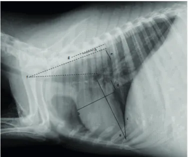
Fig.1. Thoracic radiographic image illustrating the measurement of the cardiac height (a) and width (b). The measurement of the cardiophrenic contact, the distance from the apex of the heart to the dorsal-most point of intersection of the cardiac silhouette with the diaphragm, is also illustrated (c). The me -asurement of the greatest caliber of the caudal vena cava is demonstrated (d), as well as aorta height (e), measured in the same intercostal space where the caudal vena cava di -mension was obtained. Tracheal angle, defined as the angle between the lines drowned along the dorsal border of the trachea and the ventral margin of the cranial thoracic ver -tebrae, is also illustrated (f). Length of the third to fifth tho -racic vertebrae (g) and fourth tho-racic vertebra (h) are also shown.
Table 1. Means, standard deviations (SD), range values and 95% confidence intervals (CIs) of the parameters related to heart and body sizes
Parameters Mean ± SD Range 95% CIs
Weight (kg) 41.13 ± 4.71 34.50-49.50 38.75-43.52
Length of T3 through T5 (cm) 7.53 ± 0.30 7.01-8.23 7.38-7.68
Length of T4 (cm) 2.46 ± 0.11 2.30-2.72 2.41-2.52
Cardiac height (cm) 13.83 ± 0.57 12.82-14.51 13.54-14.12
Cardiac width (cm) 8.99 ± 0.37 8.24-9.56 8.80-9.17
Cardiophrenic contact (cm) 4.55 ± 0.70 3.23-5.80 4.19-4.90
Caudal vena cava height (cm) 1.88 ± 0.19 1.58-2.17 1.79-1.97
Aorta height (cm) 2.05 ± 0.11 1.91-2.26 2.00-2.11
Tracheal angle (°) 14.36 ± 2.73 9.02-19.33 12.98-15.75
Vertebral heart scale (vertebrae) 8.99 ± 0.27 8.55-9.48 8.85-9.13 Ratio of cardiac height to length of T3 through T5 1.84 ± 0.08 1.69-1.96 1.80-1.88 Ratio of cardiac width to length of T3 through T5 1.20 ± 0.05 1.12-1.29 1.17-1.22 Ratio of cardiac height + width to length of T3 through T5 3.04 ± 0.11 2.83-3.18 2.98-3.09 Ratio of cardiophrenic contact to cardiac height 0.33 ± 0.05 0.23-0.41 0.30-0.35 Ratio of caudal vena cava height to length of T4 0.76 ± 0.08 0.62-0.88 0.72-0.81
was obtained by measuring the angle between the lines drowned along the dorsal border of the trachea and ventral margin of T3 through T5. All measurements performed in this study are illus -trated in Figure 1.
To account for differences in size of the individuals, ratios of cardiac height, cardiac width and the sum of cardiac height and width to length of T3 through T5 were calculated, as well as ratios of cardiophrenic contact to cardiac height and caudal vena cava height to length of T4.
Statistical analysis was performed by use of the statistical pro -gram SPSS 17.0 (SPSS Inc., Chicago, USA). Mean, standard devia -tion, range and 95% confidence interval (CI) about the mean were calculated for all parameters. Correlation coefficients between body weight and some variables (length of T3 through T5, length of T4 and cardiac height) were determined in order to investiga -te the exis-tence of correlation between body weight and skeletal size. P<0.05 was considered significant. Coefficients of variation of vertebral heart size and ratios of cardiac height to length of T3 through T5, cardiac width to length of T3 through T5 and the sum of cardiac height and width to length of T3 through T5 were calculated as the standard deviation/mean×100. Mean and 95% confidence interval about the mean of the coefficients of variation were determined.
RESULTS
Means, standard deviations, range values and 95% confi
-dence intervals about the mean of the variables are recor
-ded in Table 1.
Considering correlation coefficients, significant positive linear relations were identified between weight and length of T4 (r=0.852, p=0.000), weight and length of T3 throu
-gh T5 (r=0.694, p=0.000) and wei-ght and cardiac hei-ght (r=0.669, p=0.012), indicating that body size and weight are related (Fig.2).
Coefficient of variation found for the vertebral heart size was 3.05% (95% CI, 2.23 to 4.81%), ratio of cardiac height to length of T3 through T5 was 4.09% (95% CI, 2.99 to 6.44%), ratio of cardiac width to length of T3 through T5 was 3.80% (95% CI, 2.78 to 5.98%) and ratio of the sum of cardiac height and width to length of T3 through T5 was 3.47% (95% CI, 2.54 to 5.47%). The analysis showed that the vertebral heart size had the minor coefficient of varia
DISCUSSION
Subjective assessment of the heart is usually performed in radiographic evaluation of the cardiac silhouette, along with objective analysis, which provides important informa-tion about cardiac size with little interobserver variainforma-tion and aids non-experienced radiologists, who often show difficulty in distinguish cardiac abnormalities (Buchanan & Bücheler 1995, Kittleson & Kienle 1998). In objective
analysis, ratios comparing cardiac measurements to thora-cic feature reflective of body size are very useful in cardiac evaluation, as well as vertebral heart size method, since, in contrast to absolute cardiac measurements such as cardiac height and width, they minimize false positive diagnoses of cardiomegaly associated to body size, thoracic conforma-tion and magnificaconforma-tion of the radiographic image (Bucha-nan & Bücheler 1995, Mattoon et al. 2001).
Stepien et al. (1999) observed that, in ferrets, cardiac ratios appeared to result in lower measurement variation than the modified vertebral heart size method used in their study. On the contrary, in the present study, vertebral heart size showed the lowest coefficient of variation in compa-rison to the cardiac ratios, such as cardiac height and/or width to length of T3 through T5 and, therefore, it may be considered the most reliable parameter for cardiac evalua-tion in young sheep.
Vertebral heart size mean of young Bergamasca she-ep (8.99±0.27 vertebrae) was lower than the average va-lue of 10.36±0.35 vertebrae determined for 5-months-old Santa Ines individuals (Souza et al. 2012). The result was also lower than the mean values obtained for neonatal Ile de France sheep, which were 10.07±0.10, 9.97±0.09, 9.65±0.09, 9.53±0.08, 9.36±0.09 and 9.42±0.08 vertebrae in 24 hours and 7, 14, 21, 28 and 35 days of life, respective-ly (Ulian 2015). This finding may be justified by the utiliza-tion of older sheep in our study in comparison to the other ones, since Ulian (2015) found a progressive decrease in vertebral heart size in neonatal Santa Ines sheep during the progression of age. Bergamasca sheep also showed lower vertebral heart size mean than West African Dwarf goats (10.1±0.01 vertebrae) (Ukaha et al. 2013), which may be explained by the deeper thoracic conformation of the goats in comparison to sheep, since this aspect allows greater de-velopment of the cardiac axis, especially height.
Radiographic measurement of the cardiophrenic con-tact is useful in assessment of the cardiac size, since incre-ased ratios of cardiophrenic contact to cardiac height may indicate cardiomegaly in absence of diseases that leads to hypoventilation of the lung or increased intra-abdominal volume (Mattoon et al. 2001). The ratio of cardiophrenic contact to cardiac height calculated for sheep (0.33±0.05) was lower than the value found for alpaca crias (0.39±0.10) (Nelson et al. 2011), which may be explained by the exis-tence of a lower cardiophrenic contact and/or a higher cardiac height in sheep in comparison to alpacas. Greater
Fig.2. Graphs illustrating positive linear correlations between (a)
weight and length of T4 (r2=0.852, n=15, p<0.000), (b) weight and length of T3 through T5 (r2=0.694, n=15, p<0.000), and (c) weight and cardiac height (r2=0.669, n=15, p=0.012).
thoracic and/or lower intra-abdominal volumes in sheep may also justify the differences in cardiophrenic ratios sin
-ce these conditions may interfere in cardiophrenic contact measurement (Mattoon et al. 2001).
Ratio of caudal vena cava diameter to length of T4 is also a valuable parameter in radiographic evaluation of the heart, since elevated values are suggestive of right-sided congesti
-ve heart (Lehmkuhl et al. 1997, Jilintai et al. 2006). The ratio of caudal vena cava height to length of T4 found for sheep in our study (0.76±0.08) was greater than the value obtained for young cows (0.41±0.06) (Jilintai et al. 2006) and a fact that may have contributed to this variation was the use of the maximum diameter of the caudal vena cava height in the ratio of sheep and its average size in the ratio of the cattle.
Tracheal angle measurement is an indirect parameter useful in quantitative radiographic evaluation of the car
-diac size too, since a reduced tracheal angle indicates a dorsal positioning of the trachea, a radiographic sign usu
-ally evident in cardiomegaly (Mattoon et al. 2001). The si
-milarity found between the tracheal angle of young sheep (14.72±3.08°) and alpaca crias (14.24±3.57°) (Nelson et al. 2011) may be justified by the resemblance of the thoracic conformation exhibited by these two species.
As conclusion, authors suggest that the values found in this study may be used as reference for heart size in radio
-graphic evaluation of young female Bergamasca sheep and as basis for further investigations in these animals. Future researches using a greater number of animals, as well as male individuals, are need in order to determine additional information related to normal radiographic evaluation of the heart size in this specie.
Individual variability and physiologic factors such as cardiac cycle phase, circulating blood volume, total body fluid, and intra-thoracic and intra-abdominal pressure may have interfered in the measurements and ratios calculated in this study. Phase of respiration may have also affected the results despite the efforts to perform peak inspiratory radiographs.
REFERENCES
Bavegems V., Van Caelenberg A., Duchateau L., Sys S.U., Van Bree H. & De Rick A. 2005. Vertebral heart size ranges specific for whippets. Vet. Ra
-diol. Ultrasound 46:400-403.
Buchanan J.W. & Bucheler J. 1995. Vertebral scale system to measure cani
-ne heart size in radiographs. J. Am. Vet. Med. Assoc. 206:194-199. Gaschen L., Lang J., Lin S., Adé-Damilano M., Busato A., Lombard C.W. &
Gaschen F.P. 1999. Cardiomyopathy in dystrophin-deficient hypertro
-phic feline muscular dystrophy. J. Vet. Intern. Med. 13:346-356. Ghadiri A., Avizeh R., Rasekh A. & Yadegari A. 2008. Radiographic measu
-rement of vertebral heart size in healthy stray cats. J. Feline Med. Surg. 10:61-65.
Hansson K., Haggstrom J., Kvart C. & Lord P. 2005. Interobserver variability of vertebral heart size measurement in dogs with normal and enlarged hearts. Vet. Radiol. Ultrasound 46:122-130.
Harada M., Koie H., Iwaki S., Sato T., Kanayama K., Taira M. & Sakai T. 2009. Establishment of vertebral heart scale in the growth period of the Japa
-nese macaque (Macaca fuscata). J. Vet. Med. Sci. 72:503-505.
Jilintai, Hashiyama S., Gonda Y., Ishikawa H., Sato M. & Miyahara K. 2006. Radiographic evaluation of caudal vena cava size as a useful parameter for the diagnosis of heart disease in dairy cattle. J. Vet. Med. Sci. 68:995-998.
Kittleson M.D. & Kienle R.D. 1998. Small Animal Cardiovascular Medicine. Mosby, St Louis. 603p.
Kraetschmer S., Ludwig K., Meneses F., Nolte I. & Simon D. 2008. Vertebral heart scale in the beagle dog. J. Small Anim. Pract. 49:240-243. Lamb C.R., Wikeley H., Boswood A. & Pfeiffer D.U. 2001. Use of breed spe
-cific ranges for the vertebral heart scale as an aid to the radiographic diagnosis in dogs. Vet. Rec. 148:707-711.
Lehmkuhl L.B., Bonagura J.D., Biller D.S. & Hartman W.M. 1997. Radiogra
-phic evaluation of caudal vena cava size in dogs. Vet. Radiol. Ultrasound 38:94-100.
Litster A.L. & Buchanan J.W. 2000a. Radiographic and echocardiographic measurement of the heart in obese cats. Vet. Radiol. Ultrasound 41:320-325.
Litster A.L. & Buchanan J.W. 2000b. Vertebral scale system to measure he
-art size in radiographs of cats. J. Am. Vet. Med. Assoc. 216:210-214. Lord P.F. & Zontine W.J. 1977. Radiologic examination of the feline cardio
--vascular system. Vet. Clin. N. Am. 7:291-308.
Marin L.M., Brown J., McBrien C., Baumwart R., Samii V.F. & Couto C.G. 2007. Vertebral heart size in retired racing Greyhounds. Vet. Radiol. Ul
-trasound48:332-334.
Mattoon J.S., Gerros T.C. & Brimacombe M. 2001. Thoracic radiographic appearance in the normal llama. Vet. Radiol. Ultrasound 42:28-37. Nelson N.C., Mattoon J.S. & Anderson D.E. 2011. Radiographic appearance
of the thorax of clinically normal alpaca crias. Am. J. Vet. Res. 72:1439-1448.
Onuma M., Ono S., Ishida T., Shibuya H. & Sato T. 2010. Radiographic mea
-surement of cardiac size in 27 rabbits. J. Vet. Med. Sci. 72:529-531. Owens J.M. & Biery D.N. 1999. Radiographic interpretation for the small
animal clinician. 2nd ed. Williams and Wilkins, Baltimore. 308p. Sleeper M.M. & Buchanan J.W. 2001. Vertebral scale system to measure
heart size in growing puppies. J. Am. Vet. Med. Assoc. 219:57-59. Souza P.M., Rodello L., Inamassu L.R., Monteiro C.T., Babicsak V.R., Macha
-do V.M.V. & Bicu-do S.D. 2012. Radiographic evaluation of the cardiac si
-lhouette by the method of measurement VHS (vertebral heart size) in Santa Ines Boregas clinically normal. XXVII World Buiatrics Congress, XXVII World Buiatrics Congress, Lisboa. p.155.
Spasojević-Kosić L., Krstić N. & Trailović R.D. 2007. Comparison of three methods of measuring vertebral heart size in German Shepherd dogs. Acta Vet. 57:133-141.
Stepien R.L., Benson K.G. & Forrest L.J. 1999. Radiographic measurement of cardiac size in normal ferrets. Vet. Radiol. Ultrasound 40:606-610. Straub J., Pees M. & Krautwald-Junghanns M.E. 2002. Measurement of the
cardiac silhouette in psittacines. J. Am. Vet. Med. Assoc. 221:76-79. Ukaha R.O., Kene R.O.C. & Gbonko O.E. 2013. Vertebral scale system to me
-asure heart size in thoracic radiographs of West African Dwarf goats. Nig. Vet. J. 34:912-916.
Ulian C.M.V. 2015. Avaliação do desenvolvimento cardíaco neona
-tal em cordeiros. Tese de Doutorado. Available on <http://repo