Universidade de Lisboa
Faculdade de Farmácia
Research Institute for Medicines and Pharmaceutical Sciences
(iMed.UL)
Neuron Glia Biology in Health and Disease Group
MODULATION OF MICROGLIA REACTIVITY BY S100B IN
MULTIPLE SCLEROSIS
Carla Isabel Silveira Ferreira
Dissertação de Mestrado
MESTRADO EM CIÊNCIAS BIOFARMACÊUTICAS
Universidade de Lisboa
Faculdade de Farmácia
Research Institute for Medicines and Pharmaceutical Sciences
(iMed.UL)
Neuron Glia Biology in Health and Disease Group
MODULATION OF MICROGLIA REACTIVITY BY S100B IN
MULTIPLE SCLEROSIS
Carla Isabel Silveira Ferreira
Dissertação de Mestrado orientada pela Prof.ª Doutora Adelaide Maria Afonso
Fernandes Borralho e pela Doutora Andreia Pereira Barateiro
MESTRADO EM CIÊNCIAS BIOFARMACÊUTICAS
Agradecimentos
Quero começar por agradecer à Professora Doutora Dora Brites, investigadora principal do grupo Neuron Glia Biology in Health and Disease, pela forma como me acolheu e por me ter dado a oportunidade de desenvolver a minha tese de mestrado neste grupo. Além disso quero agradecer pelo sentido crítico que me incutiu e que me irá certamente ajudar no futuro.
À Professora Doutora Adelaide Fernandes, um enorme obrigada é pouco! Não há palavras que mostrem a gratidão que sinto por ti. Porque trabalhar nisto era especial para mim e deste-me a oportunidade de o fazer. Obrigada por toda a ajuda, dedicação e por todo o conhecimento que me transmitiste. Foste incansável. Um dia gostava de ser como tu!
À Doutora Andreia Barateiro, muito obrigada por tudo. Por toda a tua paciência. Por toda a ajuda que me deste ao longo deste ano e pela tua disponibilidade (até para falar de assuntos de noivas ;) ). Muito obrigada! :)
Carolina, Cátia, Cláudia e Gisela, obrigada por estarem sempre prontas a ajudar ou simplesmente para rir um bocadinho. Muito obrigada por toda a dedicação, ajuda e diversão que me proporcionaram ao longo deste ano!
Dora, companheira de almoço (desde as idas à cantina até à esplanada do F)! Obrigada por este ano. Aturaste os meus devaneios, as minhas queixas e loucuras, as minhas “coisas de noiva”… obrigada por me fazeres rir de cada vez que alguém te assustava ao ir falar contigo ou dizias “laranjas” :p, por o teu tablet ganhar personalidade e dizer-me coisas hilariantes xD e por me deixares eufórica com a notícia mais fixe deste ano :D simplesmente obrigada! :)
Maria, és um exemplo para mim! Exemplo de esforço e dedicação! Obrigada pelos momentos de diversão (tendo em conta que estava à tua frente era relativamente fácil começar a rir contigo :p) e pelos ensinamentos sobre Western Blot. Mas olha, mesmo assim o record ainda é meu! :D Torço por ti e sei que tens um futuro brilhante à tua frente!
Mafalda, partilhámos a orientadora, o fluxo durante algum tempo e até os anticorpos para WB!!! O que há a dizer?! Obrigada pela ajuda que me foste dando ao longo deste tempo ou por me aturares quando eu cá ficava até mais tarde. Sei que vais chegar longe :)
Ritinha :D lembro-me de logo nos primeiros módulos me sentar perto de ti. Depois rapidamente éramos as poderosas! Obrigada por seres assim, sempre disponível mesmo estando mais longe. Sabes que estarei sempre aqui, onde quer que estejas :)
Para ti Rui, vai um agradecimento especial! Pelas boleias todas que me deste, pelas massagens enquanto eu contava beads, por teres sempre uma palavra amiga. Não deixes de ser assim como és e tenho a certeza que vai haver muita sogra a tentar “roubar-te” :p xD
Ao Chico, por todas as conversas e cafés. Obrigada por toda a ajuda e claro, pelas correções! “A verdadeira amizade não é ser inseparáveis, mas sim estar separados sem que nada mude.” Nós somos assim! :)
À Filipa e à Diana, obrigada por aturarem a madrinha. Tenho muito orgulho em vocês! À Andreia Freitas, porque mesmo longe, estiveste sempre pronta para me ajudar e apoiar. És a melhor madrinha de curso que alguém pode ter. Desde o grupo de Genética que não nos separámos. Viste-me sorrir, viste-me chorar, viste-me vacilar… o nosso último ano uniu-nos e sei que nunca mais uniu-nos iremos separar! És especial, muito especial! Obrigada por tudo!
À Teresa e ao Nuno, ao Ricardo e à Joana, por estarem lá sempre que foi necessário :) À minha Shikha. Porque como já te disse, foi amor à primeira vista. Porque acabámos a licenciatura juntas, concorremos a mestrado juntas e vamos continuar juntas, disso tenho a certeza :) obrigada por me ouvires, por nadares comigo, por seres uma irmã, por seres como és, especial e única!
À São Piçarra, pelas correções, pelas conversas sobre Esclerose Múltipla e microglias, pelos salames e pelos tão deliciosos “S”, muito obrigada!
À Margarida, por fazer parte da minha vida! Porque para além de ser mana, melhor amiga e madrinha, me faz lutar a cada dia para deixar uma pequena marca neste mundo da Esclerose Múltipla. Esta tese é para ti, é por ti!
António, Guida, Luísa, Luís, avó Margarida e tio Teófilo, obrigada pelo apoio e disponibilidade incansáveis nestes dois anos!
Aos meus irmãos e cunhadas, obrigada por serem exemplo! Aos meus amores pequeninos, Miguel, Rui, Pedro, Ana, Sara e Rafael, obrigada por me fazerem sorrir até nas alturas de maior cansaço.
Aos meus avós, que lá do céu olham por mim.
Não pode faltar um agradecimento muito especial aos meus pais por me permitirem chegar até aqui. Por todo o vosso apoio e amor incondicional, obrigada!
Também ao Dogus, à Miaucas, ao Branco, ao Soneca, à Cinha, à Lili, à Kuka e ao Miki, pelos miminhos que foram dando :)
Por último quero agradecer a ti, Luís. Não há palavras que demonstrem o quanto te agradeço. Apoiaste-me quando as coisas não corriam bem. Festejaste comigo (e com o Olafinho!) quando as coisas correram melhor. Lutaste por este mestrado tanto quanto eu… Lutaste comigo desde o início, quando ainda nem sabia se entrava. Lutaste comigo diariamente para que chegasse até aqui. Esta vitória também é tua, é nossa! Obrigada! <3
i
Table of Contents
Abstract ... vii Resumo ... ix Abbreviations ... xi I. Introduction ... 1 1. Multiple Sclerosis ... 11.1. Clinical course of Multiple Sclerosis ... 2
1.2. The etiology of MS ... 3
1.3. Molecular mechanisms of neurodegeneration in Multiple Sclerosis ... 5
1.3.1. Free Radicals and Oxidative Stress ... 5
1.3.2. Mitochondrial dysfunction ... 6
1.3.3. Ion channel dysfunction ... 7
1.3.4. Excitotoxicity of Glutamate ... 8
1.3.5. Iron accumulation ... 9
1.3.6. Inflammatory mediators ... 9
1.4. Microglia as cellular players in Multiple Sclerosis ... 10
1.4.1. Microglial cells ... 10
1.4.1.1. Surveillant microglia ... 11
1.4.1.2. Activated microglia ... 11
1.4.1.3. Microglia in MS ... 14
2. S100B ... 15
2.1. Dual role of S100B in physiology and pathology ... 16
2.1.1. Extracellular S100B effect on microglial cells ... 18
2.2. S100B in MS ... 18
3. Experimental models to study MS pathophysiology ... 19
3.1. Novel findings on the role of S100B in the ex vivo demyelinated model ... 21
4. Aims ... 24
II. Material and Methods ... 25
1. Animals ... 25
2. Cerebellar Organotypic Slice Cultures (COSC) and its Treatment ... 25
3. Total RNA Extraction, Reverse Transcription, Semi-quantitative RealTime Polymerase Chain Reaction ... 26
4. Protein Extraction and Western Blot ... 27
5. Flow Cytometry – Fluorescence-activated cell sorting ... 28
ii
7. Statistical analysis... 29
III. Results ... 31
1. Myelin-related protein expression is recovered after S100B blockage ... 31
2. Neutralization of S100B prevents a microglia pro-inflammatory phenotype ... 32
3. Abrogation of S100B shifts microglia from a pro-inflammatory phenotype to a more neuroprotective one ... 33
4. Neutralization of S100B change microglia phagocytic ability ... 38
5. S100B inhibition tries to recover the neuron-microglia communication ... 39
IV. Discussion ... 41
iii
Figure Index
I. Introduction
Figure I. 1. Schematic representation of the evolution of disability over time in different types
of MS ... 2
Figure I. 2. Presence of microglia in active (A) or chronic (B) MS lesions. ... 14 Figure I. 3. Expression of S100B and its receptor RAGE in active (A) and chronic (B) MS lesions
... 19
Figure I. 4. S100B is markedly released upon LPC-induced demyelination of cerebellar
organotypic slice cultures. ... 21
Figure I. 5. Blockade of S100B following demyelination partially prevents loss of myelinated
fibers... 22
Figure I. 6. Blockade of S100B following demyelination apparently induces microglia migration
near to myelin debris ... 23
III. Results
Figure III. 1. S100B neutralization attenuates demyelination induced by LPC ... 31 Figure III. 2. S100B abrogation significantly decreases CD11b+/CD86+ microglia population
induced by LPC-demyelination ... 32
Figure III. 3. S100B neutralization prevents the increase of TNF-α and IL-1β expression and the
inhibition of IL-6 expression induced by LPC-demyelination ... 34
Figure III. 4. S100B neutralization prevents NLRP3 activation and inflammasome-related
molecules expression induced by LPC-demyelination ... 34
Figure III. 5. S100B neutralization prevents the increase of MHC-II, iNOS and CEBP-α expression
induced by LPC-demyelination ... 35
Figure III. 6. S100B inhibition prevents the alterations of TLR2/4 expression induced by
LPC-demyelination ... 36
Figure III. 7. S100B neutralization slightly intensifies the increase of Arg1 and FIZZ-1 expression
induced by LPC-demyelination ... 37
Figure III. 8. Inhibition of S100B neutralization diminished TGF-β and SOCS-1 expression induced
by LPC-demyelination ... 38
Figure III. 9. Blocking S100B increases the number of phagocytic cells following demyelination
iv
Figure III. 10. S100B neutralization tries to recover the neuron-microglia communication lost by
LPC-demyelination. ... 40
IV. Discussion
v
Index of Tables
I. Introduction
Table I. 1. Characteristics of different microglial phenotypes ... 12 Table I. 2. S100B target proteins involved in intracellular processes ... 17
II. Materials and Methods
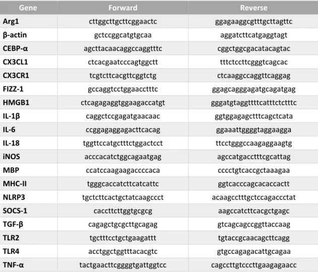
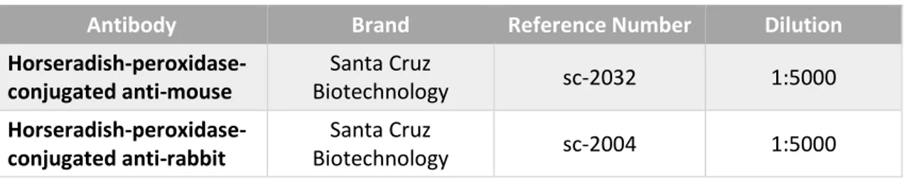
Table II. 1. Primers used for qRealTime PCR ... 27 Table II. 2. Primary antibodies used for immunoblot assays ... 28 Table II. 3. Secondary antibodies used for immunoblot assays ... 28
III. Results
Table III. 1. Demyelination or antibodies treatment on cerebellar organotypic slice cultures do
vii
Abstract
Multiple sclerosis (MS) is a neurodegenerative disease with severe effects on motor and cognitive function. Despite the evolution of knowledge in recent decades, the causes and the exact mechanisms that originate the disease are still unknown. Recent studies demonstrated that S100B protein expression is increased in MS patients and that its high levels are correlated with glial reactivity, contributing to the characteristic excessive inflammatory response of this disease. We also showed that S100B blockade does not prevent demyelination-associated activation of microglia, but it decreases the expression of pro-inflammatory markers. Interestingly, microscopic imaging suggested that upon S100B neutralization microglia moved to myelin surroundings, where they can play an important role on the clearance of myelin debris and remyelination. Thus, we decided to explore whether S100B blockade might modulate the reactivity of microglia in an ex vivo demyelinating model.
Therefore, we first evaluated whether S100B might affect demyelination. Our results corroborate a decreased expression of myelin-related protein upon demyelination, which is prevented after S100B neutralization. We also quantified the different populations of reactive microglia showing that there is an augment of M1 pro-inflammatory reactive microglial cells in consequence of demyelination, which is reduced with S100B blockade. Indeed, while upon demyelination, we verified an increase of gene expression of pro-inflammatory mediators, S100B antibody neutralization partially prevented this effect. In turn, we also observed an increase of anti-inflammatory markers usually associated to microglia M2 phenotype, but this time S100B blockade maintained their elevated gene expression. In addition, we verified that S100B neutralization although not increasing the average number of particles phagocytosed by each microglia upon demyelination, it increased the number of microglia with a phagocytic ability. Moreover, also the expression of a neuron-derived microglia calming factor, fractalkine, was enhanced by S100B neutralization suggesting the shift microglia to a damage repair phenotype.
Overall, these results suggest that by neutralizing S100B during a demyelinating event we may prevent the loss of myelin as well as the exacerbation of the inflammatory response, indicating that S100B may be a potential therapeutic target to reduce damage in demyelinating disorders associated with microglial reactivity, such as MS.
ix
Resumo
A Esclerose Múltipla (EM) é uma doença neurodegenerativa com efeitos graves a nível motor e cognitivo. Apesar da evolução do conhecimento nas últimas décadas, as causas e os mecanismos que desencadeiam a doença são ainda desconhecidos. Estudos recentes demonstraram que a expressão da proteína S100B está aumentada em doentes com EM e que estes níveis elevados estão correlacionados com a reatividade glial, contribuindo para uma resposta inflamatória excessiva, característica desta doença. Verificámos igualmente que o bloqueio de S100B não previne a ativação da microglia aquando de uma situação de desmielinização, mas diminui a expressão de marcadores pro-inflamatórios. Curiosamente, imagens de microscopia sugerem que após a neutralização de S100B a microglia movimenta-se para a zona da mielina, onde pode desempenhar um papel importante na remoção dos detritos de mielina e na remielinização. Assim, decidimos avaliar se o bloqueio de S100B pode modular a reatividade da microglia num modelo ex vivo de desmielinização.
Assim, primeiro avaliámos se a proteína S100B pode afetar a desmielinização. Os nossos resultados revelam uma diminuição da expressão génica de uma proteína associada à mielina após desmielinização, a qual é prevenida após a neutralização de S100B. Quantificámos ainda as diferentes populações de microglia reativa, mostrando que há um aumento das células microgliais com um fenótipo M1 pró-inflamatório em consequência da desmielinização, a qual é reduzida com o bloqueio de S100B. De facto, a desmielinização leva a um aumento da expressão génica de mediadores pró-inflamatórios, enquanto a neutralização de S100B previne parcialmente este efeito. Por seu lado, observámos igualmente um aumento dos marcadores anti-inflamatórios associados a um fenótipo microglial M2, mas o bloqueio de S100B manteve a sua elevada expressão génica. Verificámos ainda que a neutralização de S100B apesar de não aumentar o número médio de partículas fagocitadas por cada microglia após desmielinização, elevou o número de células microgliais com capacidade fagocítica. Curiosamente, observámos igualmente que a expressão de um fator derivado dos neurónios que acalma a reatividade microglial, a fractalkina, estava aumentada após neutralização de S100B, sugerindo que a microglia possa ter adquirido um fenótipo mais adequado para a reparação do dano.
De uma maneira geral, estes resultados sugerem que ao neutralizar o S100B podemos prevenir a desmielinização bem como uma resposta inflamatória exacerbada, indicando que a S100B pode ser um potencial alvo terapêutico para reduzir o dano em doenças desmielinizantes associadas à reatividade microglial, tais como a EM.
xi
Abbreviations
AMPA α-amino-3-hydroxy-5-methyl-4-isoxazolepropionic acid
Arg1 Arginase 1
BBB Blood-brain-barrier
Ca2+ Calcium
CEBP-α CCAAT-enhancer binding protein α
CD Cluster of differentiation
CNS Central nervous system
COSC Cerebellar organotypic slices culture
CSF Cerebrospinal fluid
CX3CL1 CX3C chemokine ligand 1
CX3CR1 CX3C chemokine receptor 1
DIV Days in vitro
DNA Deoxyribonucleic acid
EAAT Excitatory amino acid transporter
EAE Experimental Autoimmune Encephalomyelitis
Fe2+ Iron
FIZZ-1 Resistin-like alpha ou found in inflammatory zone
F4/80 EGF-like module-containing mucin-like hormone receptor-like 1
HMGB1 High-mobility group box 1
IL Interleukin
iNOS Inducible nitric oxide synthase
K+ Potassium
LPC Lysophosphatidylcholine or lysolecithin
MBP Myelin basic protein
MFG-E8 Milk fat globule factor-E8
MHC-II Major histocompatibility complex, class II
MMP Metalloproteinase
MS Multiple sclerosis
mRNA Messenger ribonucleic acid
mtDNA Mitochondrial DNA
xii
NF-KB Nuclear factor kappa-light-chain-enhancer of activated B cells
NLRP3 NOD-like receptor family, pyrin domain containing 3
PLP Proteolipid protein
PPMS Primary-progressive multiple sclerosis
PRMS Progressive-relapsing multiple sclerosis
qRealTime PCR Quantitative real-time polymerase chain reaction
RAGE Receptor for Advanced Glycation Endproducts
RNA Ribonucleic acid
RNS Reactive nitrogen species
ROS Reactive oxygen species
RRMS Relapsing-remitting multiple sclerosis
SOCS Suppressor of cytokine signaling
SPMS Secondary-progressive multiple sclerosis
TGF-β Transforming growth factor-β
Th T helper
TNF-α Tumor necrosis factor-α
1
I. Introduction
1. Multiple Sclerosis
Multiple sclerosis (MS) is a chronic inflammatory and neurodegenerative disorder of the Central Nervous System (CNS) which leads to the development of focal demyelinated plaques in the white matter (Lassmann, 2011). Furthermore, this disease is characterized by reactivation of antigen-presenting cells, microglial activation, production of cytotoxic mediators and recruitment of systemic immunocompetent cells that leads to a generalized neural tissue damage (Gonsette, 2008).
Generally, MS starts in young adulthood with neuroinflammation, characterized by the CNS infiltration of immune cells across the Blood-Brain-Barrier (BBB), resulting in focal demyelinated plaques formation and axonal damage (Compston and Coles, 2008; Lassmann, 2011; Stadelmann et al., 2011). Depending on plaque location, the symptomatology may be very different (Friese et al., 2014; Lassmann et al., 2012). The immune cell invasion may lead to a permanent activation of macrophages and microglia in parenchyma that result in demyelination and neurodegeneration (Compston and Coles, 2008; Napoli and Neumann, 2010). Therefore, the neurodegeneration which starts with acute lymphocytic inflammation may progress towards chronic inflammation (Ciccarelli et al., 2014).
Demyelination is the total or partial loss of the myelin sheath around axons, thus compromising the efficient conduction of action potentials (Love, 2006). This loss may be a consequence of several factors, including inflammatory processes or viral infections that damage myelin sheaths, which are constituted by two main proteins, proteolipid protein (PLP) and myelin basic protein (MBP); or the cells that synthetize them, oligodendrocytes in the CNS and Schwann Cells in the Peripheral Nervous System (PNS) (Lassmann, 2011; Love, 2006). Axonal myelination is a crucial factor for correct signal transmission and any damage in myelin sheaths may have serious consequences at cognitive and motor levels (Berger and Reindl, 2007;
I. Introduction
2
Lassmann, 2011). Thus, in order to delay disease progression it is important to reduce this demyelination, which is a hallmark of MS, or to promote the remyelination process.
1.1.
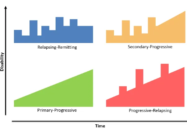
Clinical course of Multiple Sclerosis
In MS patients usually experience a first neurologic event that is sustained for at least 24 hours. As showed in Figure I. 1, there are 4 types of MS named in accordance with the clinical course of the disease over time: Relapsing-Remitting Multiple Sclerosis (RRMS); Primary-Progressive Multiple Sclerosis (PPMS); Secondary-Primary-Progressive Multiple Sclerosis (SPMS) and Progressive-Relapsing Multiple Sclerosis (PRMS).
The RRMS is the most common form of MS. Initially, about 85% of patients are diagnosed with RRMS. This form of MS is characterized by active lesions and temporary relapses that can be partially or completely reversible. The majority of this patients with RRMS progress to a secondary progressive phase (Lindberg and Kappos, 2006), being designated as SPMS, a phase characterized by irreversible deficits and neurodegeneration which are steadily increasing, with or without the occurrence of relapses and remissions (Lindberg and Kappos, 2006).
3 A small percentage of MS patients (~10%) have PPMS that is characterized by a slow and continuous progression without relapses (Lindberg and Kappos, 2006). The least common form (~5%) is PRMS that is characterized by a steady worsening of neurologic function, with occasional relapses but no complete remissions, with or without recovery. Once PRMS is progressive from onset, it may be diagnosed as PPMS and subsequently changed to PRMS, when a relapse occurs (Lassmann et al., 2012; Milo and Miller, 2014).
In the course of MS it is possible to distinguish different types of focal plaques of demyelination: active, chronic active lesions slowly expanding, inactive lesions and remyelinated shadow plaques. The classic active plaques are characterized by high inflammation with perivascular infiltrates of lymphocytes and macrophages, a complex architecture being commonly detected during the acute or relapsing–remitting stages. The chronic active lesions are considered to comprise approximately 50% of the lesions in progressive stage of disease. The center of this type of lesion usually does not have myelin nor presents remyelination signals, but shows axonal loss. Furthermore, lesions are surrounded by an area of microglial activation and initial tissue injury. The inactive lesions are the most frequent type in all stages of the disease and present signals of an inefficient remyelination, lack of myelin and axonal loss. In addition, the lymphocytic infiltrates and microglial activation are reduced. Finally, the shadow plaques result from remyelination and normally present low axonal injury and thinly remyelinated axons. This remyelination occurs during the acute inflammation, following myelin debris removal by phagocytosis, but it may also be detected in the progressive phase (Lassmann, 2010; Lassmann et al., 2012; Love, 2006).
Once the disease has an early and strong inflammatory component, treatments targeting the inflammatory insult have been shown to be effective mostly in the relapsing stage. However, in the progressive phase of disease, the anti-inflammatory or immunomodulatory treatments used so far showed no clinical relevance (Lassmann, 2011). Thus, a key step for the treatment of progressive MS is the development of new therapies for inflammatory and neurodegenerative components of the disease.
1.2.
The etiology of MS
The etiology of MS is not fully understood yet, but in the majority of patients, the disease progression is characterized by acute relapses (RRMS) leading to a progressive and irreversible accumulation of neurological deficits (SPMS). Relapses are the manifestations of inflammatory demyelinating lesions in the CNS (Charil and Filippi, 2007; Compston and Coles, 2008; Love,
I. Introduction
4
2006). In a primordial phase, the inflammation is a transitory feature and is followed by remyelination that contributes to total or partial clinical remission of the symptomatology. However, this remyelination may not be complete and, over time, repeated acute insults and the failure of reparative system may lead to extensive microglial activation associated with irreversible axonal and neuronal loss resulting into neurodegeneration (Charil and Filippi, 2007). For many years, the focus of MS research had been on inflammatory white matter pathology, once the disease was initially considered to be an immune-mediated demyelinating disorder. However, there are results that show axonal loss occurring in early phase of disease (Lindberg and Kappos, 2006). These evidences suggest a strong neurodegenerative component, which contributes to pathogenesis and reveal that MS may be an inflammatory demyelinating and neurodegenerative disease that affects all the CNS (Herz et al., 2010). One hallmark of MS are in fact demyelinated lesions in white matter related with axonal degeneration and immune cells infiltration. The focal demyelinated plaques, present in the grey and white matter at all stages of the disease, are infiltrated by populations of immune cells and immune mediators such as T cells, B cells, macrophages and microglia, as well as cytokines, chemokines and other toxic agents (Lassmann et al., 2012; Napoli and Neumann, 2010). The infiltration of immune cells is a consequence of BBB disruption, partly due to matrix metalloproteinases (MMPs). The expression of several MMPs, particularly MMP-2 and -9, are also altered in microglial cells of MS lesions (Könnecke and Bechmann, 2013; Rosenberg, 1995).
Besides demyelination, immune cell-related inflammation is critical for neuronal damage due to pro-inflammatory neurotoxic substances release and consequent damaging processes. In these patients, the inflammatory lesions consist of perivascular and parenchymal infiltrates of lymphocytes and macrophages (Lassmann et al., 2012). While in active lesions, there are low levels of T cells at sites of the initial tissue injury, the ongoing tissue damage is associated with the infiltration of macrophages and the activation of resident microglia. In addition, the brains of these patients also display global changes as widespread inflammation, microglial activation, astrocytic gliosis and slight demyelination and axonal loss in normal-appearing white matter. These changes together with widespread loss of tissue volume observed in the cortex result in brain atrophy with ventricles dilatation (Herz et al., 2010; Lassmann et al., 2012).
5
1.3.
Molecular mechanisms of neurodegeneration in Multiple Sclerosis
As a result of inflammatory processes many molecular changes occur in CNS during MS, namely the secretion of neurotoxins that induce immune responses with important roles in homeostasis and neuronal metabolism. These responses have different functions depending on the time of exposure. At short-term, the immune response has a crucial role in tissue defense but, at long-term, immune cells induce stress responses (Friese et al., 2014). The molecular pathways involved in MS neurodegeneration are very complex due to the heterogeneity of this disease. Although most of these mechanisms are also associated with other neurodegenerative diseases, the extensive primary demyelination and preservation of axons is specific for MS. It is therefore necessary to understand the pathogenesis of MS and identify the mechanism that specifically affects myelin and the cells responsible for its production, contributing to widespread primary demyelination (Lassmann, 2013). After this first myelin destruction due to inflammation, other known mechanisms may be responsible for axonal loss, worsening MS clinical course.
1.3.1. Free Radicals and Oxidative Stress
Oxidative stress reflects an imbalance between the reactive oxygen species (ROS) production and the ability to detoxify this reactive species and repair the damage (Mao and Reddy, 2010). Although it cannot be generalized, there are evidences that in some patients with MS, oxidative stress may be the principal mechanism implicated in the pathogenesis of disease (van Horssen et al., 2008).
Oxidative stress is able to damage the cells by promoting the oxidation of cellular components, as lipids, proteins and nucleic acids (mainly mitochondrial DNA, mtDNA), which consequently leads to cell death (Mao and Reddy, 2010). Specifically, it was verified an accumulation of oxidized DNA and lipids within lesions in all stages of the disease. However, in active lesions there is a greater indication of oxidative damage than in inactive lesions (predominate in the progressive stage of MS), which presents a low signal (Haider et al., 2011). As already described, the excessive inflammatory environment in demyelinating lesions is favorable to ROS and reactive nitrogen species (RNS) formation, and this increased levels of reactive species may compromise the antioxidant defenses in our organism, particularly in the lesions (Mao and Reddy, 2010). In support of these data, it was observed an increased expression of enzymes responsible for free radical production (e.g. myeloperoxidase, nicotinamide adenine dinucleotide phosphate oxidase, xanthine oxidase) in active lesions,
I. Introduction
6
mainly in areas of initial tissue injury (Fischer et al., 2012). The presence of superoxide and peroxynitrite, which are produced due to ROS and RNS reaction, as well as the increased levels of DNA oxidation within plaques shows that the generation of reactive species may have an extremely toxic effect in neuronal and glial cells (Mao and Reddy, 2010). In support of these evidences, the presence of oxidized DNA and lipids in apoptotic oligodendrocytes and dystrophic axons also indicates the key role of ROS in demyelination and neurodegeneration (Lassmann et al., 2012).
Despite the fact that anti-oxidative activity is not different in MS patients, comparatively with healthy controls, there are evidences that sulfhydryl groups, which have antioxidants properties, are decreased in MS patients (Mao and Reddy, 2010). However, some antioxidant enzymes (e.g. superoxide dismutases, catalase, peroxiredoxins) are upregulated in active MS lesions, which may indicate an active defense mechanism to reduce cellular damage caused by ROS (van Horssen et al., 2008).
Oligodendrocytes are particularly sensitive to higher levels of these reactive species, comparatively with astrocytes and microglia. This fact may result from their reduced ability for antioxidant defense, rendering them more prone to oxidative stress toxicity which leads to oligodendrocyte death and consequent demyelination. Furthermore, ROS and RNS can also damage the own myelin sheath and promote its clearance by macrophages and microglia. In an early stage of MS, the oxidative stress appears to be triggered by activated microglia but, in a progressive stage, it may be intensified by further factors (Friese et al., 2014; Lassmann, 2013; Mao and Reddy, 2010).
1.3.2. Mitochondrial dysfunction
Mitochondria has as principal function to provide energy to cells in the form of ATP. It participates in many cellular processes, including fatty acid oxidation, apoptosis and calcium homeostasis. So, the high energy needed by CNS render it slightly vulnerable to mitochondrial damage.
A serious consequence of high levels of ROS is mitochondrial dysfunction, which may result from different mechanisms. The released free radicals and modifying proteins can disrupt mitochondrial function by interfering with some components of respiratory chain and promoting mtDNA damage (Ellwardt and Zipp, 2014; Lassmann et al., 2012). These mitochondrial changes may explain pathological features of MS lesions as demyelination and remyelination impairment, destruction of thin-calibre axons, differentiation arrest of
7 oligodendrocyte progenitor cells, oligodendrocyte apoptosis and astrocyte dysfunction (Haider et al., 2011; Lassmann et al., 2012).
Mitochondria plays several crucial functions in different pathways including oxidative energy metabolism, where the most of the ATP is synthesized. Therefore, it is easy to understand that the impairment of mitochondria, besides leading to the production of more reactive species that will exacerbate the tissue injury (Mao and Reddy, 2010), will also induce energy failure. Indeed, it is clear that mitochondrial injury and consequent energy failure is a very important factor that drives to MS tissue injury (Lu et al., 2000; Witte et al., 2010).
The first evidence that the mitochondrial damage has some role in MS lesions demonstrated a compromised NADH dehydrogenase activity as well as an increase of complex IV activity within lesions (Lu et al., 2000). Active lesions show significant changes in proteins of the mitochondrial respiratory chain and, in addition, deletions in mitochondrial DNA are present in neurons, especially in the progressive stage of the disease.
Concerning oligodendrocytes, the mitochondrial damage results on release of apoptosis-inducing factor (AIF), which translocates into the nucleus and induces DNA damage. With the damage, one polymerase (poly ADP-ribose polymerase) is activated in an attempt to repair the injury, however, this leads to further energy deficiency (Lassmann et al., 2012).
There are also suggestions that activated microglia play an important function in mitochondrial dysfunction, namely in MS. Activated microglia are responsible for ROS and NO production that, besides damaging mtDNA, can lead to the inhibition of oxidative phosphorylation pathway disrupting the ATP production and increasing ROS formation (Witte et al., 2010, 2014). Additionally, there has been reported a reduction in PGC-1α (peroxisome proliferator-activates receptor gamma coactivator-1α), a transcriptional co-activator and regulator of mitochondrial function, in MS cortex. This decreased in PGC-1α levels coinciding with both reduced expression of subunits involved in oxidative phosphorylation pathway and decreased expression of several mitochondrial antioxidants (Witte et al., 2013). Therefore, mitochondria has a decreased capacity to produce ATP and detoxify oxidative stress, which can compromise its efficient functioning.
1.3.3. Ion channel dysfunction
The intracellular environment is very important for maintaining neuronal functions. Abnormal expression of Na+ channels, acid-sensing Na+ channels, glutamate receptors and
I. Introduction
8
context, the ion channel dysfunction has a great impact on neurons and axons, which might even lead to their degeneration and death in progressive phase of MS (Friese et al., 2014; Lassmann et al., 2012; Mao and Reddy, 2010). As already mentioned, in response to an inflammatory stimulus, energy imbalance and demyelination may lead to activation, dysfunction and altered distribution of ion channels, inducing downstream mechanisms. These mechanisms are mostly responsible for Ca2+ accumulation and apparently may be the promoters of neurotoxicity and
trigger of innumerous enzymes activation, which compromise both the normal mitochondrial functioning and axonal transport and result in additional increase of Ca2+levels. Moreover, the
wrong distribution of voltage-gated Ca2+ channels (VGCCs) in demyelinated fibers may lead to
an abnormal influx of Ca2+, which contributes to axonal death (Friese et al., 2014).
The Na+ channels are responsible for the acceleration of the saltatory conduction in
myelinated axons, which is the propagation of an action potential from one node of Ranvier to another, along a myelinated fiber. When into the axon, Na+ is exchanged for K+ by Na+/K+-ATPase,
and this ion exchange is important for axonal polarization. This enzyme is responsible for correcting Na+ and K+ levels and for preventing a pathological influx of Na+ in axons, however, in
pathological conditions, inflammatory mediators are released and cause the Na+/K+-ATPase
failure and consequent mitochondrial injury (Mao and Reddy, 2010). It has also been shown that under hypoxia, this Na+/K+ pump activity are inhibited and that reactive species increase its
degradation (Waxman, 2008; Young et al., 2008).
1.3.4. Excitotoxicity of Glutamate
Glutamate has been found in MS lesions at high concentrations (Srinivasan et al., 2005). It is one of the most important excitatory neurotransmitter of the CNS and modulates ion homeostasis into the cells through several receptors including N-methyl-D-aspartate (NMDA) and α-amino-3-hydroxy-5-methyl-4-isoxazolepropionic acid (AMPA)/kainate receptors. The glutamate-mediated excitotoxicity is an important connection between neuroinflammation and neurodegeneration, once high levels of glutamate stimulate its receptor and lead to deregulation in ion homeostasis contributing to neurotoxicity associated with axonal, oligodendroglial and myelin damage or even cell death (Ellwardt and Zipp, 2014; Takaki et al., 2012). So, excessive levels of glutamate, which are released by activated immune cells including activated microglia, may contribute to the lesion development in MS by overstimulation of ionotropic receptors (Pitt et al., 2000). This involvement in lesion development is supported by results that demonstrate a reduction of neurological disability and axonal damage, and an
9 increase of oligodendrocyte survival with AMPA/kainate receptor antagonist treatment. Thus, these results show that AMPA/kainate-mediated glutamate excitotoxicity has an important role in CNS damage in the animal model of MS, the experimental autoimmune encephalomyelitis (EAE) and probably also in the course of MS (Pitt et al., 2000).
Also the activation of microglia seem to be involved in the impairment of glutamate transporters. Domercq and collaborators showed that high ROS levels released by activated microglia inhibit glutamate uptake by oligodendrocytes, resulting in extracellular glutamate increase (Domercq et al., 2007). In addition, it has further been demonstrated that activated microglia are correlated with focal loss of excitatory amino acid transporters, EAAT1 and EAAT2. They also verified alterations in the mechanisms of glutamate uptake only in the presence of activated microglia (Vercellino et al., 2007). Once EAATs are fundamental in both the maintenance of low extracellular glutamate levels and in prevention of excitotoxicity as well, activated microglia appear to have an important role in excitotoxicity present in MS.
1.3.5. Iron accumulation
It is known that iron accumulates in healthy human brain with age increase. This fact may be relevant once progressive phase of MS usually starts between 40 and 50 years of age (Lassmann et al., 2012).
Iron, which is mainly stored in oligodendrocytes, is crucial for normal brain metabolism, including for myelination, however, it may generate ROS (Friese et al., 2014; Hametner et al., 2013). As consequence of oxidative stress, activated microglia release H2O2 that diffuses into
oligodendrocytes with Fe2+ accumulation and forms toxic radicals leading to cell death. The Fe2+
released from these dying oligodendrocytes is taken up by microglial cells, which subsequently become dystrophic. These dystrophic microglia release more Fe2+ that results in more oxidative
tissue injury. So, iron accumulation within oligodendrocytes is also a relevant mechanism that further contribute to neurodegeneration in MS (Ciccarelli et al., 2014; Lassmann et al., 2012).
1.3.6. Inflammatory mediators
Myelin sheaths loss occurs due to the migration of auto-reactive immune cells through the BBB that attack myelin components. Within the CNS, activated T-cells together with activated microglia, macrophages and astrocytes, release pro-inflammatory cytokines, creating a pro-inflammatory environment which can lead to neurodegeneration. The accumulation of
I. Introduction
10
these pro-inflammatory mediators, which are described in section 1.4.1.2, amplify both inflammatory and immune response contributing to demyelination and then neurodegeneration (Dendrou et al., 2015; Glass et al., 2010; Lassmann and van Horssen, 2011; Vogel et al., 2013). Besides all these inflammatory contributors, including high levels of S100B, the protein in which we are interested, stimulate the release of further inflammatory mediators, meaning that it has a function on exacerbating the inflammatory response (Bianchi et al., 2010; Villarreal et al., 2014).
1.4.
Microglia as cellular players in Multiple Sclerosis
As mentioned above, in all phases of MS, active tissue injury is associated with inflammatory infiltrates. In addition, several immune cells including activated microglia are observed in lesions borders. These cells express cytokines and enzymes involved in the production of ROS and RNS (Fischer et al., 2012), having a crucial effect on MS pathogenesis.
1.4.1. Microglial cells
Microglia are the tissue macrophages of the brain and the main form of immune defense in the CNS constituting around 10% of the cells in this system. They are members of the innate immune system and respond to danger signals, initiating an acute inflammatory response, within the CNS (Goldmann and Prinz, 2013; Jack et al., 2005). These cells have functions similar to those of other tissue macrophages such as phagocytosis, antigen presentation and secretion of cytokines (Herz et al., 2010). Microglia act quickly after an insult in order to restrain the damage and promote recovery. However, besides promoting neuroprotection and stimulating tissue repair, activated microglial cells can exacerbate an inflammatory status and trigger neurotoxic pathways, which may lead to a progressive neurodegeneration (Correale, 2014).
An important characteristic of microglia are their extensive branches, which allows that these cells continuously patrol the CNS parenchyma (Benarroch, 2013; Olah et al., 2011a). This characteristic makes microglial cells the first line of defense in CNS (Correale, 2014; Giunti et al., 2014; Olah et al., 2011a). As consequence of brain injury, resident microglia change their surveillance phenotype to an “activated” morphology, which are associated with different phenotypes highly dependent on the type, intensity and duration of their exposure to stimuli (Benarroch, 2013; Correale, 2014; Perry et al., 2010). These different microglial phenotypes may
11 be defined based on morphological, molecular and functional characteristics (Kettenmann et al., 2011).
1.4.1.1. Surveillant microglia
Under healthy conditions, microglial cells display a surveillant/patrol or M0 phenotype, which are characterized by a ramified morphology, a slow turnover rate and low expression of surface molecules. This apparently “quiescent” microglia are constantly scanning their environment for exogenous or endogenous signals (Giunti et al., 2014; Kettenmann et al., 2011), and are ready to rapidly switch to the “activated” state, after injury occurrence (Kettenmann et al., 2011).
Surveillant microglia phenotype seems to be preserved through interactions between some receptors and their respective ligands expressed in neurons, such as CD200-CD200R and fractalkine (CX3CL1)-CX3CR1, which are described as “off signals” (Correale, 2014; Jones and Lynch, 2014; Perry et al., 2010). A study that reveals the presence of a microglia mainly activated in CD200 knockout mice comparatively with control mice, clearly demonstrated the importance of these interactions in the surveillant phenotype (Hoek, 2000). Besides this, neurons are also responsible for several neurotrophic factors release (e.g. nerve growth factor and brain-derived neurotrophic factor) that keep the microglial cells at rest (Perry and Teeling, 2013).
1.4.1.2. Activated microglia
Any disturbance, which may indicate a potential danger to the CNS changes microglial morphology, gene expression and their functional behavior (Correale, 2014; Kettenmann et al., 2011). Through their branches, microglia sense variations in their microenvironment, recognize danger signs and consequently become activated. Morphologically, microglia retract their branches changing their shape to an amoeboid form (Correale, 2014). Moreover, microglia become motile and go to the lesion following chemotactic gradients. In addition, CNS injury induces microglia proliferation that provide more cells for protection and repair of tissue homeostasis (Benarroch, 2013; Kettenmann et al., 2011).
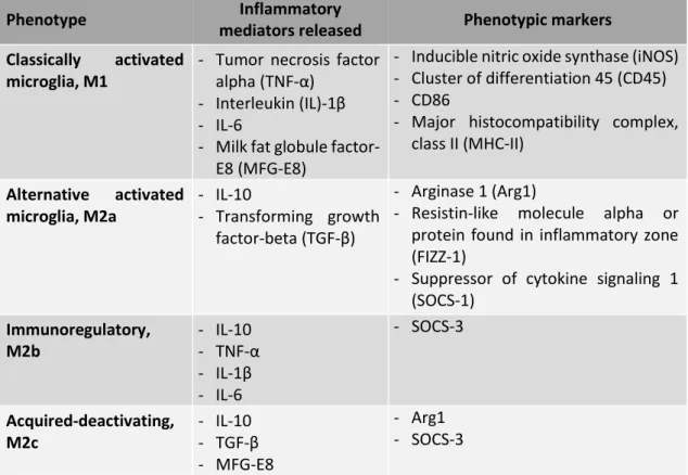
Besides all these alterations, microglia display molecular alterations such as upregulation of some surface markers and specific proteins, presenting a classically activated (M1) or an alternative activated (M2) phenotype (Chhor et al., 2013; Correale, 2014; Giunti et al., 2014; Goldmann and Prinz, 2013). M1 consists of a pro-inflammatory phenotype mainly
I. Introduction
12
associated with cytotoxic response, which is responsible for mediating innate immune responses but also adaptive immune responses. The innate immune response may be triggered by damage-associated molecular patterns (DAMPs), misfolded proteins and other proteins released from injured neurons, which in turn activates toll-like receptors (TLR), in microglial cells. In response to this interaction, TLRs activate downstream signaling cascades leading to transcriptional activation of nuclear factor kappa-B (NF-KB) and subsequent acute inflammation.
This feature is characterized by the production ROS, that leads to neuronal death, engagement of NLRP3 inflammasome, expression of altered enzymes and surface markers and release of pro-inflammatory cytokines, as shown in Table I. 1 (Benarroch, 2013; Chhor et al., 2013; Correale, 2014; Goldmann and Prinz, 2013). The activation of inflammasome by DAMPs enhances the inflammatory response. Also the high-mobility group box 1 (HMGB1), a protein that is secreted by damage neurons for signaling the cell damage, can interact with TLRs, among others, further activating microglia and exacerbating the NF-KB signaling cascade (Brites and Vaz, 2014). Additionally, the adaptive immune response is triggered by interferon gamma, which is released from T helper cells type 1 (Th1), and in response to that, microglia become antigen-presenting cells and release pro-inflammatory mediators as effectors of adaptive immunity (Benarroch, 2013).
Table I. 1. Characteristics of different microglial phenotypes (compiled from Brites and Vaz, 2014; Correale, 2014;
David and Kroner, 2011; Liu et al., 2008)
Phenotype Inflammatory
mediators released Phenotypic markers
Classically activated microglia, M1
- Tumor necrosis factor alpha (TNF-α)
- Interleukin (IL)-1β - IL-6
- Milk fat globule factor-E8 (MFG-factor-E8)
- Inducible nitric oxide synthase (iNOS) - Cluster of differentiation 45 (CD45) - CD86
- Major histocompatibility complex, class II (MHC-II) Alternative activated microglia, M2a - IL-10 - Transforming growth factor-beta (TGF-β) - Arginase 1 (Arg1)
- Resistin-like molecule alpha or protein found in inflammatory zone (FIZZ-1)
- Suppressor of cytokine signaling 1 (SOCS-1) Immunoregulatory, M2b - IL-10 - TNF-α - IL-1β - IL-6 - SOCS-3 Acquired-deactivating, M2c - IL-10 - TGF-β - MFG-E8 - Arg1 - SOCS-3
13 M2 microglia phenotype plays a crucial role in repair and healing of tissues, once microglia secrete extracellular matrix proteins and growth factors, and promote phagocytosis of cellular and myelin debris contributing for the remyelination process. Moreover, these microglia are also known for their involvement in synapse repair and remodeling (Correale, 2014). M2 microglial phenotype is induced by signals from apoptotic cells, as heat shock protein 60 (Hsp60), that activates triggering receptor in myeloid cells-2 (TREM-2), or by anti-inflammatory cytokines such as interleukin (IL)- 4 and IL-13, which are released from Th2 helper cells (Benarroch, 2013; Olah et al., 2011b). Thus, this phenotype seems to be beneficial compared with M1 however, a prolonged activation of this M2 phenotype may be harmful for preventing axonal growth (Brites and Vaz, 2014). In addition, there are three different subclasses of M2 microglia, M2a, M2b and M2c, which are induced through polarizing signals and have different functional properties. M2a is an alternative activation repair/remodeling phenotype, which is recruited for phagocytosis and inflammation repair and induced by IL-4 and IL-13, and express specific markers, as presented in Table I. 1 (Brites and Vaz, 2014; Chhor et al., 2013; Varnum and Ikezu, 2012). Moreover, M2a microglia release inflammatory mediators that trigger an anti-inflammatory response and promote tissue repair (Correale, 2014; Goldmann and Prinz, 2013). The immunoregulatory or M2b phenotype is stimulated through immune complexes, TLR agonists and IL-1R ligands. Although M2b microglia release IL-10, an anti-inflammatory cytokine, they also release some pro-inflammatory cytokines (Table I. 1), which suggests that M2b microglial cells may act in and modulate different conditions of inflammation (Chhor et al., 2013; David and Kroner, 2011). The specific markers of M2b phenotype are SOCS-3 and IL-1R antagonist (IL-1Ra) (Chhor et al., 2013). The acquired-deactivating or M2c phenotype is induced by IL-10, TGF-β, glucocorticoids and enhances anti-inflammatory marker expression (Table I. 1) while decreasing pro-inflammatory cytokine levels (Chhor et al., 2013; Varnum and Ikezu, 2012). Besides modulating the anti-inflammatory polarization through the downregulation of pro-inflammatory markers, SOCS-1 also regulates M2 phenotype, since the expression of SOCS-1 is increased in M2 phenotype (Davey et al., 2006; Guedes et al., 2013; Wilson, 2014).
As already mentioned, an important neuroprotective function of microglial cells is phagocytosis. M1 and M2c microglia can produce MFG-E8 that recognizes the phosphatidylserine (PS) exteriorized by apoptotic cells and triggers a signaling cascade that stimulates the phagocytic process of dying cells (Brites and Vaz, 2014).
I. Introduction
14
1.4.1.3. Microglia in MS
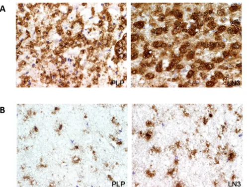
The role of microglia in MS is controversial. Several evidences indicate that classically activated microglia promotes neuroinflammation and demyelination in MS and in EAE, contributing to CNS injury. To elucidate the function of microglial cells, it has been shown that the inhibition of microglial activation results in a delayed EAE onset (Bogie et al., 2014; Giunti et al., 2014; Heppner et al., 2005). In addition, microglial activation has been observed in post-mortem brain tissue of MS patients as well as the presence of clusters of activated microglia within the normal-appearing white matter (Giunti et al., 2014; van Horssen et al., 2012). Our group also observed a large population of activated microglia/macrophages and PLP-positive macrophages in active MS lesions in contrast to chronic MS lesions (Barateiro et al., 2015) (Figure I. 2).
Figure I. 2. Presence of microglia in active (A) or chronic (B) MS lesions. Frozen sections of autopsied brain samples
of MS patients were immunostained for PLP to detect white matter and for HLA-DR MHC class II clone LN3 to detect macrophages/microglial cells. (A) Active MS lesions were outlined by PLP staining and LN3 immunoreactivity. Magnification ×43. In contrast, (B) chronic MS lesions showed decreased PLP and LN3 immunostaining. Magnification ×43. (Adapted from Barateiro et al., 2015).
However, all activation states of microglia may be present in disease indicating that there are multiple populations of microglia occurring in the course of a demyelinating episode (Goldmann and Prinz, 2013). Thus, microglia can also present an alternative activated phenotype in EAE or MS, with the ability to phagocyte apoptotic cells and myelin debris, a crucial step for tissue regeneration. In this context, the role of TREM-2, a phagocytic stimulator, was evaluated
15 and it has been demonstrated that this receptor is upregulated in EAE, facilitating the debris clearance. Moreover, its blockade results into EAE exacerbation with more infiltrates and demyelination in the parenchyma (Piccio et al., 2007).
A study that evaluated the role of microglia/macrophages polarization in remyelination showed that a switch from an M1- to an M2-phenotype is essential for the remyelination process. Particularly, they observed that this change occurs at the time of oligodendrocytes differentiation from oligodendrocyte progenitor cells, which have been recruited into the lesion. Thus, they demonstrated that M2 microglia are necessary for oligodendrocyte differentiation and maturation, which are very important for the efficient remyelination (Miron et al., 2013).
Given that microglial cells present distinct roles along disease progression, it is important to understand how they exert a beneficial role in MS with the objective to modulate this reactivity to a more neuroprotective phenotype during the course of the disease.
2. S100B
S100 proteins are part of a low-molecular-weight and acidic proteins family that are known to contain two distinct EF-hand helix-loop-helix calcium-binding sites (Donato et al., 2013). This family is subdivided into three subgroups according to their action: (i) only exert intracellular regulatory effects, (ii) have intracellular and extracellular functions, or (iii) play extracellular regulatory effects (Donato et al., 2013). S100B is a 10.5 kDa member of this family, with both intracellular and extracellular functions, that is expressed in some cell types from different tissues and that appears to be expressed at highest levels, in the CNS, namely by glial cells (Adami et al., 2001; Michetti et al., 2012). Usually, this protein exists within cells as a homodimer but it can also form heterocomplexes when associated with S100A1 monomer (Donato et al., 2009).
Due to important functions that S100B presents in proliferation, migration, differentiation and apoptosis, it plays a critical role during brain development acting as a neurotrophic factor, by furthering neurite outgrowth and neuronal survival for low/physiological levels (nM) (Donato et al., 2013; Koppal et al., 2001).
On the other hand, it was also demonstrated that S100B expression is increased in many tumors as well as in the aging brain and in the brain of patients affected by different pathologies as Alzheimer's disease, HIV infection, chronic epilepsy and other brain pathological conditions as MS (Donato et al., 2009; Hein Née Maier et al., 2008). Once these high concentrations of
I. Introduction
16
S100B are present in several brain pathologies, this protein may be considered a biomarker of brain damage (Donato et al., 2013).
2.1.
Dual role of S100B in physiology and pathology
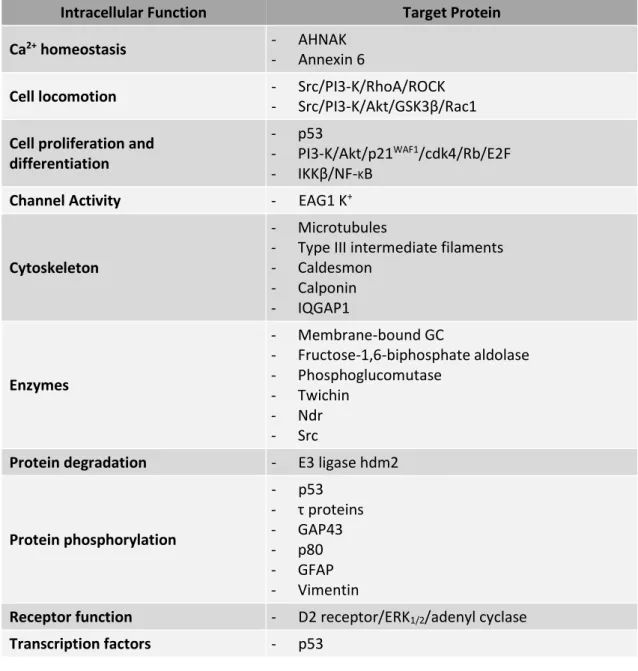
As mentioned above, S100B is a Ca2+ sensor, so, upon binding to Ca2+, S100B changes its
conformation leading to hydrophobic patch exposure to the solvent and its interaction with target proteins (Sorci et al., 2013). Several of these target proteins have been already identified and its interaction with S100B is involved in important intracellular processes (Table I. 2). Accordingly, S100B acts as a stimulator of proliferation and migration as well as an inhibitor of apoptosis and differentiation. Moreover, S100B is involved in the regulation of energy metabolism, transcription, and protein phosphorylation, as well as in Ca2+ homeostasis (Donato
et al., 2009; Sorci et al., 2010).
It is known that astrocytes are the main cell type in CNS that express and secrete S100B, however, other S100B-expressing cells can release the protein in case of damage or necrosis (Sorci et al., 2010). Therefore, besides having a regulatory function within the cytoplasm where it is expressed, S100B can act as a signal molecule in extracellular space given that it can be released by damaged cells (Rustandi et al., 2000; Shashoua et al., 1984; Sorci et al., 2010). Once released, S100B may have a beneficial or harmful action. There are evidences that this release may be mostly dependent on the presence and activation of its receptor RAGE (Receptor for Advanced Glycation Endproducts), which engagement may stimulate further S100B release (Donato et al., 2009; Sorci et al., 2013).
RAGE is a member of the immunoglobulin-like cell surface receptor superfamily composed by a cytosolic domain responsible for signal transduction, a transmembrane domain which anchors it in the membrane, a variable binding-domain and two constant domains (Ostendorp et al., 2007; Sparvero et al., 2009). This receptor is able to transduce inflammatory stimuli and the effects of neurotrophic and neurotoxic factors and therefore, S100B emerged as a damage-associated protein that regulate inflammation-related events and play a role in pathophysiology of neurodegenerative disorders and inflammatory brain diseases (Bianchi et al., 2011; Donato et al., 2013; Zhang et al., 2011b).
17 Table I. 2. S100B target proteins involved in intracellular processes (adapted from Donato et al., 2013)
Intracellular Function Target Protein
Ca2+ homeostasis - AHNAK
- Annexin 6
Cell locomotion - Src/PI3-K/RhoA/ROCK
- Src/PI3-K/Akt/GSK3β/Rac1
Cell proliferation and differentiation
- p53
- PI3-K/Akt/p21WAF1/cdk4/Rb/E2F
- IKKβ/NF-KB
Channel Activity - EAG1 K+
Cytoskeleton
- Microtubules
- Type III intermediate filaments - Caldesmon - Calponin - IQGAP1 Enzymes - Membrane-bound GC - Fructose-1,6-biphosphate aldolase - Phosphoglucomutase - Twichin - Ndr - Src
Protein degradation - E3 ligase hdm2
Protein phosphorylation - p53 - τ proteins - GAP43 - p80 - GFAP - Vimentin
Receptor function - D2 receptor/ERK1/2/adenyl cyclase
Transcription factors - p53
Besides having different functional roles, the extracellular form of S100B, which has more effects in CNS, presents different effects on neurons, astrocytes and microglia, depending on the concentration (Donato et al., 2013). At physiological levels the protein displays trophic effects on neurons promoting neuron survival and growth as well as microglia quiescence, although high concentration of S100B display inflammatory effects and activate pro-apoptotic pathways (Reali et al., 2005). In particular, high levels of S100B can stimulate the nitric oxide synthesis by astrocytes and microglia leading to neuronal and astrocyte apoptosis. Moreover, high S100B stimulates the release of cytokines contributing to brain inflammatory response (Bianchi et al., 2011).
I. Introduction
18
2.1.1. Extracellular S100B effect on microglial cells
The high expression levels of S100B are normally associated to astrogliosis in the course of neurodegenerative diseases. These levels might be the result of neuronal and glial cells pathology given that the protein is released by damaged oligodendroglial or astroglial cells, which have will then activate microglial cells responsible for the innate immune response (Sorci et al., 2010). However, expression levels of S100B exerts two different roles in microglia depending on concentration.
At physiological concentrations, S100B can prevent microglia activation via STAT3 pathway and may also act as a signaling trophic protein that promotes a more protective phenotype of microglia (Zhang et al., 2011b). On the other hand, high S100B concentrations, in the presence of bacterial endotoxin or interferon-γ (IFN-γ), play a main role on microglia activation, exacerbating brain inflammatory response. This activation was shown to be mediated by stimulation of iNOS leading to an increase of nitric oxide release (Adami et al., 2001; Bianchi et al., 2007).
In addition, there is an evidence that S100B stimulates the microglia migration via RAGE-dependent mechanism. It was demonstrated that once S100B accumulates in the extracellular space after brain damage, the increased S100B levels might contribute to intensify the inflammatory response by activating microglia and stimulating their migration (Bianchi et al., 2011).
2.2.
S100B in MS
Increased S100B levels were first detected in cerebrospinal fluid (CSF) of MS patients (Michetti et al., 1979). Another study revealed that S100B levels in CSF were higher in patients with RRMS than in patients with SPMS (Bartosik-Psujek et al., 2011). We confirmed that both in CSF and in serum samples from RRMS patients there is a significant increase of S100B production at the time of diagnosis (Barateiro et al., 2015). S100B increase may exacerbate the release of inflammatory mediators and subsequently contribute to neuronal death (Hu et al., 1996). These results indicate S100B as a potential biomarker for MS diagnosis and prognosis, possibly helping to distinguish between relapsing remitting and progressive phases of MS.
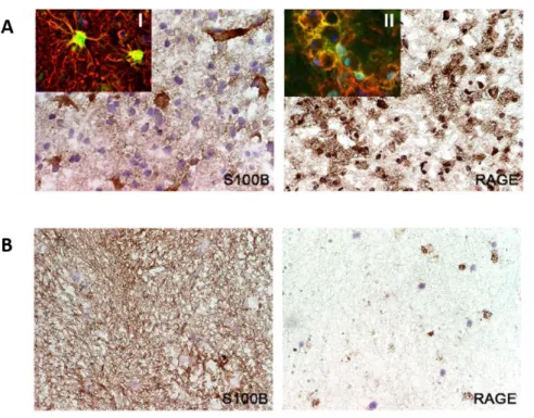
One study reported the presence of S100B in acute lesions of post-mortem brain tissue of patients with RRMS (Petzold et al., 2002). In similar samples, our recent work demonstrated that S100B is overexpressed both in active and chronic lesions, mainly by astrocytes (Figure I. 3).
19 In addition, it showed that in chronic lesions S100B is still diffusely expressed into the demyelinated area (Barateiro et al., 2015). Interestingly, those active MS lesions also showed an increase of RAGE expression that co-localized with microglia/macrophages cells, while almost no RAGE was found in chronic lesions that are almost depleted of microglia/macrophages (Figure I. 3).
Figure I. 3. Expression of S100B and its receptor RAGE in active (A) and chronic (B) MS lesions. Frozen sections of
autopsied brain samples of MS patients were immunostained for S100B and RAGE. (A) S100B and its receptor RAGE are markedly expressed in active MS lesions by astrocytes and activated macrophages/microglia, respectively. Magnification x40. Insets show the co-localization of (I) glial fibrillary acidic protein (GFAP, red), an astrocytic marker, with S100B (green) and the co-localization of (II) LN3 (red), a marker of activated macrophages/microglia with RAGE (green). Magnification x63. (B) S100B but not RAGE is continuously expressed in chronic MS lesions. Magnification x40. (Adapted from Barateiro et al., 2015).
Given the increased expression of S100B during MS episodes, the role of S100B in different neurodegenerative diseases and its apparent involvement in microglia activation, it is of utmost importance to assess whether by modulation of S100B function it is possible to change the reactivity of microglia to a more neuroprotective phenotype during the course of MS.
3. Experimental models to study MS pathophysiology
Failure to understand the neurodegenerative mechanisms involved in MS and therapeutic inefficacy of diverse treatments led to development of multiple experimental
I. Introduction
20
models that mimic the hallmarks of the disease. However, the complexity of MS still hinders the development of the perfect model for this disease (Murta and Ferrari, 2013).
The EAE, a model characterized by inflammation, demyelination and neurodegeneration, is one of the most widely used in vivo model to study MS that involves the immunization of genetically susceptible animals with a myelin protein inducing brain inflammation and destruction of myelin. However, EAE models fail to expect the clinical efficacy in patients (Mathew et al., 2013; Ransohoff, 2012). Moreover, in vivo models entail an expensive cost, as well as ethical problems and, therefore, alternative models should be used whenever possible.
For decades, organotypic slice cultures were used in CNS research due to three-dimensional architecture, maintenance of contact between different cells and the presence of all the cells of CNS which play a key role in lesion recovery (Denic et al., 2011; Gähwiler, 1984). Organotypic slice culture is a more complex model to study cell-cell interactions, being named ex vivo, and can be prepared from different brain regions, such as cerebellum, hippocampus, striatum, cortex and spinal cord, (Birgbauer et al., 2004; Gähwiler, 1984; Stoppini et al., 1991). The preferential region used in MS research, compared with other CNS regions is cerebellum due to the abundance of white matter and the well-known pattern of myelin tracts (Zhang et al., 2011a).
This method was initially used for electrophysiological studies, in 1941, but the process of myelination was only reported in 1956, in cerebellar slices (Hild, 1956; Levi and Meyer, 1941). In 2004, lysophosphatidylcholine (LPC) was used to demyelinate rat cerebellar slices, which was followed by the reappearance of myelin sheaths, suggesting remyelination (Birgbauer et al., 2004). Thus, LPC can be used as a good molecule to induce demyelination in cerebellar organotypic slice cultures (COSC), providing a model that allows the study of demyelination and remyelination in an ex vivo model. This model offers advantages compared with other in vitro models, once it can mimic the multicellular complexity as well as the structure and functionality of in vivo conditions (Cho et al., 2007). Besides preserving glial cells contribution into myelination-associated processes, organotypic slice model excludes the systemic immune system interactions that would render the model more complex (Miron et al., 2010). Thus, this ex vivo model is an attractive proposal for MS study and the assay of potential new therapeutic strategies.
21
3.1.
Novel findings on the role of S100B in the ex vivo demyelinated model
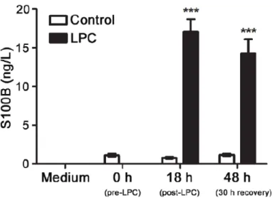
Recently, our group showed that S100B is highly released upon LPC-induced demyelination in COSC (Figure I. 4), mostly by astrocytes, in parallel to a massive gliosis (Barateiro et al., 2015).
Figure I. 4. S100B is markedly released upon LPC-induced demyelination of cerebellar organotypic slice cultures.
Cerebellar organotypic slice cultures were exposed to LPC at 7 days in vitro during 18 h and allowed to recover for 30 h. Samples for detection of S100B secretion were collected before the incubation (0 h), at 18 h post-incubation with LPC and at 48 h, i.e. after 30 h of recovery. Results are mean ± SEM. ***p<0.001 vs. Control. (Adapted from Barateiro et al., 2015).
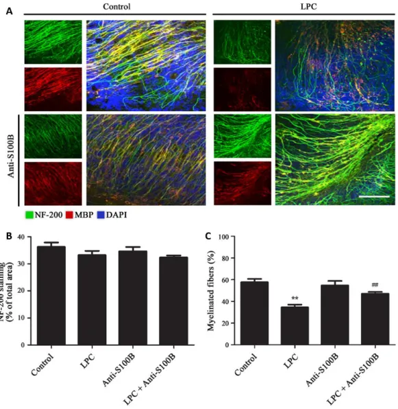
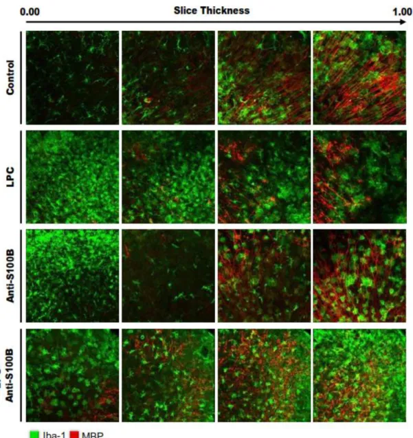
Curiously, when S100B was therapeutically neutralized using a specific antibody, we could observe a reduced demyelination (Figure I. 5), as well as reduced astrogliosis. Nevertheless, although the density of microglia in the slice showed no apparent differences upon anti-S100B co-treatment, the release of cytokines was markedly reduced suggesting a potential inhibition of microglia pro-inflammatory response (Barateiro et al., 2015).