ISOLATION AND CHARACTERIZATION OF MILK COAGULATING GASTRIC
ENZYMES FROM BIGEYE TUNA
(THUNNUS OBESUS)
JOAQUIM F. DA PONTE TAVARES
&J O S ~
A.
BETTENCOURT BAPTISTFA
TAVARES, JOAQUIM F. PONTE & JOSE A. BETTENCOURT BAPTISTA 1996. Isolation and characterization o f milk coagulating gastric enzymes f r o m bigeye tuna
( T h ~ n n u x o h e r a r ) ArquipPingo. L i f e and M a r i n e Sciences 14A: 75-84. P o n t a Delgada. I S S N 0873-4704.
Six proteolytic active fractions were separated from bigeye tuna stomachs by preparative
IEF rotofor, and purified by gel permcation and ion-exchange chromatography. The prescncc of NaCl in thc extraction process markedly enhanced the yield of tuna gastric enzyme. Molecular wcight of the most active pure fraction by SDS-PAGE corresponded to a 31.000 daltons. The amino-acid composition of thc same fraction differed slightly from literature values. and its themostability determined by DSC, indicated peak denaturation temperature of 61.85" C. The tuna protease activity at 14-26" C was Icss tempcraturc depcndent than commercial rcnnet and completely lcst the activity above 40" C of temperature. TAVARES, JOAQUIM F. PONTE & JOSE A. BETTENCOURT BAPTISTA 1996.
Isolamento e caracterizaqZo d e enzimas g i s t r i c o s coaguladores d e leite extraidos d e patudo (Thlrnnus obesus). ArquipPlago, CiCncias Bioldgicas e M a r i n h a s 14A:
75-84. P o n t a Delgada. I S S N 0873-4704.
Seis fracq6es proteoliticas activas de estdmagos de atum foram separadas com o recurso ao "Rotofor IEF" e purificadas pelas ticnicas cromatogrificas dc permeaqjio dc gcl c dc permuta ibnica. A utilizaqjio de NaCl aumcntou o rendimcnto do process0 dc cxtrac@o. 0 peso molecular da fracqjio pura mais activa, detcrminado pela tCcnica "SDS-PAGE". foi dc 31.000 daltons. A composi~jio amino-hcida obtida da mesma fracqjio diferiu da referidn na litcratura e a sua termocstabilidade, dctcrminada por "DSC", evidenciou urn pico d e degradaqjio h tcmperatura de 61.85 "C. A actividade da protease extraida dc estbmagos de atum. i s tcrnpcraturas dc 14-26 "C, mostrou-se menos dependente da temperatura do que a d o "rcnnct" comercial e pcrdcu toda a sua actividade acima de 4 0 "C.
Jonquim F. Ponre Tnvnres, Unii~ersihcle (10s A ~ o r e s , Rua rln iMc7e rle Deus, 5S, PT-9500 Potltu Delgadn. A ~ o r e s , Porlugnl. - Jose' A. Retrer~coccri Bnpiism, Depnri~~zeni o f M o l e c ~ d n r and Medical Genetics, Universiiy of Toronto, Toronto, Ontario M5S IA8, Cnrlada. Presem address: Universidnde dos A ~ o r e s , Run dn Mrle de Derrs, 58, PT-9500 Ponin Delgndn,
I N T R O D U C T I O N
T h e increased production o f fish offal in the fish industries presents a growing pollution and disposal problem, and consequently, t h e major concerns o f t h e world leading fish processing and exporting countries, i s the management and d e v e l o p m e n t o f n e w products using underutilized fish. T h e r e h a s also been a n increased interest in
t h e search for rennet substitutes. a s genetically engineered recombinant enzymes, b e c a u s e o f a decline in t h e n u m b e r o f calves slaughtered and a n increase in d e m a n d o f proteases f o r c h e e s e production (DE-KONING e t al. 1978). R e n n e t substitutes o f microbial origin h a v e been accepted b y the industry, although they h a v e n o t been totally satisfactory (ERNSTROM 1974). and plant proteases generally a p p e a r to give rise t o a softer
curd than calf rennet (OOSTHUIZEN & SCOTT- BLAIR 1963). Another alternative could be the extraction of milk clotting enzymes from fish stomach mucosa. In addition, the potential exists for establishing a new local food-related industry, which could be considered either, in less developed countries, as an alternative to produce rennet substitutes for domestic use, or eventually, to export for cheese-producing nations. At the same time the utilization of a waste material to produce a value-added product could likely increased overall fish processing. Availability of an inexpensive rennet would encourage cheese manufacturers to convert milk into a more stable and preserved food product (EDELSTEN & JENSEN 1970; GREEN & STACKOPOOLE 1975). The purpose of the present study was to isolate and characterize the major active protease from bigeye tuna.
MATERIALS
Spray dried non-fat dry milk was purchased from new Dundee Creamery, New Dundee, Ontario, Canada. Commercial rennet was provided by Dairy and Food Laboratories (Waukesha, Wiscosin). Protease substrate gel tablets, chemicals for isoelectric focusing (Bio-Lyte 3/10 ampholytes) and molecular weight calibration standards for SDS gel electrophoresis, were provided by Bio-Rad, Richmond CA. DEAE- Sephadex A-50 and Sephadex G-100 were obtained from Pharmacia. MW 12,000-14,000 cut-off tubing of 45 mm diameter (spectralpor standard cellulose tubing) was purchased from Spectrum Medical Industries, California. Unless otherwise specified, all reagent grade chemicals were obtained from E. Merck, Darmstadt, Germany, and Sigma Chemicals Company, St. Louis, MO, USA.
Bigeye tuna (Thunnus obesus, 30-50 kgtfish), caught from fishery zones in the North Atlantic Ocean in proximity of the Azores Islands, was obtained from the University of the Azores. The stomachs were removed from the fish after it was caught, frozen rapidly, and stored at - 20' C.
METHODS
EXTRACTION OF TUNA FISH PEPSINOGENS The stomachs, were partially thawed, split, cleaned, and briefly rinsed 3 times in tap water. The inner mucosa linings were peeled away from outer muscular layer and chopped in small pieces, using a sharp knife. The material was homogenised, using a combination meat grinder~waring commercial blender, with equal weight of distilled water (DW), containing 25% W/W of total weight (tissue plus DW) of NaCl, and maintained at 4O C. On the following day, the homogenate was stirred for 1 min. at O°C, using a Polytron tissue homogenizer, and the solid material was removed by centrifugation, utilizing a Beckman L8-70 ultracentrifuge unit at 35,000 x g, for 60 minures at 4" C. The pellet was twice more extracted as described above and the clear supernatants, called "crude pepsinogen", were combined, concentrated approximately 10 fold, using a Biichi rota-evaporator at room temperature or partially freeze dried in a Labconco lyophilizer, and stored at -20" C for further purification.
ISOELECTRIC FOCUSING AND ELECTROPHORESIS The crude pepsinogen was exhaustively, about six times, dialysed (spectrolpor standard cellulose membrane tubing of 45 mm i.d., MW cut-off 12,000-14,000) against distilled water, for 48 hrs. at 4" C, to remove the substantial amount of NaCl. This procedure insures that the nominal pH range of the ampholyte will extend over the full length of the focusing chamber, and then the maximum voltage can be applied. A narrow range ampholyte (bio-Lyte 3/10, Bio-Rad, Richmond, CA) was mixed with desalted tuna crude pepsinogen to a final concentration of 2% (wlv). The sample volume (50 ml) was fractionated using a Bio-Rad preparative IEF rotofor apparatus, originally designed by EGAN et al. (1984) and adapted by Bio-Rad Laboratories, equipped with Bio-Rad computer in order to control the electrophoresis
power of 12 watts. Tlie temperature
of
the system was maintained at 4" C with a circulating wntcr bath. The differe~~t enzymes from c r d e pepsinogen werc se:>arated according ta( . l ~
.isoePectric point (Ip), in :I reproducibhc .pII
gradient. However, runpholytes can intc~,felz w i h some assays, amino acids analyses, or subsequent purifications steps. I n order to rernovc ~ \ i e arnpholytes, each one of the most active fractions was again dialysed into a high salt solution, such as 1 M NaCl to strip electros!atically ljound
ampholytes from enzylncs by icn exchanil,i:, ;!nd
then against distilled water prior to lyophi1iza:ion. The molecular weight components of the active fractions, obtained ii-on1 rotofor, '~vere estimated by polyacrylaniidc gel clectrophor~esis, in presence of 2.5% of sodiunl L ' S ~ C C Y I sulphate and 5% 2.-mercaptoethanol, by compnrisora w i ~ h a calibration mixture, according to die PhastS).stetn Development Technique (Pharnncia). Electrophoresis was done wi!h Fttast gel graclicnt 8-25 using phast gels buffer strips. Illotccular weight standards for SDS-PAGE gel were: phosphorilase b (91,000), ovalbumin (67,000). carbonic anhydride (31,000), soybean trypin inhibitor (20,100), and a-lactalbumin (11,400). After electropl~oresis, gels werc fixed with 12.5% trichloroacetic acid for 15 nlin. and stained with Coomassiz brilliant blue G250 for 1 11 (DIIzeL et al. 1972). The molecular \veiglits w c x also confirmed by a Becklnan I-IPLC System, model 324 on a (60 cm x 7.5 nlm i.d.) Syherosel TSK 3000 SW column, equilibrated and e!uted with 0.15 M KH2P04
+
0.15RI
Na2S04, 9.t pH 5.0, versus molecular weight standards.ASSAY OF PKOTEOLYTIC ACTIVITY
T o determine the total activity of the tuna protease extracts, the zynlogen was activated prior to use, by adding 0.1 N I-ICI to pH 3.0-4.0, holding this pH at room temperature for 60 minutes, and readjusting the pH to 5.0 with 0.1 N NaOH (BREWER et nl. 1984).
The activity of the bigeye tuna protease was assayed by the measurement of milk coagulation time versus a known liquid standard rennet of 230 IMCUIml (20% chymosin, 80% bovinc pepsin in
:I solution c f 16% NaCl and 0.59'0 sodium
bemonte), usiog a Nalneter Viscon~eter (Nameter Co, Edison b?.J.), equipped with 1BM compatible conip!lter v i ~ an analog-to-digital converter, which nieasuxs the increasing of milk viscosity due to clot fmnmion. Fresh skim milk powder (97% T.S.) :d pII 6.3, was reconstituted to 12%
( \ ~ / v ) in 10 IPM CaC12 (BEKRIDGE 1952, 1955), and stored ovtmight at 4" C. This substrate was walmed and maintained, at coagulation temperature of 30" C. for one hour prior to tuna enzyme extirct or standard rennet addition (suitably dilcted in order to produce visible clotting i l l a:)prosirmtely 6 minutes). The milk
clotting t i n e was also measured, using the apparatus demibed by SOILIMER & MATSEN. (1935), a r J ~nodifiell by RAND & ERNSTROM (1961.). TI.$ protzass act~vity, dete~inined in MClJIml, \vss carried out at 30° C in 25 ml portions o l substrate, in wide mouthed bottles. One ml oq cliluted enzyme was added, and the clotting ti:w, in minutes, was measured by the number of revolutions (20 rpm). The appearance of the 1'11st \isible flakes of curd on the moving glass surS:ice was taken as the end point of the dcterrni~~:!tisn. All the assays were performed in triplicate in:!uding the blanks.
Another method used to evaluate the proteolytic activity of tuna gastric enzymes was the Bio-Rr:i'h protease substrate gel tablets, that provided i~ r q i d and a convenient process for
plcparing agar dil'fi~sion plates. The tablets produce a I % agar gel containing n bovine casein preparation ill a Tris buffered physiological saline solution at pH 7.2. Protease diffusion into the substrate gcl was accompanied by digestion of the casein, and t l ~ c s i x of the transparent ring around the samples wells in a turbid gel measure the enzyme proteolytic activity.
G E L PERMEATION CHROMATOGKAPIIY
The most x t i v e fractions, determined by Bio- Rad's protcase substrate gel tablets, from crude pepsinogen rotofor separation, were combined,
and furth.;.; purified by gel permeation cliroiiiatography, on a Sephadex G-100 (30 cm x 15 nu11 id.) column, from Pharmacia, eluted with
50 rnM Tris-Chloride buffer, at pH 7.0. The sample, dissolved in 3 ml of the eluent buffer, was loaded on the column, after its equilibration, and the effluent was collected every 5 minutes. Elution was carried out, using a Isco peristaltic pump at a flow rate of 0.5 mllmin and the detection was accomplished with an optical unit model UA-5 (280 nm) recording absorbance monitor (1x0, Lincoln, NE). Alternate fractions were assayed for proteolytic activity and the pooled active fractions were combined, immediately dialysed against distilled water and lyophilized.
Ion-exchange technique take advantage of the anionic properties of aspartic proteases. The combined fractions from gel permeation chromatography were repurified on (30 cm x 18 mm i.d.) DEAE-Sephadex A-50 column, from Pharmacia, pre-equilibrated with 20 mM Tris- Chloride buffer at pH 7.0. After sample loading, the column was eluted at a rate of 0.5 mllmin with the starting buffer, followed by a step gradient with 0.25 M NaCl in 20 rnM Tris-Chloride buffer, pH 7.0, as an eluent. A second step gadient, was finally applied using 0.50
M
NaCl in 20 mM Tris- Chloride buffer, until the end of the separation, maintaining the same pH. Fractions were collected every 15 minutes and the estimation of the enzyme was determined according to ANSON (1938) by the absorbance at 235 and 280 nms, using a Shimadzu UV-VIS spectrophotometer, model 260. The pooled material was assayed for proteolytic activity, and the active fractions were combined, dialysed, and lyophilized. The final purification was performed by high performance liquid chromatography, with a HEMA BIO- DEAE column (10 cm x 8 mm i.d.) using a linear gradient of 0-100% B in 30 minutes between A = 20 mM TrisICl (pH 7.0) and B = A+
0.5 N NaCl (pH 7.0). at a flow rate of 0.5 mllmin. The purity of the collected material was confirmed by diode array UV detector, comparing the UV-profile, at the upslope, top and downslope of the peak.The amino acid composition of the purified tuna protease fraction after hydrolysis, were determined using the Beckman DABS Amino Acid Analysis Kit, according to NOLAN
Rc
DOVlCHr (1 987). This methodology is applied for both primary and secondary amino acids, using 4-
dimethylaminoazobenzene-4-sulfonyl (dabsyl) chloride (2 mglml, in acetone) in the presence of 0.5 ml of 0.5 M sodium bicarbonate (final pH adjusted to 8.5-9.0). The kit included an amino acid calibration standards and the analysis were carried out in a Beckman HPLC system, model 324. An Ultrasphere C18 column (250
x
4.6mm i.d.) was used with a gradient elution, according to Beckrnan DABS-Amino Acid Kit, between (A)10 mM citric acid and 4% dimethylformamide (DMF), pH 6.50 +I- 0.05 and (B) 300 ml of "A" plus 700 ml Acetonitrile-with 4% DMF, with detection at 436 nm.
RESULTS AND DISCUSSION
The yield of extraction, increased with increasing amount of sodium chloride from 15% to 25% (wlw). According to
TAVARES
et al. (in press), salt concentration of15%
(wlw) slightly accelerated activation, whereas of 25% (wlw) markedly enhanced the yield of tuna protease, compared with 0.20 M sodium phosphate at pH 7.2 (BREWER et al. 1984). An extraction of 25% (wlw) NaCl at pH 5.0, from a tuna stomach of 343 g produced a total recovered yield of 482 MCU.Separations, based on the isoelectric properties, have been successful in obtaining active aspartic proteases from several sources (EGAN et al. 1984). The method can be used, as long as the enzyme is both stable and soluble at its isoelectric point. The bioactivity, after the
separation, was maintained, possibly because the proteases were in solution at low temperature (4" C), probably in their native conformation, and organic solvents were not used. The fraction from the first IEF rotofor separation showing the highest activities, in Bio-Rad protcase substrate tablets, within pH range of 3.94 to 6.50, were combined to be reapplied to the rotofor cell for further separation.
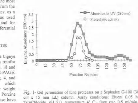
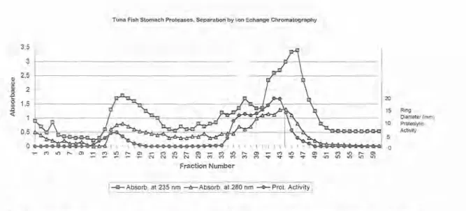
The most active fractions (# 18 and # 19) from the second crude pepsinogen IEF rotofor separation, were combined and applied to a gel permeation column. The eluted fractions, numbered 50 to 90, containing the protease free of small contaminant molecules, were pooled dialysed and lyophilized, (Fig. I). The resulting enzymatic material were repurified by ion- exchange chromatography using the starting buffer until fraction # 23, followed by a step gradient until fraction # 34 and a final step gradient until the end of the separation, fraction #
52. The most active fraction emerged as a medium peak with its centre at fraction # 41. A small enzyme peak (or shoulder), containing potential proteolytic activity, preceeded the main peak, and emerged from the fractions #s 35-39 (Fig. 2). The combined material, was submitted to
The molecular weights determined by HPLC
-
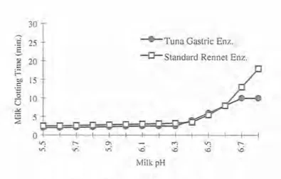
size-exclusion chromatography were higher than those determined by SDS-PAGE. Long rod- shaped molecules can show this effect to difficulty of the molecules in penetrating the pores of the gel structure packing. A similar difference between molecular weights determined by SDS-PAGE and gel filtration was also found with gastric protease from seal (SHAMSUZZAMAN & HAARD 1984). COMPARISON OF MILK CLOTTING ENZYMES The effects of proteolytic activity of tuna gastric and commercial rennet enzymes on milk clotting were measured at different pHs by relative clotting time. The milk coagulation time for both had a pH dependence profile similar for pH values ranging 5.5-6.3, but the first one was less sensitive to losses of activity above pH 6.4 (Fig. 5).The amino acid composition of the most active pure fraction, isolated from tuna gastric proteases, summarized in Table 1, was compared with data published by TANJI et al. (1988). Bigeye tuna HPLC final purification, and the most
active fraction that emerged from the column between 18-24 minutes, as a single symnletrical peak, was used for amino-acid composition and for thermostability studies by differential scanning calorimeter (Fig. 3).
MOLECULAR WEIGHT ESTIMATES
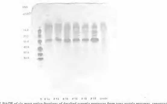
The molecular weights of the bigeye tuna prolease fractions, from rotofor separations (#s 13, 14, 15, 16, 18 and 19), were estimated by SDS-PAGE. The results are shown in Fig. 4, and exhibited a strong band, which corresponds to a molecular weight close to 3 1,000 daltons. Porcine pepsin and cod gastric protease have molecular weights slightly higher (SQUIRES et al. 1985).
+ ~ b s o r t i o n in U V (280 nrn) Proteolytic activity
h
Fraction NumberFig. 1- Gel permeation of tuna proteases on a Sephadex 6-100 (30 crn x 15 mm i.d.) column. Assay conditions: Eluent 0.05 M TrisIChloride, pH 7.0, temperature 4" C., flow rate 0.5 mllmin., detection by UV (280 nrn).
Tuna Fish Stomach Proteases. Separation by ILon Echange Chromatography
Ring
Dlameter (mm)
Proteolytic Activity
F A b s o r b . at 235 nrn -&-Absorbb at 280 nrn -0-Prot. Activity]
Fig. 2- Ion-exchange chromatography of tuna proteases on a DEAE cellulose (30 cm x I 8 mm i.d.) column. Assay conditions: 20 mM TrisIChloride, pH 7.0 until fraction # 23. Step gradient with 0.25 M NaCl in 20 mM TrisIChloride, pH 7.0 until fraction # 34 and a final step gradient with 0.5 M NaCl in 20 m M TrisIChloride, pH 7.0 until the end (fraction # 52). Flow rate 0.5 mllmin., and detection by UV (280 nm).
differred slightly somewhat in containing less serine, cysteine, isoleucine, and more basic amino acids, lysine and arginine, possibly
because of different environments
-
E 0.8 were the fish was caught. The amino sacid composition of the various o 00 proteases were also compared by 0.6 SQUIRES et al. (1985), that found Q) o
similar differences between gastric
f
0.4 proteases from the same species.g
40
THERMOSTABILLITY
2
0.2Recent studies, on the circular 0.0
dichroism (CD) spectra of enzymes, 0 5 10 I 5 20 25 30 Time(min.)
have born out the conclusions that
the enzymatic properties, such as Fig. 3 HPLC-IEC of tuna protease fraction on HEMA BIO-DEAE (1 0 thermostability and specificity, are cm x 8 mm i.d.) column. Assay conditions: Mobile phase: A = 20 mM determined to a large extent by the TrisICl (pH 7.0) and B = A
+
0.5 N NaCl (pH 7.0). Linear gradient primary structure of the particular elution of 0-100% B in 30 minutes. Flow rate 0.5 rnl/min., andenzyme. detection by UV (280 nm).
The influence of assay temperature on clotting One approach to study kinetic stability of the time is summarized in Fig. 6. Data shows that in aspartic proteases, is a technique called low temperature renneting milk (14-26" C) for differential scanning calorimeter (DSC). It is used cheese making, less tuna protease would be to measure the temperature and heat flow required to facilitate renneting than calf rennet. associated with transitions in materials, as a
s # 1 3 # ? 6 6 15 # . I 6 # 18 # 19 m u d e
Fig. 4- SDS-PAGE of six most active fractions of desalted aspartic proteases from tuna gastric enzymes, separated using IEF with a range 3-10 ampholyte. The gel was loaded with 0.5 pl of the sample quantity (3 mgtrnl, dry weight). Lane "S" contained low molecular weight standards: alpha-lactalbumin (14,400), trypsin inhibitor
(20,100), carbonic anhydride (31,000), ovalbumin (67,000) and phosphorilase-b (94,000). Stained with coomassie blue.
Table 1
Amino Acid composition of the tuna gastric protease fraction compared to data published in literature (TANJI
et a]. 1988).
Tuna Mole % Mole %
protease (Experiment) (Literature values)
ASP 10.7 8.55-12.62 Thr 6.7 5.10-8.06 Ser 7.2 9.10-13.85 Glu 11.2 6.39-12.83 Pro 3.6 3.60-5.28 GIY 10.7 8.90-1 1.73 Ala 9.9 4.78-12.50 CYS 0.0 0.56-1.97 V a1 6.2 6.25-10.06 Met 1.9 1.11-2.57 Ile 3.9 4.28-7.08 Leu 7.3 4.44-9.78 TYr 2.6 3.07-6.30 Phe 3.5 3.89-6.25 His 2.4 0.3 1-2.50 LYS 6.7 0.3 1-3.06 Arg 5.7 0.62-3.90
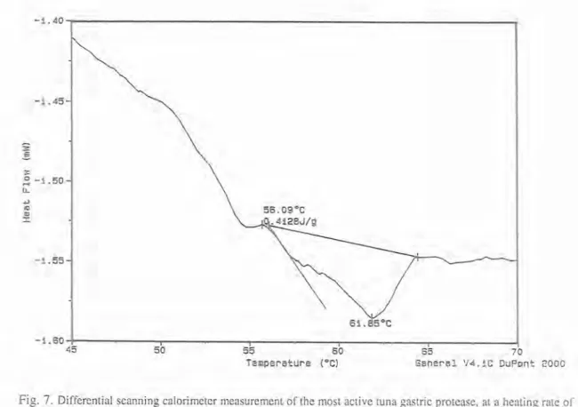
function of time and temperature. Such measurements provide quantitative and qualitative information about physical and chemical changes, that involve endothermic or exothermic processes, or changes in heat capacity. Using this methodology the purified tuna protease was dialysed, liophylized and diluted to a concentration of 10 mg/ml. Sample volume of 10 yl was hermetically sealed in aluminun pan, and 10 yl of distilled water was sealed in the reference pan. The activation energy of denaturation was generated using the Arrhenius expression. A sigmoidal baseline option was used, since a substantial change in heat capacity occurred during the process. DSC thermogram of the most active tuna gastric enzyme fraction, indicates peak denaturation temperature of 61.85" C, and the activation energy of denaturation was 0.4128 Jlg, (Fig. 7).
CONCLUSIONS
1. Tuna gastric enzyme induced clotting of milk, via initial limited proteolysis of one of the casein
- b ~ u n a Gastric Enz. *Standard Rennet Enz.
Fig. 5- Comparison of milk clotting activity of tuna protease relative to commercial rennet at different pH values. Assays conditions were 12% milk solids. 10 rnM CaCl,, and incubation temperature of 30°C. Data are means of triplicate determinations.
-
Standard Rennet Enz.I
-
Tuna Gastric Protease/
Milk Temperatura ( C )
Fig. 6- The influence of assay temperature on the clotting time. Assay conditions were
15%
milk solids. 10 mM CaC12. pH 6.3. and incubation temperature in the range 14-50" C. Data are means of triplicate determinations.fractions (k-casein), and subsequent aggregation of casein micelles.
2. It is our belief that there exists an enormous potential for the recovery and use of gastric proteases from bigeye tuna fish. In spite of the potential diversity of microbial enzymes, very few species are used to produce industrial enzyme, because the cost of getting a microorganism stringently evaluated and accepted as safe is substantial.
3. The finding that bigeye tuna protease can more efficiently clot milk at low temperature, indicates its usefulness in cold renneting milk.
4. Bigeye tuna protease and commercial rennet where similarly effective at mild temperatures, but the former enzyme can be inactivated above 40" C. This phenomenon represents an advantage in some experiments (e.g., prevention of oxidised flavour in milk), since residual enzyme can be eliminate by pasteurization, and would not be
Fig. 7. Differential scanning calorimeter measurement of the most active tuna gastric protcase, at a heating rate of
5" C/min.. from 45 to 70°C. Trace was average of three scans.
present to cause subsequent hydrolysis of milk proteins. Thermostability studies, lend support to the theory of the evolutionary adaptation t o various thermal environments. Generally with the enzymes, there is a direct relationship between the habitat temperature of the organism, from which the enzyme is derived, and its thermal stability.
5. T h e milk pH influence on the milk coagulation by tuna gastric enzyme had a p H dependence profile similar to that of calf rennet for p H values, ranging 5.5-6.3, but the first.,,. one was less sensitive to losses of activity above pH 6.4.
6. T h e colby Cheese manufactured with bigeye tuna fish protease was tested by a sensory panel constituted by 100 consumers. T h e data evaluated by analysis of variance was already published by
TAVARES e t al. (1993).
REFERENCES
ANSON, M.L. 1938. The estimation of pepsin. tripsyn. papain and cathepsin with haemoglobin. Jour~zal of General Plzysiology 22: 1-79.
BERRIDGE, N.J. 1952. An improved method of observing the clotting of milk containing rennin.
Jour17al of Daity Research 19: 328-329.
BERRIDGE, N.J. 1955. Purification and assay of rennin.
Methods in Enzynrology 2:69-77.
BREWER, P.. N. HELBIG & N.F. HAARD 1984. Atlantic Cod Pepsins - Characterization and use as a Rennct Substitute. Canadian Institute. Jo~tr~znl of Food Science Technology 17: 38-43.
DE-KONNING. P.J., P.J. VAN ROOIJEN & S. VISSER 1978. Application of a syntetic hexapeptide as a standard substrate for the determination of the activity of chymosin. Netherland Milk Dair?, Journal 32: 232-244.
DIEZEL, N., G. KIPPERSCHLAGGER & E. HOFFMAN 1972. An improved procedure for protein staining in polyacrylamide gels with a new type of Coomassie brilliant blue. Analytical Biochemistry 48: 617-620. EDELSTEN, D. & J.S. JENSEN 1970. Paper presented at 18th International Dairy Congress. Sydney I. E., 280pp.
EGAN, N.B., W. THORMANN, G.E. T ? ~ T Y & M. BIER 1984. Separation of ampholytes from focused proteins by rotofor cell. Electrophoresis 83: 547-550. ERNSTROM, C.A. 1974. Milk clotting enzymes and cheese chemistry. In WEBB B.H., A.H. JOHNSON, & J. ALFORD (Eds). Fundamentals of Dairy Chemistry. A VI Publication, Westport, CI. U.S.A. GREEN, M. L. & A. STACKOPOOLE 1975. The
preparation and assessment of a suitable Mucor pusillus Lindt proteinase swine pepsin mixture for Cheddar cheese-making. Journal of Dairy Research. 42: 297-312.
NOLAN, T.G. & N.J. DOVICHI 1987. HPLC analysis of amino acids dabsyl chloride derivatives. Analytical chemistry 59: 2803-2805.
OOSTHUIZEN, J.C. & G.W. SCOTT-BLAIR 1963. Journal of Dairy Research 30: 197-198.
RAND, A.G.Jr. & C.A. ERNSTROM 1964. Effect of pH
and Sodium Chloride on Activation of Prorennin. Journal of Dairy Science 47: 1 1.
SHAMSUZZAMAN, K. & N.F. HAARD 1984. Canadian. Journal of Biochemistry 62: 699-708.
SQUIRES, E.J., N.F. HAARD & A.W. FELTHAM 1985. Gastric Proteases of Greenland Cod Gadus ogac. I1 Strutural Properties. Biochemical Cell Biology 64: 215-222.
SOMMER, H.H. & H. MATSEN 1935. The relation of mastitis to rennet coagulability and curd strength of milk. Journal of Dairy Science 18: 741.
TANJI, M., T. KAGEYAMAT & K. TAKAHASHI 1988. Tuna Amino Acids Squences. Depart. of Biophysics and Biochemistry, Faculty of Sciences, University of Tokyo, Tokyo 113 Japan.
TAVARES, J.F.P., A. HILL, R. YADA& E. GULLET 1993. Characterization of colby cheese made with fish enzymes. Arquipklago. Life and Marine Sciences
1 1 : 65-72.
TAVARES, J.F.P..J.A.B. BAPTISTA & M.F. MARCONI Iin press. Milk coagulating enzymes of tunafish waste as Rennet substitute. Journal of Food Science and Nutrition 48.