Potential Carrier for Drug Delivery into Melanoma
Metastatic Cells
Domenica Capasso1., Ivan de Paola2., Annamaria Liguoro3., Annarita Del Gatto2
, Sonia Di Gaetano2, Daniela Guarnieri4, Michele Saviano5, Laura Zaccaro2*
1Department of Pharmacy, University of Naples ‘‘Federico II’’, Naples, Italy,2Institute of Biostructures and Bioimaging -CNR, Naples, Italy,3Diagnostic and Molecular Pharmaceutics -Scarl, Naples, Italy,4Center for Advanced Biomaterials for Health Care @ CRIB- Italian Institute of Technology, Naples, Italy,5Institute of Crystallography -CNR, Bari, Italy
Abstract
avb3 integrin is an important tumor marker widely expressed on the surface of cancer cells. Recently, we reported some biological features of RGDechi-hCit, anavb3 selective peptide antagonist. In the present work, we mainly investigated the pro-apoptotic activity of the molecule and its ability to penetrate the membrane of WM266 cells, human malignant melanoma cells expressing high levels ofavb3 integrin. For the first time we demonstrated the pro-apoptotic effect and the ability of RGDechi-hCit to enter into cell overexpressing avb3 integrin mainly by clathrin- and caveolin-mediated endocytosis. Furthermore, we deepened and confirmed the selectivity, anti-adhesion, and anti-proliferative features of the peptide. Altogether these experiments give insight into the biological behavior of RGDechi-hCit and have important implications for the employment of the peptide as a new selective carrier to deliver drugs into the cell and as a therapeutic and diagnostic tool for metastatic melanoma. Moreover, since the peptide shows a pro-apoptotic effect, a great perspective could be the development of a new class of selective systems containing RGDechi-hCit and pro-apoptotic molecules or other therapeutic agents to attain a synergic action.
Citation:Capasso D, de Paola I, Liguoro A, Del Gatto A, Di Gaetano S, et al. (2014) RGDechi-hCit:avb3 Selective Pro-Apoptotic Peptide as Potential Carrier for Drug Delivery into Melanoma Metastatic Cells. PLoS ONE 9(9): e106441. doi:10.1371/journal.pone.0106441
Editor:A Ganesan, University of East Anglia, United Kingdom
ReceivedMay 8, 2014;AcceptedJuly 30, 2014;PublishedSeptember 23, 2014
Copyright:ß2014 Capasso et al. This is an open-access article distributed under the terms of the Creative Commons Attribution License, which permits unrestricted use, distribution, and reproduction in any medium, provided the original author and source are credited.
Data Availability:The authors confirm that all data underlying the findings are fully available without restriction. All relevant data are within the paper and its Supporting Information files.
Funding:This study was supported by FIRB MIUR "RENAME" 2010-RBAP114AMK and Programma Operativo Nazionale Ricerca e Competitivita` 2007-2013 PON 01_02388. The funders had no role in study design, data collection and analysis, decision to publish, or preparation of the manuscript.
Competing Interests:The authors have declared that no competing interests exist. * Email: lzaccaro@unina.it
.These authors equally contributed to this work.
Introduction
The avb3 receptor is a member of the integrin family, heterodimeric membrane glycoproteins, with a prominent role in angiogenesis and metastatic dissemination [1,2]. Interaction between avb3 integrin and the extracellular matrix (ECM) proteins has been identified as the most important survival system for nascent vessels, by controlling different cellular functions, including survival, proliferation, migration and apoptosis [3,4]. Since this integrin is expressed at high levels on the surface of many cancer cells [5,6,7] as well as tumor-associated endothelial cells [8], it has become an important target in the development of new anticancer strategies.
Integrinavb3 performs its function by interacting with several ECM proteins containing the RGD motif, recognized by membrane-bound adhesion molecules, playing a key role as cell adhesion mediator [4]. Peptides containing this motif show potent anti-adhesion effects, since they compete for the integrin-matrix interaction and show anti-proliferative, antichemotactic and pro-apoptotic effects [9,10].
In the last twenty years, a large number of avb3 antagonists, including antibodies, small molecules, peptidomimetics, and cyclic RGD peptides, have been developed with the aim of selectively inhibitingavb3-mediated processes [11,12,13]. Most importantly, Kessler and colleagues in 1996 reported the development of cRGDf[N(Me)]V, anavb3/avb5 antagonist known as Cilengitide [14] that was in phase III clinical study as anti-angiogenic drug for glioblastoma therapy [15,16,17,18]. Unfortunately, very recently (News/Press release from Merck, February 25, 2013) it was announced that the Phase III trial of the investigational integrin inhibitor Cilengitide did not meet its primary endpoint of significantly increasing overall survival when added to the current standard chemoradiotherapy regimen (temozolomide and radio-therapy). Furthermore, neither Cilengitide nor all known antag-onists are able to discriminate betweenavb3 and other type of integrins.
fundamental feature for the design of new systems with reduced side effects and dosage. In agreement with in vitro findings, imaging studies on human glioblastoma U87MG also indicated that RGDechi-hCit allows selective visualization ofavb3 expres-sion [20]. Furthermore, very recently we showed the ability of RGDechi-hCit to significantly inhibit some intracellular pathways acting as an avb3 integrin inhibitor, and its role as an antiangiogenic agentin vitroand invivo[21].
In melanomas, a number of studies have shown malignant transformation to be associated with a general up-regulation of cell adhesion molecules [22,23]. In particular, it is well-known that over-expression of avb3 heterodimer is often associated in melanoma with the transition to a metastatic phenotype [24,25]. Indeed, while in normal epithelial cells the avb3 heterodimer is expressed at very low levels or is even absent, it is highly over-expressed in MM (metastatic melanoma) cells, and this increased expression is correlated with the degree of tumor progression.
Importantly, MM is highly resistant to conventional radio and chemotherapies and remains a disease of poor prognosis, with a survival times of a few months. Different chemotherapeutic agents, alone or in combination, have been employed over the last few years resulting, unfortunately, in limited activity with relatively low response rates [26,27], and providing so far only a small impact on overall survival. As chemoresistance remains a serious concern for melanoma therapy, a great challenge is the development of molecules, to use alone or in combination with other drugs, having the ability to interfere with systems involved in angiogenesis and metastatic dissemination processes and so be remarkably effective in shrinking melanoma tumors.
Very recently [28], we evaluated thein vitroantitumor efficacy of RGDechi-hCit peptide on melanoma cell lines differently expressing avb3 integrin. The data obtained showed that RGDechi-hCit induces a significant inhibition of proliferation only on the WM266 cell line, in accordance with its very high surface expression ofavb3.
On the basis of these promising data and taking into account the key role played by integrinavb3 in melanoma progression, the aim of this paper was to thoroughly investigate the biological behavior of RGDechi-hCit on the WM266 metastatic cell line to reinforce its potential as an anticancer drug and as carrier for drug delivery. In particular, adhesion, binding, uptake, proliferation and apoptosis studies by flow cytometry and confocal microscopy were performed.
Materials and Methods
Peptide Synthesis, Cyclization and Labelling
Polypropylene reaction vessels and sintered polyethylene frits were supplied by Alltech Italy. MeIm, MSNT, TFA and scavengers were purchased from Fluka; NovaSyn TGA resins, coupling reagents and all amino acids were from Novabiochem. DIPEA was purchased from Romil; piperidine, PhSiH3 and
Pd(PPh3)4, NBD-Cl and FITC from Sigma-Aldrich.
RGDechi-hCit (c(KRDGe)MDDPGRNPHHocitGPAT-OH)
and the scrambled sequence (Ac-KPGRGHNDPDPGHocitDeM-HAT-OH) were synthesized in solid phase by Fmoc chemistry basically as previously reported [19], but introducing some synthetic improvements and a different Lys functionalisation procedure [29]. Briefly, assembly of fully protected peptides was carried out manually in a polypropylene reaction vessel fitted with a sintered polyethylene frit using NovasynTGA resin (0.21 mmol/ g) and all standard amino acids, except for D-Glu. The first amino acid was bound to the support by treatment with Fmoc-Thr(tbu)-OH (5 equiv)/MSNT (5 equiv)/MeIm (3.75 equiv) in DCM for
3 h. The Fmoc protecting group was removed by treatment with 30% piperidine in DMF (3610 min). Coupling reactions of the
following amino acids were performed by using 10 equiv of Fmoc protected derivative activated in situ with HBTU (9.8 equiv)/ HOBt (9.8 equiv)/DIPEA (20 equiv) or COMU or HATU (9.8 equiv)/DIPEA (20 equiv) in DMF for 30 min. The coupling efficiency was assessed by the Kaiser test. Each step was followed by resin washings (365 min). For RGDechi-hCit, before the final Fmoc deprotection, selectivea-carboxyl deprotection of the D-Glu residue from the allyl group was carried out by treatment of the peptidyl resin with PhSiH3(24 equiv)/Pd(PPh3)4(0.25 equiv) in
DCM (2630 min). The cyclisation between aNH of D-Glu and
aCO of Lys was performed with PyBop (1.5 equiv)/HOBt (1.5 equiv)/DIPEA (2 equiv) in DMF for 5 h at millimolar pseudo-dilution. For both peptides Fmoc-Lys(ivDde)-OH was employed as the last amino acid with the orthogonal ivDde protecting group in order to selectively remove it onto the resin and carry on the following functionalisation with the NBD-Cl (4-Chloro-7-nitro-1,2,3-benzoxadiazole) or FITC (fluorescein isothiocyanate) ivDde group which was removed from Lys by using a solution of 2% hydrazine in DMF (1062 min); coupling reactions were per-formed with 3 equiv of NBD-Cl or FITC and 6 equiv DIPEA in DMF overnight. Finally the peptides were cleaved off the resin and deprotected using a mixture of TFA/H2O/EDT/TIS
(94:2.5:2.5:1v/v/v/v) for 3 h. The resins were then filtered, and the white solid peptides were obtained by precipitation from cold anhydrous diethyl ether. The crude products were purified by preparative RP-HPLC on the Shimadzu LC-8A system, equipped with an UV-Vis detector SPD-10A using a Phenomenex Jupiter Proteo column (21.26250 mm; 4mm; 90 A˚ ) and a linear gradient
of H2O (0.1% TFA)/CH3CN (0.1% TFA) from 5 to 70% of
CH3CN (0.1% TFA) in 30 min at a flow rate of 20 mL/min. The
purified peptides were analysed and characterised by an ESI-LC-MS instrument (ThermoFinnigan) equipped with a diode array detector combined with an electrospray ion source and a quadrupole mass analyzer, using a Phenomenex Jupiter Proteo column (4.606150 mm; 4mm; 90 A˚ ) at a flow rate of 0.8 mL/min
and the same gradient used for the purification step.
Cell lines and culture conditions
Human adenocarcinoma cells line (HeLa) (ATCC U.S.) were grown in DMEM supplemented with 10% fetal bovine serum (FBS), 2 mM glutamine, 100 U/mL penicillin and 100mg/mL streptomycin (Euroclone, Italy). Human metastatic melanoma cells line (WM266), kindly provided by Dr. Gentilcore (IRCCS-Fondazione Pascale, Naples Italy), were grown in RPMI, supplemented with heat-inactivated 10% fetal bovin serum (FBS), 1% glutamine, 100 U/mL penicillin and 100mg/mL streptomycin. The cells were maintained in humidified air containing 5% CO2at 37uC.
FACS analysis foravb3 integrin
intensity were obtained from the histogram statistic of CellQuest software.
Cell adhesion and detachment assays
NUNC MaxiSorp 96 well plates (Dasit Sciences, Italy) were coated overnight at 4uC with vitronectin, fibronectin or collagen type I (Millipore, USA), at 10mg/mL concentration diluted in
PBS, pH 7.4; then a blocking step for nonspecific binding was performed with 1.5% BSA in PBS for 1 h at room temperature. WM266 cells were suspended and mixed in Hank’s balanced salt solution (HBSS: 50 mM HEPES, 1 mg/ml BSA, 1 mM CaCl2,
1 mM MgCl2, 1 mM MnCl2, pH 7.4, Sigma Aldrich, Italy) with
peptides (50mM) or anti-avb3 antibody (10mg/mL) for 30 min at 4uC and then plated on pre-coated plates (1.56104cells/well). Non
adherent cells were gently removed by repeated washings. After 1 h of incubation, the adherent cell number was evaluated by crystal violet (Sigma Aldrich, Italy) assay, which correlates optical density with cell number [30]. Briefly, cells were washed with PBS and fixed by adding 10% formalin solution. After 15 min cells were washed with deionized water and stained with 100ml of
0.1% crystal violet solution in water for 30 min. Excess dye was removed by washing with deionised water and plates were air-dried prior to bound dye solubilisation in 200mL of 10% acetic acid. The optical density of dye extracts was measured directly in plates at 595 nm (BioRad microplate reader model 680). The mean value 6 SE of adherent cells for each treatment was expressed as relative percentage of cell numbervscells not treated (control). Statistical differences were determined by Student’s t test, unpaired, two-sided. All experiments were performed in triplicate and repeated at least 3 times; a p value less than 0.05 was considered to be significant.
Cell proliferation assay and apoptosis
WM266 cells were seeded at 46103cells/mL in 96-well plates in RPMI 10% FCS. After 24 h the cells were starved (4% FCS in RPMI) for 4 h and incubated with increasing concentrations (10, 25 and 50mM) of peptides. The proliferation was evaluated after 24 h using crystal violet assay.
The apoptosis assay was analysed on WM266 cells seeded at 26105cells/well in a 6 well plate and starved as described above. Next, the cells were incubated at 37uC with different peptides at 50mM concentration, and apoptosis was analysed after 16 h by
staining with annexin V/FITC and Propidium iodine (PI) (eBioscience, USA). Briefly, after incubation, the untreated and treated cells were detached with Accutase solution (eBioscience), harvested and washed with cold PBS. Subsequently, the cells were treated following the manufacturer’ instructions. The percentage of cell undergoing apoptosis or necrosis was quantified using a flow cytometer (Becton Dickinson, USA) equipped with Cell Quest software.
Cell binding and uptake of peptides by FACS
WM266 cells were detached with 0.1 mM EDTA in PBS, washed and incubated with peptides. In detail, 2.56105cells were treated with 10mM NBD-labeled RGDechi-hCit or 10mM scrambled peptide, in 100mL of 20 mM CaCl2/HBSS at room
temperature in slow agitation for 30 min. Then, the cells were washed in 20 mM CaCl2/HBSS and resuspended in the same
buffer to perform FACS analysis. The NBD-labeled peptides were excited at 465 nm and fluorescence was measured at 530 nm. After the analysis, 5mL of dithionite (Sigma Aldrich, Italy) stock solution (1 M freshly prepared in 1 M Tris-HCl pH 10) was added to cells at 4uC for 5 min and the fluorescence of internalised peptides was detected by FACS.
Cellular uptake of peptides by confocal microscopy
WM266 and HeLa cells were seeded on a 35 mm diameter Fluorodish Cell Culture Dish (World Precision Instruments, Inc., Florida) at the concentration of about 56104cells/mL. 24 h after
seeding, cells were incubated with FITC-RGDechi-hCit and FITC-RGD scrambled at the final concentration of 50mM in HBSS buffer for 30 min at 37uC. Cells were rinsed twice with PBS to remove peptide excess and fixed with 4% paraformaldehyde at room temperature. Samples were then observed with a confocal microscope (Leica SP5) with a 636oil immersion objective with a
488 nm wavelength laser line and transmitted light.
Indirect immunofluorescence
For co- localization experiments, after peptide incubation, cells were first rinsed twice with PBS to remove non-internalised peptide and fixed with paraformaldehyde 4% for 20 min. The cells were washed for 10 min in 10 mM PBS/20 mM glycine, permeabilised with 10 mM PBS/20 mM glycine containing 0.005% saponin for 7 min, and blocked with PBS/20 mM glycine 1% albumin.avb3 integrin was performed by using mouse anti-human integrinavb3 primary antibodies (Chemicon) and Alexa-fluor 568 goat anti-mouse secondary antibodies (Molecular Probes, Invitrogen). Endocytic vesicles were localised by incubat-ing samples first with mouse anti-clathrin monoclonal (ABR) and rabbit anti-caveolin 1 (Abcam) primary antibodies and then with Alexa-fluor 568 goat anti-mouse and anti-rabbit secondary antibodies (Molecular Probes, Invitrogen), respectively. Lysosomes were localised with rabbit anti-LAMP 2 polyclonal (Abcam) primary antibodies and with 568 goat anti-rabbit secondary antibodies (Molecular Probes, Invitrogen).
All samples were then observed with a confocal microscope with a 636oil immersion objective. Co-compartimentalisation between
the peptide and the endocytic markers was analysed with ImageJ analysis software plugin.
Inhibition of clathrin- and caveolin-mediated endocytosis
Hypertonic challenge was carried out by incubating the WM266 cells with 0.45 M sucrose for 20 minutes at 37uC [31] in order to inhibit the clathrin-mediated endocytosis. Treatment aimed at inhibiting caveolae-mediated endocytosis was evaluated by incubating cells with filipin at 5mg/mL for 30 minutes at 37uC. After both treatments, cells were incubated with 50mM
RGDechi-hCit solution in HBSS buffer for 30 minutes at 37uC. Then, cells were washed twice with PBS. Some samples were fixed with paraformaldehyde for the confocal microscopy observations and other ones were lysed with 1% Triton 6100 in PBS for the
quantitative analysis. To this aim, the intensity fluorescence of cell lysates was measured at an excitation wavelength of 488 nm by a spectrofluorimeter (Perkinelmer). Results reported as percentage of cellular uptake were normalized with respect to non-treated control cells (expressed as 100%).
Results
Synthesis of functionalized RGDechi-hCit and scrambled peptides
D-Glu5 residue from the allyl group was carried out by the treatment of the peptidyl resin with a solution of PhSiH3/
Pd(PPh3)4in DCM [33]. The cyclisation between theaNH group
of Lys1 and the aCO group of D-Glu5 was performed with PyBop/HOBt/DIPEA [34] in DMF.
For both peptides, Fmoc-Lys(ivDde)-OH was employed as the last amino acid with the orthogonal ivDde protecting group in order to selective remove it onto the resin and carry out the subsequent functionalisation with NBD-Cl or FITC. The ivDde group was removed from Lys by using a solution of 2% hydrazine in DMF. The coupling reactions were performed using NBD-Cl or FITC and DIPEA in DMF overnight. Final deprotection and cleavage from the resin were achieved with TFA and scavengers. The peptides were purified by preparative RP-HPLC; the purity and identity of the peptides were confirmed by analytical RP-HPLC and ESI-LC-MS mass spectrometry. The used synthetic conditions permitted the increase of the overall yield of the peptide up to 30%. This is a good result considering the chimeric nature of a molecule having both a cycle and linear motif and a lysine residue for its functionalization in view of its specific applications.
Expression ofavb3 integrin on human melanoma cells (WM266) and human adenocarcinoma cells (HeLa)
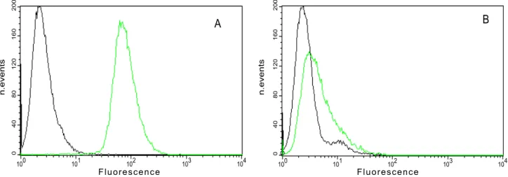
Evaluation of the expression levels of avb3 integrin on the membrane surface of WM266 and HeLa cell lines was performed by flow cytometric analysis using the monoclonal antibody LM609. To maintain surface marker integrity, cells were detached using 0.1 mM EDTA in PBS. Data showed that WM266 cells expressedavb3 at high level, while HeLa cells displayed low levels of avb3. Specifically, the relative percentage of fluorescence is about 5% on HeLa cells and 85% on WM266 (Figure 1). Therefore we used WM266 and HeLa cell lines as positive and negative controls foravb3 integrin expression, respectively.
Effect of RGDechi-hCit on WM266 adhesion
The influence of RGDechi-hCit on the adhesion of WM266 cells seeded onto vitronectin (a matrix able to recognise bothavb3 and avb5 integrins), fibronectin (recognising avb3) or collagen type I (not recognising eitheravb3 oravb5 integrins) [35,36,37] was investigated in a specific cells adhesion buffer [38] containing
fundamental bivalent ions for integrin activity, such as Mg2+
or Ca2+
, to perform the assay under optimal conditions.
In order to prevent receptor internalisation, cells were preincubated at 4uC in adhesion buffer with peptides or monoclonal antibody anti avb3 integrin and then seeded into plates previously coated with different matrices. As shown in Figure 2A, RGDechi-hCit decreased the adhesion of WM266 plated onto vitronectin (,70%) and fibronectin (,40%), but did not have significant effect on adhesion to collagen type I. Similar data were obtained with cilengitide used as positive control and with the monoclonal antibody LM609 used to demonstrate the specificity ofavb3 -dependent adhesion.
Incubation of cells with scrambled peptide, used as negative control, did not decrease WM266 adhesion onto matrix proteins. Furthermore, it is worth noting that the inhibition of adhesion by RGDechi-hCit occurred in a concentration-dependent manner giving an IC50 of 25.5mM and 53mM for vitronectin and fibronectin, respectively (Figure 2B).
In addition, the effect of RGDechi-hCit on cell detachment from vitronectin was tested 1 h after attachment of cells to vitronectin, the peptide was added and a significant cell detachment was observed (50%); an analogous effect was obtained using Cilengitide (60%) and antibody LM609 (50%), while using a scrambled peptide, no significant effect was observed (Figure 3), further confirming the RGDechi-hCit specific activity onavb 3-dependent adhesion.
RGDechi-hCit inhibits proliferation and induces apoptosis in WM266 cells
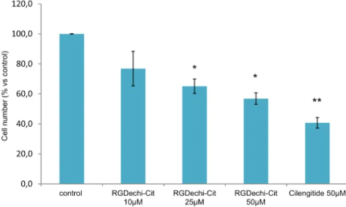
To evaluate cell proliferation, starved WM266 were incubated with Cilengitide (50mM) or with RGDechi-hCit peptide at different concentrations (10-50mM) for 24 h. The inhibition of cell proliferation results in a dose-dependent manner. At the higher concentration used, RGDechi-hCit peptide induced a significative inhibition of proliferation (,45%) in respect to the untreated cells (Figure 4) and comparable to the Cilengitide (,60%)
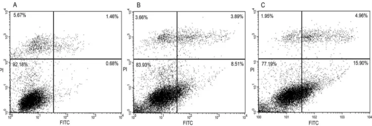
In the same conditions of starvation, after 16 h of treatment with RGDechi-hCit or Cilengitide (both 50mM), apoptosis was evaluated with annexin V-FITC/PI double staining by flow cytometry analysis. Starved WM266 cells, treated with RGDechi-hCit, exhibited 8.5% early apoptotic cells, 3.9% late apoptotic and
Figure 1. Analysis of expression ofavb3 integrin in WM266 and HeLa cells by flow cytometry.Pre-confluence cells (WM266 cells (A), HeLa cells(B)) were detached by 0.1 mM EDTA in PBS and incubated with antiavb3 antibody (green curve) or isotype (black curve) and then with secondary antibody FITC conjugate.
3.7% of necrotic cells; whereas using Cilengitide, 16.0% early apoptotic cells, 5.0% late apoptotic and 2.0% of necrotic cells were obtained (Figure 5). The same experiment conducted on starved cells in the absence of peptides displayed 0.7% early apoptotic cells, 1.4% late apoptotic and 5.6% necrotic cells.
RGDechi-hCit cellular uptake by flow cytometry analysis and confocal microscopy
The uptake of NBD-RGDechi-hCit into WM266 cells was determined by flow cytometry. The trans-bilayer distribution of NBD-peptides was determined by comparing the fluorescence intensity before and after addition of sodium dithionite, an essentially membrane-impermeant molecule, which irreversibly
suppresses the fluorescence of the accessible NBD-moiety localised on the external cell surface [39]. The NBD-RGDechi-hCit (10mM) was incubated with WM266 cells for 30 min at 37uC and then its quenching by dithionite treatment, was measured. To avoid dithionite diffusion in the biological membranes, this reaction was performed at 4uC. The addition of dithionite determined a strong increase of fluorescence compared to the intrinsic fluorescence of the cells. This result showed that a remarkable percentage of peptide (,90%) was internalised. In the same experimental conditions, NBD-scrambled peptide was used to exclude the non-specific fluorescence signal (data not show).
To confirm the peptide cell penetration and to collect information about the mechanism of internalisation in WM266 cells, confocal studies were also performed.
Figure 2. Inhibition of adhesion of WM266 on different extracellular matrix coated plates (A).Cells were pre-incubated with peptides (50mM) or anti-avb3 antibody (10mg/mL) for 30 min at 4uC and then seeded on extracellular matrix pre-coated plates. Cell adhesion was evaluated after 1 h of incubation using crystal violet reagent. The results are presented as the percentage of adherent cells respect to the control (untreated cells) and are expressed as means6SE of three independent experiments performed in triplicate. Statistical significance was analyzed using Student’s t test, unpaired, two-sided (*p,0.05). RGDechi-hCit dose-effect on WM266 cell adhesion (B). Cells were pre-incubated with increasing concentrations of RGDechi-hCit for 30 min at 4uC and then seeded on vitronectin or fibronectin (10mg/mL) pre-coated plates at 37uC. The cell adhesion was evaluated after 1 h of incubation using crystal violet reagent. The results are presented as the percentage of adherent cells respect to the control (untreated cells) and are expressed as mean6DS from three independent experiments performed in triplicate.
doi:10.1371/journal.pone.0106441.g002
Figure 3. Detachment of WM266 cells from vitronectin coated plates.Cells were plated onto vitronectin (10mg/mL) pre-coated wells and were allowed to adhere for 1 h. Peptides (50mM) or anti-avb3 antibody (10mg/mL) were added and the incubation was continued for 2 h. Cell adhesion was determined by crystal violet assay. Results are presented as percentage of adherent cells respect to the control (untreated cells) and are expressed as means 6 SE of three independent experiments performed in triplicate. Statistical significance was analyzed using Student’s t test, unpaired, two-sided (* p,0.05). doi:10.1371/journal.pone.0106441.g003
Figure 4. Cytotoxicity assay.WM266 were treated with starvation conditions (4% FCS) for 4 h and then peptides at different concentra-tions were added and incubated for 24 h. The proliferation was evaluated using crystal violet assay. The results are presented as the percentage of proliferating cells respect to the control (untreated cells) and are expressed as means6SE of three independent experiments performed in triplicate. Statistical significance was analyzed using Student’s t test, unpaired, two-sided (* p,0.05, **p,0.01).
FITC-RGDechi-hCit was efficiently internalised after 30 min of incubation at 37uC (Figure 6A). Indeed, confocal images show that the peptide was localised within cell cytoplasm and mainly distributed into discrete spots suggesting its confinement in vesicular structures. Conversely, no cellular uptake was observed for FITC-scrambled peptide in WM266 cells (Figure 6B) and also for FITC-RGDechi-hCit in HeLa cells (Figure 6C) not expressing
avb3 integrin receptor.
In order to investigate the mechanism of RGDechi-hCit peptide uptake, co-localisation experiments by using anti-avb3 integrin antibody were carried out. Figure 7A shows a partial co-localisation of FITC-RGDechi-hCit withavb3 integrin along cell boundaries (white arrows), suggesting a possible interaction between the peptide and the receptor at the cell membrane level. Endocytosis of avb3 integrin was described to occur through clathrin-dependent [40] or caveolae endocytosis [41]. Therefore, to determine whether RGDechi-hCit peptide uptake followed the same endocytic pathways, we performed indirect immunofluores-cence analyses using anti-clathrin and anti-caveolin 1 antibodies. Results showed a partial co-compartimentalisation of RGDechi-hCit both with caveolin 1 (Figure 7B) and clathrin (Figure 7C) proteins. A clear co-compartimentalisation of RGDechi-hCit with LAMP2 (Lysosome-Associated Membrane Proteins, whose func-tion is mainly related to the protecfunc-tion, maintenance, and adhesion of the lysosome) was found, indicating an accumulation
of this peptide in lysosomes (Figure 7D), as a possible consequence of receptor-mediated endocytic pathway.
To further elucidate the mechanism of RGDechi-hCit peptide uptake, WM266 cells were treated with sucrose and filipin which inhibit clathrin- and caveolin-mediated endocytosis, respectively. Confocal images showed a decrease of intracellular peptide fluorescence after both treatments compared to non-treated cells (Figure 8 A–F). A quantitative analysis indicated a reduction of about 40% and 20% RGDechi-hCit cellular uptake upon sucrose and filipin treatments, respectively (Figure 8 G). These results confirmed that the uptake of RGDechi-hCit peptide partially involves clathrin- and caveolin-mediated endocytic mechanisms.
Discussion and Conclusion
Several ECM proteins, enclosing the RGD motif, play a crucial role in integrin-mediated cell adhesion and consequently, in cell proliferation, survival and migration [4,42,43]. So far a huge variety of RGD peptides and mimetics are reported in the literature as potential candidates for tumor imaging, cell-targeting and therapy [44,45,46,47] thanks to their ability to recognise integrin receptors and eventually to internalise into different cell types, including endothelial and melanoma cells [42,48,49,50,51]. Many studies have identified marked differences in the surface expression and distribution of integrins in malignant tumor cells [52]. In particular avb3 integrin is expressed strongly on the
Figure 5. Apoptosis analyses with annexin V-FITC/PI double staining on WM266 cells.Untreated cells (A); cilengitide treated cells (B); RGDechi-hCit treated cells (C). Upper left quadrant: necrotic cells; upper right: advanced apoptotic cells; lower left: viable cells; lower right: early apoptotic cells. These pictures are representative of three independent experiments.
doi:10.1371/journal.pone.0106441.g005
Figure 6. Merge of confocal and transmitted light images of peptide cellular uptake.WM266 cells after 30 min incubation with 50mM RGDechi-hCit (A) and scrambled peptide(B). HeLa cells after 30 minute incubation with 50mM RGDechi-hCit (C). Green fluorescence indicates peptides. Magnification bar: 20mm.
surface of malignant melanoma cells and angiogenic blood vessels, but weakly on pre-neoplastic melanomas and quiescent blood vessels [30,38]. So a high expression ofavb3 integrin seems to be correlated to the increase of the metastatic potential of the melanoma cell line [19,28,39,40].
Over the last few years we focused our attention on the study of the biological features of a highly selective chimeric avb3 antagonist named RGDechi-hCit. Very recently [28], we reported some data on in vitro antitumor efficacy of RGDechi-hCit on melanoma cell lines differently expressing avb3 integrin. In particular, the data showed that the peptide induces a significant inhibition of proliferation only on the WM266 cell line, in accordance with its very high surface expression ofavb3.
Here we mainly investigated the pro-apoptotic activity of RGDechi-hCit peptide and its ability to penetrate the WM266 cell membrane. Simultaneously we deepened the selectivity, anti-adhesion, and anti-proliferative features of the peptide. Interesting-ly, for the first time we demonstrated the ability of RGDechi-hCit to enter cells overexpressingavb3 integrin. This exciting result opens the way to consider this peptide as a new potential carrier to deliver drugs into the cell also confirming its selectivity.
In detail, to reinforce the evidence regarding the receptor selectivity of the molecule, we investigated the adhesion effect of RGDechi-hCit on WM266 cells, using vitronectin, fibronectin and collagen I as different matrix proteins. Remarkably, we observed that peptide activity depends on the matrix used; it evidently inhibits cell adhesion to vitronectin and somewhat to fibronectin in contrast to the effect on collagen I, which does not interact withav integrin and protects cells from the RGDechi-hCit anti-adhesion activity. The higher effect observed on cells seeded on vitronectin with respect to fibronectin is very likely to be ascribed to the different level of receptor expression on the WM266 cell surface, where the presence of theavb3 receptor dominates over that of
avb5 (avb3:avb5, 10:1 ratio, data not shown).
Since vitronectin interacts withavb3 as well asavb5 receptors while fibronectin interacts witha5b1,aIIbb3 andavb3, we have strong reasons to believe that the different behavior of RGDechi-hCit on the various matrices is due to the capability of the WM266 cells to bind fibronectin most efficiently, ascribable to the presence of other integrins not directly interacting with RGDechi-hCit. The inhibition of adhesion by RGDechi-hCit occurs in a concentra-tion-dependent manner for vitronectin and fibronectin and the ability to detach adherent cells from vitronectin is very similar to that of the anti-avb3 antibody, reinforcing our findings that the action of the peptide is integrin specific. Additional experiments
Figure 7. Co-localization areas (white arrows) of RGDechi-hCit peptide (green) withavb3 integrin (red)(A), caveolin 1 (red)(B), clathrin (red)(C) and LAMP 2 (red)(D) in WM266 cells after 30 minute incubation.(A, C, E, G) fluorescence images; (B, D, F, H) transmitted and fluorescence image overlapping. Magnification bar: 20mm.
doi:10.1371/journal.pone.0106441.g007
confirmed our previous data [28] that RGDechi-hCit significantly inhibits the growth of melanoma cells. The anti-proliferative effect of the peptide on cancer cells is comparable to Cilengitide used as positive control and to other RGD–based systems reported in the literature [9,10].
Interestingly, studies by FACS indicated that RGDechi-hCit is also able to induce apoptosis in a similar way to Cilengitide on other cell lines [53] and in accordance with other reports indicating that cells detached by treatment with RGD molecules go on to apoptosis. This outcome enhances the value of the RGDechi-hCit peptide even if the principal criterion for its success was the evidence of its cell entry ability. For this reason, particular attention was paid to the investigation of the mechanism of internalisation both by flow cytometric and confocal microscopy studies. Generally, RGD-based peptides are able to cross the cell membrane both by passive diffusion [54,55] and receptor specific mechanism. Here we reported that our peptide is able to enter cells overexpressing avb3 integrin and shows a partial co-localisation with anti-avb3 integrin antibody, suggesting very likely anavb3 receptor-mediated uptake.
Actually, indirect immunofluorescence analysis highlighted a partial co-compartimentalisation of RGDechi-hCit both with caveolin 1 and clathrin proteins. This result was confirmed by using inhibitors of clathrin- and caveolin-mediated endocytosis which induce a clear reduction of peptide cellular uptake. What is more, an evident co-compartimentalisation of RGDechi-hCit with LAMP2 was observed by the accumulation of peptide in lysosome, clear consequence of a receptor-mediated endocytic pathway. Altogether these findings strongly suggest the uptake of RGDechi-hCit occurs mainly through the internalisation of avb3 receptor
viaan endocytic pathway. Further investigations will be necessary to confirm such data.
The observed uptake poses the question regarding the mechanism of apoptosis induced by RGDechi-hCit. Studies on RGD peptides have led to several models of antagonist-induced cell death including a type of caspase-dependent apoptosis known as anoikis [42,56] due to the cells loosing adherence and subsequent detachment caused by interaction with integrins. In our case, the apoptosis could be ascribed both to anoikis and/or to activation of an intracellular pathway consequent to the peptide entrance into the cell. Further studies will be needed to address likely intracellular targets mediating the apoptosis.
All results here reported give some convincing evidence that RGDechi-hCit is able to penetrate cells mainly byavb3 integrin-dependent endocytosis and could be considered a novel carrier to deliver selectively drugs into the cell. What is more, we discovered that the peptide displays an apoptotic effect per se. This consideration and the previous data on the ability to inhibit some intracellular pathways [21], suggest that RGDechi-hCit can be potentially considered a trueavb3 integrin inhibitor.
In conclusion, a great challenge to evaluate the achievable application of RGDechi-hCit molecule in vivo is the development of new selective systems containing both our peptide and pro-apoptotic molecules or different therapeutic agents to induce a synergic action.
Author Contributions
Conceived and designed the experiments: DC ADG LZ. Performed the experiments: DC IdP AL DG. Analyzed the data: DC IdP AL ADG SDG DG MS LZ. Wrote the paper: DC SDG ADG MS LZ.
References
1. Hynes RO (2002) Integrins: bidirectional, allosteric signaling machines. Cell 110: 673–687.
2. Zaccaro L, Del Gatto A, Pedone C, Saviano M (2007) Integrin receptor family: promising target for therapeutic and diagnostic applications. Current Topics in Biochemical Research 9: 45–56.
3. Brooks PC, Clark RA, Cheresh DA (1994) Requirement of vascular integrin alpha v beta 3 for angiogenesis. Science 264: 569–571.
4. Giancotti FG, Ruoslahti E (1999) Integrin signaling. Science 285: 1028–1032. 5. Gladson CL, Cheresh DA (1991) Glioblastoma expression of vitronectin and the
alpha v beta 3 integrin. Adhesion mechanism for transformed glial cells. The Journal of clinical investigation 88: 1924–1932.
6. Liapis H, Adler LM, Wick MR, Rader JS (1997) Expression of alpha(v)beta3 integrin is less frequent in ovarian epithelial tumors of low malignant potential in contrast to ovarian carcinomas. Human pathology 28: 443–449.
7. Albelda SM, Mette SA, Elder DE, Stewart R, Damjanovich L, et al. (1990) Integrin distribution in malignant melanoma: association of the beta 3 subunit with tumor progression. Cancer research 50: 6757–6764.
8. Zhaofei L (2008) Integrin avb3-Targeted Cancer Therapy. Drug Development Research 69: 329–339.
9. Aguzzi MS, D’Arcangelo D, Giampietri C, Capogrossi MC, Facchiano A (2011) RAM, an RGDS analog, exerts potent anti-melanoma effects in vitro and in vivo. PloS one 6: e25352.
10. Oliveira-Ferrer L, Hauschild J, Fiedler W, Bokemeyer C, Nippgen J, et al. (2008) Cilengitide induces cellular detachment and apoptosis in endothelial and glioma cells mediated by inhibition of FAK/src/AKT pathway. Journal of experimental & clinical cancer research: CR 27: 86.
11. Zaccaro L, Del Gatto A, Pedone C, Saviano M (2009) Peptides for tumour therapy and diagnosis: current status and future directions. Current medicinal chemistry 16: 780–795.
12. Chen K, Chen X (2011) Integrin targeted delivery of chemotherapeutics. Theranostics 1: 189–200.
13. Millard M, Odde S, Neamati N (2011) Integrin targeted therapeutics. Theranostics 1: 154–188.
14. Dechantsreiter MA, Planker E, Matha B, Lohof E, Holzemann G, et al. (1999) N-Methylated cyclic RGD peptides as highly active and selective alpha(V)beta(3) integrin antagonists. Journal of medicinal chemistry 42: 3033–3040. 15. Reardon DA, Neyns B, Weller M, Tonn JC, Nabors LB, et al. (2011)
Cilengitide: an RGD pentapeptide alphanubeta3 and alphanubeta5 integrin inhibitor in development for glioblastoma and other malignancies. Future oncology 7: 339–354.
16. Tabatabai G, Weller M, Nabors B, Picard M, Reardon D, et al. (2010) Targeting integrins in malignant glioma. Targeted oncology 5: 175–181. 17. Yin AA, Cheng JX, Zhang X, Liu BL (2013) The treatment of glioblastomas: a
systematic update on clinical Phase III trials. Critical reviews in oncology/ hematology 87: 265–282.
18. Scaringi C, Minniti G, Caporello P, Enrici RM (2012) Integrin inhibitor cilengitide for the treatment of glioblastoma: a brief overview of current clinical results. Anticancer research 32: 4213–4223.
19. Del Gatto A, Zaccaro L, Grieco P, Novellino E, Zannetti A, et al. (2006) Novel and selective alpha(v)beta3 receptor peptide antagonist: design, synthesis, and biological behavior. Journal of medicinal chemistry 49: 3416–3420. 20. Zannetti A, Del Vecchio S, Iommelli F, Del Gatto A, De Luca S, et al. (2009)
Imaging of alpha(v)beta(3) expression by a bifunctional chimeric RGD peptide not cross-reacting with alpha(v)beta(5). Clinical cancer research: an official journal of the American Association for Cancer Research 15: 5224–5233. 21. Santulli G, Basilicata MF, De Simone M, Del Giudice C, Anastasio A, et al.
(2011) Evaluation of the anti-angiogenic properties of the new selective alphaVbeta3 integrin antagonist RGDechiHCit. Journal of translational medicine 9: 7.
22. Fogel M, Mechtersheimer S, Huszar M, Smirnov A, Abu-Dahi A, et al. (2003) L1 adhesion molecule (CD 171) in development and progression of human malignant melanoma. Cancer letters 189: 237–247.
23. Haass NK, Smalley KS, Li L, Herlyn M (2005) Adhesion, migration and communication in melanocytes and melanoma. Pigment cell research/ sponsored by the European Society for Pigment Cell Research and the International Pigment Cell Society 18: 150–159.
24. Marshall JF, Rutherford DC, Happerfield L, Hanby A, McCartney AC, et al. (1998) Comparative analysis of integrins in vitro and in vivo in uveal and cutaneous melanomas. British journal of cancer 77: 522–529.
25. McGary EC, Lev DC, Bar-Eli M (2002) Cellular adhesion pathways and metastatic potential of human melanoma. Cancer biology & therapy 1: 459–465. 26. Jilaveanu LB, Aziz SA, Kluger HM (2009) Chemotherapy and biologic therapies
for melanoma: do they work? Clinics in dermatology 27: 614–625.
27. Garbe C, Peris K, Hauschild A, Saiag P, Middleton M, et al. (2010) Diagnosis and treatment of melanoma: European consensus-based interdisciplinary guideline. European journal of cancer 46: 270–283.
29. Monfregola L, De Luca S (2011) Synthetic strategy for side chain mono-N-alkylation of Fmoc-amino acids promoted by molecular sieves. Amino acids 41: 981–990.
30. Gillies RJ, Didier N, Denton M (1986) Determination of cell number in monolayer cultures. Analytical Biochemistry. 159: 109–113.
31. Hansen SH, Sandvig K, van Deurs B (1993) Clathrin and HA2 adaptors: effects of potassium depletion, hypertonic medium, and cytosol acidification. The Journal of cell biology 121: 61–72.
32. Fields CG, Lloyd DH, Macdonald RL, Otteson KM, Noble RL (1991) HBTU activation for automated Fmoc solid-phase peptide synthesis. Peptide research 4: 95–101.
33. Coste J, Le-Nguyen D, Castro B (1990) PyBOP: a new peptide coupling. 34. reagent devoid of toxic byproduct. Tetrahedron Lett 31: 205–208.
35. Davis GE (1992) Affinity of integrins for damaged extracellular matrix:avb3 binds to denatured collagen type I through RGD sites. Biochem Biophys Res Comm 182: 1025–1031.
36. Kanda S, Kuzuya M, Ramos MA, Koike T, Yoshino K, et al. (2000) Matrix metalloproteinase and alphavbeta3 integrin-dependent vascular smooth muscle cell invasion through a type I collagen lattice. Arteriosclerosis, thrombosis, and vascular biology 20: 998–1005.
37. Barczyk M, Carracedo S, Gullberg D (2010) Integrins. Cell and tissue research 339: 269–280.
38. Seiffert D, Smith JW (1997) The cell adhesion domain in plasma vitronectin is cryptic. The Journal of biological chemistry 272: 13705–13710.
39. Angeletti C, Nichols JW (1998) Dithionite quenching rate measurement of the inside-outside membrane bilayer distribution of 7-nitrobenz-2-oxa-1,3-diazol-4-yl-labeled phospholipids. Biochemistry 37: 15114–15119.
40. De Deyne PG, O’Neill A, Resneck WG, Dmytrenko GM, Pumplin DW, et al. (1998) The vitronectin receptor associates with clathrin-coated membrane domains via the cytoplasmic domain of its beta5 subunit. Journal of cell science 111 (Pt 18): 2729–2740.
41. Alam MR, Dixit V, Kang H, Li ZB, Chen X, et al. (2008) Intracellular delivery of an anionic antisense oligonucleotide via receptor-mediated endocytosis. Nucleic acids research 36: 2764–2776.
42. Maubant S, Saint-Dizier D, Boutillon M, Perron-Sierra F, Casara PJ, et al. (2006) Blockade of alpha v beta3 and alpha v beta5 integrins by RGD mimetics induces anoikis and not integrin-mediated death in human endothelial cells. Blood 108: 3035–3044.
43. Plow EF, Cierniewski CS, Xiao Z, Haas TA, Byzova TV (2001) AlphaIIbbeta3 and its antagonism at the new millennium. Thrombosis and haemostasis 86: 34–40.
44. Rosenow F, Ossig R, Thormeyer D, Gasmann P, Schluter K, et al. (2008) Integrins as antimetastatic targets of RGD-independent snake venom compo-nents in liver metastasis [corrected]. Neoplasia 10: 168–176.
45. White DE, Muller WJ (2007) Multifaceted roles of integrins in breast cancer metastasis. Journal of mammary gland biology and neoplasia 12: 135–142. 46. Ellerby HM, Arap W, Ellerby LM, Kain R, Andrusiak R, et al. (1999)
Anti-cancer activity of targeted pro-apoptotic peptides. Nature medicine 5: 1032– 1038.
47. Andersen MH, Svane IM, Becker JC, Straten PT (2007) The universal character of the tumor-associated antigen survivin. Clinical cancer research: an official journal of the American Association for Cancer Research 13: 5991–5994. 48. Adderley SR, Fitzgerald DJ (2000) Glycoprotein IIb/IIIa antagonists induce
apoptosis in rat cardiomyocytes by caspase-3 activation. The Journal of biological chemistry 275: 5760–5766.
49. Matsuki K, Sasho T, Nakagawa K, Tahara M, Sugioka K, et al. (2008) RGD peptide-induced cell death of chondrocytes and synovial cells. Journal of orthopaedic science: official journal of the Japanese Orthopaedic Association 13: 524–532.
50. Zaffaroni N, Pennati M, Daidone MG (2005) Survivin as a target for new anticancer interventions. Journal of cellular and molecular medicine 9: 360–372. 51. Grossman D, Altieri DC (2001) Drug resistance in melanoma: mechanisms, apoptosis, and new potential therapeutic targets. Cancer metastasis reviews 20: 3–11.
52. Mizejewski GJ (1999) Role of integrins in cancer: survey of expression patterns. Proceedings of the Society for Experimental Biology and Medicine Society for Experimental Biology and Medicine 222: 124–138.
53. Lautenschlaeger T, Perry J, Peereboom D, Li B, Ibrahim A, et al. (2013) In vitro study of combined cilengitide and radiation treatment in breast cancer cell lines. Radiation oncology 8: 246.
54. Castel S, Pagan R, Mitjans F, Piulats J, Goodman S, et al. (2001) RGD peptides and monoclonal antibodies, antagonists of alpha(v)-integrin, enter the cells by independent endocytic pathways. Laboratory investigation; a journal of techni-cal methods and pathology 81: 1615–1626.
55. Aguzzi MS, Fortugno P, Giampietri C, Ragone G, Capogrossi MC, et al. (2010) Intracellular targets of RGDS peptide in melanoma cells. Molecular cancer 9: 84.