http://dx.doi.org/10.1590/bjb.2014.0085 Original Article
Reproduction, development and habits of the large turkey
louse Chelopistes meleagridis (Phthiraptera: Ischnocera)
under laboratory conditions
Maturano, R.
a*
and Daemon, E.
aaPrograma de Pós-graduação em Ciências Biológicas - Comportamento e Biologia Animal – PPGCB-CBA,
Universidade Federal de Juiz de Fora – UFJF, Rua José Lourenço Kelmer, s/n, Campus Universitário Bairro Martelos, CEP 36036-330, Juiz de Fora, MG, Brazil
*e-mail: ralphmaturano@gmail.com
Received: August 27, 2012 – Accepted: April 11, 2013 – Distributed: August 31, 2014
(With 4 figures)
Abstract
The bionomy of Chelopistes meleagridis off the host was observed with the aim of better understanding the aspects of this species’ life cycle. For this purpose, C. meleagridis adults were collected and maintained under controlled
conditions to reproduce (35°C and RH > 80%), with turkey feathers as the food source. From the offspring of these
lice, the development of 150 individuals was observed from the egg to the adult phase. These eggs were divided into two groups of 75 each. After hatching, one group was given a diet composed of feathers while the other received
feathers plus skin of the host turkey (Meleagris gallopavo). The “feather + skin” diet resulted in the greatest number
of adults, so this diet was given to the next generation of lice reared in vitro, starting from the first instar, to observe
their fertility, fecundity and longevity. High reproduction rates were found in relation to other lice of the Ischnocera sub-order, particularly the number of eggs per day and number of eggs produced per female over the lifetime (means
of 2.54 and 26.61 eggs, respectively, for wild females and 2.11 and 29.33 eggs for laboratory-reared females). The inclusion of skin in the diet was a determining factor for development to the adult stage, since 48% of the lice fed this
diet reached that stage, versus 1.3% that reached maturity fed only with feathers. The development time of the males
and females was similar (mean of 29.38 days), without any difference in the sexual proportion of the adults.
Keywords:Chelopistes meleagridis, feather lice, laboratory conditions, life history, longevity.
Reprodução, desenvolvimento e hábitos de
Chelopistes meleagridis
(Phthiraptera: Ischnocera) em laboratório
Resumo
A bionomia de Chelopistes meleagridis fora do hospedeiro foi observada com o objetivo de compreender aspectos relacionados ao ciclo de vida desta espécie. Para isto, adultos de C. meleagridis foram coletados e colocados em
condições controladas (temperatura de 35°C e umidade relativa superior a 80%) para se reproduzir, oferecendo-se
pena como alimento. Da prole destes adultos, foi observado o desenvolvimento de 150 indivíduos desde o ovo até a fase adulta. Para 75 destes, foi oferecida a dieta composta de pena, enquanto para os outros 75 a dieta foi composta de pena e pele do hospedeiro (peru, Meleagris gallopavo). Ao verificar que a dieta “pena + pele” foi a que resultou no
maior número de adultos, foram observadas a fertilidade, fecundidade e a longevidade de piolhos criados in vitro desde o primeiro ínstar alimentados com esta dieta. Valores altos relacionados à reprodução desta espécie foram encontrados em relação a outros piolhos da subordem Ischnocera, destacando-se: número de ovos produzidos por dia e número de ovos produzidos por fêmeas durante a vida (médias de 2,54 e 26,61 ovos, respectivamente, para fêmeas selvagens e 2,11 e 29,33 ovos, respectivamente, para fêmeas criadas in vitro.). A inclusão de pele na dieta foi determinante para
o desenvolvimento até o estágio adulto, uma vez que 48% dos piolhos alimentados com essa dieta atingiram a fase adulta. Quando foi oferecido apenas pena, 1,3% dos piolhos atingiram a maturidade. O tempo de desenvolvimento de
machos e fêmeas foi semelhante (média de 29,38 dias) sem haver diferença na proporção sexual dos adultos.
1. Introduction
Knowledge of the biology of chewing lice that parasitise
birds, first provided by the studies of Osborn (1890a, b), advanced greatly in the last century, through studies published by various researchers detailing aspects of the life cycle of diverse species (Martin, 1934; Wilson, 1934, 1939; Conci, 1952). These studies, carried out in the laboratory, provide the basic data for understanding the parasite-host interaction and consequently the impacts caused on the host birds. Constant abiotic conditions allow obtaining maximum biotic potential values, information
that is important to determine the risk factors based on the infestation levels as well as to make inferences
about the population structure under natural conditions (Gupta et al., 2007).
The majority of studies of the biology of chewing lice have focused on species that parasitise domesticated birds and thus are of economic importance to poultry producers, such as parasites of Gallus gallus (Linnaeus, 1758): Menacanthus stramineus (Nitzsch, 1818), Lipeurus caponis
(Linnaeus, 1758) and Goniocotes gallinae (De Geer, 1778);
and parasites of Columba livia (Gmelin, 1789): Columbicola columbae (Linnaeus, 1758) and Colpocephalum turbinatum
(Denny, 1842). However, there are still many voids in the
knowledge about the biology of some poultry louse species
of economic importance (Emerson, 1979; Kettle, 1995).
In this respect, little is known about the bionomical
aspects of Chelopistes meleagridis (Linnaeus, 1758), a
parasite of domesticated turkeys [Meleagris gallopavo
(Linnaeus, 1758)] (Price and Graham, 1997; Guimarães et al., 2001). The only reports are those of Qureshi (1957), evaluating the survival of adults off the host, and Conci
(1952), reporting no success in rearing this species in vitro. Therefore, the aim of the present study was to establish a method for in vitro rearing of C. meleagridis, to permit obtaining data on the reproduction, development and habits of this species under laboratory conditions.
2. Material and Methods
Chelopistes meleagridis adults were collected from
turkeys bred on farms located in the municipalities of
Juiz de Fora and Matias Barbosa, Minas Gerais state,
Brazil. These specimens were identified by the key of Emerson (1962). The experiments were conducted in the Laboratório de Artrópodes Parasitos of the Universidade Federal de Juiz de Fora.
2.1. Bioassay 1 – Evaluation of the diet of Chelopistes meleagridis and in vitro development from offspring of wild adults
In the laboratory, the lice were sexed according to Clay
(1941) and then placed in Petri dishes lined with filter paper (5 couples per dish). The dishes were then placed in the dark in a climate-controlled chamber at 35 °C and relative
humidity greater than 80%. The lice were provided with
turkey breast feathers that had been previously frozen to kill any mites and eggs present. Breast feathers were chosen
because this is a site commonly infested by the species. The feathers were observed daily under a stereoscopic microscope and the eggs were removed and transferred
individually to Petri dishes lined with filter paper, also
containing feathers. From these eggs, two groups were formed, with 75 specimens each, with the following diets:
1 – turkey feathers; 2 – turkey feathers + turkey skin. In diet 1, the eggs were placed on a turkey breast feather fragment while in diet 2 the eggs were also offered skin fragments taken from the epidermis, in an adaptation of
the methodology of Hopkins and Chamberlain (1972). The
skin fragments were first dehydrated in a drying chamber
at 60 °C for 12 hours. After being cooled, the fragments were placed on the feathers. Both groups were maintained in a climate-controlled chamber under the same conditions as the adults. The lice were maintained individualised from egg to death. When necessary, the dish bottoms were cleaned with tweezers to remove fecal material.
The lice were observed daily to record the biological
data: time of hatching, day of molting to the first nymphal
instar, second instar, third instar and adult stage, sex and longevity.
2.2. Bioassay 2 - In vitro reproductive biology Chelopistes meleagridis adults (wild and laboratory-reared)
To evaluate the reproduction of wild adults, 30 couples were formed from adults gathered from naturally infested
turkeys. These were placed individually in Petri dishes lined with filter paper and turkey feathers. The temperature and
humidity conditions were the same as in Bioassay 1. Each feather was observed daily to obtain data on longevity of the males and females and number of eggs laid per day. When the male died before the female, a new male, from the same collection, was placed in the dish to assure obtaining the data on the biotic potential of the female. In these cases, the data on male longevity refer to the males utilised at the start of the experiment. When the female died before the male, the male was observed daily until death to compute the average longevity.
Each day of oviposition, the barbules with the eggs
attached were removed with fine-tipped scissors and
placed on sheets of blue paper to facilitate observation of
the eggs. Each egg was identified as to the parent couple
and oviposition day. The paper sheets with the eggs were
kept in a chamber under the same conditions as for the
adult lice. At the end of the incubation period, the eggs were observed under a stereoscopic microscope to tabulate the data on hatching of nymphs.
To assess the reproduction of the lice reared in vitro, they were fed the diet of Bioassay 1, which had resulted in the largest number of adults. A new collection of adults was carried out for this generation and the eggs produced
were placed in Petri dishes (five per dish) and then kept in
On the first day after molting to the adult stage, 30 couples were placed in Petri dishes (one couple per dish). The
procedures for observation of longevity, fecundity and fertility were the same as for the wild adults.
3. Statistical Analysis
The Bioestat (Ayres et al., 2007) version 5.0 program was used to calculate the statistical measures. The survival time, longevity of adults, fecundity and fertility were compared between the groups by the t-test for parametric data and the Mann-Whitney test for nonparametric data,
in both cases a 5% significance (p<0.05). The Spearman
test was also applied to obtain the correlations between age of adults and fecundity and fertility.
4. Results
4.1. Bioassay 1
With diet 1, only one individual among the 75 developed
to adult form, while with diet 2, 36 of the 75 eggs (48%)
reached the adult stage (Table 1). The average egg incubation time was 3.81 days, ranging from 3 to 5 days. All 150
eggs chosen were viable. For diet 1, the first instar lasted an average of 7.72 days, differing significantly from the nymphs fed with diet 2 (9.04 days). Due to the low number
of nymphs of the second and third instars obtained with diet 1, there was no statistical analysis for these stages (Table 1).
The second instar on diet 2 had shorter average duration
(7.42 days) than the first and third instars (9.41 days), with a significant difference in both comparisons. The average
longevity of the adult stage was 27.33 days, but with great
amplitude (51 days). The average time for development
from oviposition to adult was 29.38 days, with a minimum of 24 and maximum of 38 days (Table 1).
The data on the males and females that reached the adult stage on diet 2 are presented in Table 2. All told,
there were 19 females and 17 males, with no significant
difference in sexual proportion. The mean development time of both sexes to the adult stage was 29 days, again
with no significant difference. However, the longevity differed significantly between the sexes, with the females
living longer (Table 2).
4.2. Bioassay 2
The data on the reproductive parameters for the wild and laboratory-reared adults are presented in Table 3. For the wild lice, the males lived longer on average than the females, with maximum times of 30 and 46 days for females and males, respectively, a difference that was
significant. Among the 30 couples formed, 29 females laid
eggs, within a maximum period of 25 days. The average number of eggs laid per day was approximately 2, with lower and upper limits of 0 and 12 eggs. The average hatching rate considering each couple was 91.33%, ranging from 60.83% to 100%. For all the eggs irrespective of the parental pair, the development of 812 eggs was observed, with a nymphal hatching rate of 81.28%.
For the adults reared in vitro, the longevity of the males was greater than that of the females (25.33 and
21.10 days, respectively), but the difference was not significant. Of the 30 couples, 27 females laid eggs, with
an average of 29.33 and range from 2 to 79 eggs. The average number of eggs laid per day was 1.79, varying from 0 to 10. The average pre-oviposition period was 8.07 days, ranging from 6 to 17 days. The oviposition period lasted on average 15.4 days, with limits of 6 and 17 days.
Table 1. Mean duration (in days) of the life stages of Chelopistes meleagridis and development to adult stage in the laboratory
(35 °C, RH>80% and escotophase).
Stage
Diet
Feather Feather + skin
Limits Mean ± SD (Number of lice) Limits Mean ± SD (number of lice)
1st instar 6-10 7.72A ± 1.27 (18) 6-17 9.04Ba ± 2.11 (67)
2nd instar 8-9 8.5 ± 0.7 (2) 3-15 7.42b ± 2.37 (47)
3rd instar - 9 (1) 7-14 9.41a ± 1.74 (36)
Adult - 7 (1) 2-53 27.33 ± 15.04 (36)
Egg-Adult - 34 (1) 24-38 29.38 ± 0.53 (36)
Means followed by equal letters do not differ statistically at a significance level of 5%. Capital letters are comparisons in line. Small letters are comparisons in columns.
Table 2. Number of males and females, development time and longevity of Chelopistes meleagridis reared in the laboratory
(35 °C, RH> 80% and escotophase) obtained with the feather + skin diet from 75 eggs.
Females Males
Limits Mean ± SD Limits Mean ± SD
Number of adults - 19A - 17A
Development time (egg – adult) 24-38 29.21A±3.72 25-35 29.58A±2.64
Longevity of adult 3-53 33.00A±3.29 2-45 21.00B±13.5
Egg – Death 28-80 62.21A±15.31 29-78 50.59B±14.98
The average hatching rate considering each couple was 87.66%, with a low of 54.54% and high of 100%. In the total of 880 eggs evaluated, the nymphal hatching rate was 89.31% (Table 3).
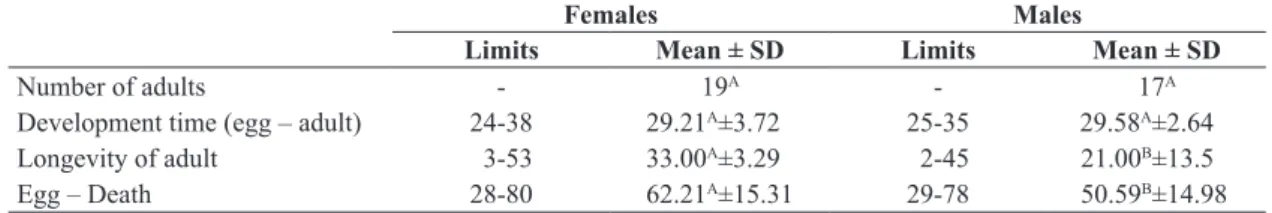
Figure 1 shows the pace of oviposition of the wild females. The number of eggs laid decreased with a declining number of couples, as expected. There was also a decline in the number of eggs per couple starting on the sixth day, indicating that the females’ fecundity diminished with increasing age. There was a negative correlation between
female age versus number of eggs per couple (Spearman’s coefficient = –0.0785; p<0.05).
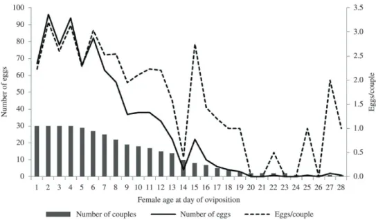
The hatching percentage according to the age of the wild females on the oviposition day is presented in Figure 2. As can be seen, this percentage declined until the 21st day, indicating a fall in fertility. There was a negative
correlation between female age at oviposition and hatching
rate (Spearman’s coefficient = –0.726; p<0.01).
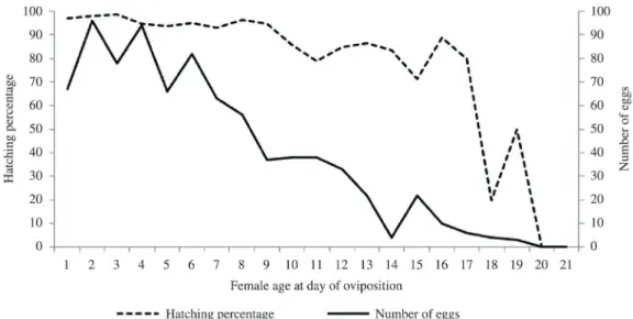
Figure 3 shows the pace of oviposition of the laboratory-reared females. They started laying on the 6th day of adult
age and continued doing so until the 38th day, with a decline
in egg production as of the 19th day, although there were
peaks on three subsequent days. There was a negative
correlation between the number of eggs per couple in
relation to age (Spearman’s coefficient = –0.611; p<0.01).
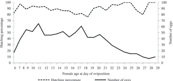
With respect to hatching percentage with time, there was no tendency for decrease or increase (Figure 4),
confirmed by the correlation test between the hatching rate over time, where the result was not significant (Spearman’s coefficient = 0.259; p>0.05)
5. Discussion
Unlike the results reported in the literature for other
avian Ischnocera, C. meleagridis depended on the inclusion
of skin in the diet to develop to the adult stage (Martin,
Table 3. Longevity, fertility and fecundity of wild and laboratory-reared Chelopistes meleagridis couples kept in a climate-controlled chamber at 35 °C, RH> 80% and escotophase.
Wild adults Laboratory-reared adults
Limits Mean ± SD
(number of lice) Limits
Mean ± SD (number of lice)
Longevity of males (days) 5-46 19.63Aa±10.82 (30) 1-44 25.33Ba ± 11.09 (30)
Longevity of females (days) 5-30 12.20Ab±5.66 (30) 2-38 21.1Aa ± 8.01 (30)
Number of eggs/ couple 3-54 28.00A±13.68 (29) 2-79 32.59A ± 20.68 (27)
Number of eggs/ couple.day 0-12 2.54A ± 2.10 (29) 0-10 2.11B ± 1.71 (27)
Pre-oviposition period (days) - - 6-17 8.07 ± 2.18 (27)
Oviposition period (days) 3-25 9.82A±5.10 (29) 6-17 15.40B ± 6.72 (27)
% hatching/ couple 60.86-100 91.33A±10.13 (29) 54.54-100 87.66A ± 15.28 (27)
% hatching (total) - 81.28 (812) - 89.31 (880)
Means followed by equal letters do not differ statistically at a significance level of 5%. Capital letters are comparisons in line. Small letters are comparisons in columns.
Figure 1. Rhythm of oviposition of wild Chelopistes meleagridis females kept under laboratory conditions (35 °C, RH> 80%
1934; Conci, 1956; Gupta et al., 2007). This dependence
was already present in the first instar, when only 24% of
the nymphs fed only with feathers molted to the second instar (Table 1). This took less time than for the nymphs
fed on feathers + skin, which was the best diet. The low
ecdysis rate repeated for the nymphs of the second instar,
suggesting that the ingestion of skin is fundamental in
these two stages. This tendency was not observed for the nymphs of the third instar due to the fact that only one nymph fed the feather-only diet reached the adult stage. However, the individuals of all instars and the adults that
were offered skin as part of the diet fed on this item.
Of the individuals fed with feather and skin, 48%
reached the adult phase, with the highest mortality rates occurring in the second and third instars (30% and 23%
respectively) (Table 1). However, the percentage of survivors
was sufficient to maintain the colony in the laboratory. Due
to failed attempts to breed C. meleagridis, Conci (1952) speculated that this species does not only feed on feathers.
The requirement to ingest skin by Ischnocera is known for
members of the Trichodectidae family, which are mammal parasites (Hopkins and Chamberlain, 1969; Hopkins et al., 1976). Regarding bird parasites, particularly those of the Philopteridae family, some authors state that they feed on
Figure 2. Hatching percentage of nymphs and number of eggs laid by wild Chelopistes meleagridis females in relation to female age on the day of oviposition. Days on which fewer than 5 eggs were produced (as of the 21st day) were disregarded.
Figure 3. Rhythm of oviposition of laboratory-reared Chelopistes meleagridis females kept under laboratory conditions
skin, although this does not appear to be essential, since in
the published studies of in vitro rearing only feathers were given (Wilson, 1934; Wall and Shearer, 2001; Johnson and Clayton, 2003; Saxena et al., 2009). The fact that
C. meleagridis ingests skin in its diet mainly composed
of feathers indicates these lice must spend part of their
time on the host bird’s skin rather than only the feathers.
According to Rai and Lakshminarayana (1980), lice can
specifically infest feathers of certain sites, so that attempts
to breed them in vitro with feathers of the host species
taken from different sites than the preferred ones can fail. Just as feather site, the skin site can be a limiting factor in
the distribution of C. meleagridis on the host body. From an economic standpoint, the presence of C. meleagridis
on the skin of turkeys can cause irritations, with negative
impacts on weight gain and egg production, so that the economic losses caused can be underestimated.
With regard to the development, the incubation time and duration of the instars of C. meleagridis are similar
to the figures observed for other Ischnocera species kept
at 35 °C (Table 1) (Gupta et al., 2007; Saxena et al., 2007, 2009; Agarwal, et al., 2011). Some virgin females produced eggs, but none were viable. This ability of virgin females to lay unviable eggs was also observed by Wilson (1939) and Agarwal (1967) for L. caponis and Falcolipeurus frater
(Giebel, 1874), respectively. Although in this study it took
approximately 30 days for the eggs to develop into the adult stage, the period for sexual maturation of the adults plus the time for females to produce eggs (pre-oviposition
period) delay the next generation in plus eight days, as
observed in Bioassay 2 (Table 3). The sexual ratio of the lice that reached the adult stage was 1:1, in contrast to studies involving collection of this species from natural infestations, where a ratio biased toward females has been observed (Table 3) (Qureshi, 1957; Lane et al., 2006).
The differences in the reproduction of lice bred in vitro
in relation to those found in the wild can be due to the difference in age between the two groups (Table 3). For example, the oviposition period of those bred in vitro was longer than those captured in the wild. In counterpart, the average number of eggs laid per day by the wild females was higher. As a consequence of this, the average number of eggs produced per couple between the groups did not differ
significantly, although the females bred in vitro produced a larger number of eggs. Agarwal (1967), comparing the fecundity of F. frater between wild and laboratory-bred females, found lower egg production by the latter group. He speculated that this reduction was due to the nutritional
deficiency while breeding the nymphs in the laboratory,
which apparently did not occur in the present experiment. The survival time between males and females varied according to the reproduction. The wild females in Bioassay 2 survived on average for a shorter period than the males, while the survival period of the laboratory-bred females was the same as for the males. In contrast, the females in Bioassay 1 maintained as virgins until death lived longer than the males (Tables 1 and 3). A possible explanation for this fact is the investment cost of reproduction, which is higher for females (Loof, 2011). Qureshi (1957) observed that at 37 °C, wild C. meleagridis adults survived off the host for seven days on feathers and three days in the absence of this food source. In the present study, age
negatively influenced the fecundity of the females in both
groups (Figures 1 and 3). The same finding was reported by Agarwal (1967), Gupta et al. (2007) and Saxena et al.
(2007) for F. frater, Brueelia amandavae Rekasi and
Saxena, 2005 and G. gallinae, respectively. Nevertheless, only the wild females suffered a negative impact of time on
egg viability, which can reflect the effect of stress caused by the change from the natural to artificial environment
(Figures 2 and 4).
Figure 4. Hatching percentage of nymphs and number of eggs laid by laboratory-reared Chelopistes meleagridis females in relation to female age on the day of oviposition. Days on which fewer than 5 eggs were produced (as of the 29th day) were
The size of a female insect is one of the factors that determines the potential fecundity (Björkman et al., 2009), which can explain the high number of eggs produced by
C. meleagridis females in comparison to other Ischnocera species, since C. meleagridis females are relatively large
(3.24 to 3.54 mm) (Clay, 1941; Naz et al., 2003) in comparison with other species of this suborder.
Based on knowledge of the biology of lice, it is possible
to formulate control strategies (e.g., place to apply insecticide,
interval between applications), avoid risk factors (e.g., crowding of hosts, high humidity) and make inferences about the infestation levels. Since the data obtained in the
present study are similar to those for natural populations, the time for a C. meleagridis generation is approximately 30 days. Therefore, disregarding extrinsic factors (e.g., defensive and immunological behaviors of the host, abiotic
factors) and assuming that the values obtained for the first
in vitro generation repeat in subsequent generations, the population of C. meleagridis grows at a rate of 12.5 times
per generation. As a consequence of this, confined birds
are subject to high infestations by this louse when it is
present or introduced, since the time to raise a turkey to
slaughter is around 140 days, thus aggravating the potential economic losses caused by this parasite.
Acknowledgements
The junior author (RM) is grateful to CAPES (Coordenação de Aperfeiçoamento de Pessoal de Nível Superior) for financial support. We are in debt to all those people and
entities that encouraged us to continue studying the biology of living organisms.
References
Agarwal, GP., 1967. Studies on the bionomics and life history of Falcolipeurus frater (Giebel: 1874). Indian Journal of Zootomy, vol. 8, no. 1, p. 21-40.
Agarwal, GP., Ahmad, A., Rashmi, A., Arya, G., Bansal, N.
and Saxena, AK., 2011. Bio-ecology of the louse, Upupicola
upupae, infesting the Common Hoopoe, Upupa epops. Journal of Insect Science, vol. 11, no. 46, p. 1-9. http://dx.doi. org/10.1673/031.011.4601. PMid:21861650
Ayres, M., AyreS-JUNIOR, M., Ayres, DL. and Santos, AAS.,
2007. Bioestat: Aplicações estatísticas nas áreas das ciências biomédicas. Belém: Sociedade Civil Mamirauá. 364 p.
Björkman, C., Gotthard, K. and Pettersson, MW., 2009. Body size. In VINCENT, HR. and RING, TC. (Eds.). Encyclopedia of insects (Second Edition). San Diego: Academic Press. p. 114-116. http://dx.doi.org/10.1016/B978-0-12-374144-8.00038-2.
Clay, T., 1941. A new genus and species of Mallophaga. Parasitology, vol. 33, no. 01, p. 119-129. http://dx.doi.org/10.1017/
S0031182000024318.
Conci, C., 1952. L’allevamento in condizioni sperimentali dei Mallofagi I. - Cuclotogaster heterographus Nitzsch. Bolletin dei Musei e Instituto Biologia. Universitaire di Genova, vol. 24, no. 150, p. 17-40.
Conci, C., 1956. L’allevamento in condizioni sperimentali dei
Mallofagi : III. - Columbicola c. columbae (Linnaeus, 1758).
Bolletin dei Musei e Instituto Biologia. Universitaire di Genova, vol. 26, no. 162, p. 47-70.
Emerson, KC., 1962. Mallophaga (Chewing lice) occurring on the turkey. Journal of the Kansas Entomological Society, vol. 35, no. 1, p. 196-202.
Emerson, KC., 1979. Lice in my life. Arlington: Emerson, 106 p.
Guimarães, JH., Tucci, EC. and Barros-Battesti, DM., 2001. Ectoparasitos de importância veterinária. São Paulo: Plêiade/
FAPESP. 218 p.
Gupta, N., Kumar, S. and Saxena, A., 2007. Intrinsic rate of natural increase of Brueelia amandavae (Ischnocera, Phthiraptera)
populations infesting Indian red avadavat. Biologia, vol. 62, no. 4, p. 458-461. http://dx.doi.org/10.2478/s11756-007-0088-2.
Hopkins, DE. and Chamberlain, WF., 1969. In vitro colonization
of the goat biting lice, Bovicola crassipes and B. limbata. Annals of the Entomological Society of America, vol. 62, no. 4, p. 826-828.
Hopkins, DE. and Chamberlain, WF., 1972. In vitro colonization of
the cattle biting louse, Bovicola bovis. Annals of the Entomological Society of America, vol. 65, no. 3, p. 771-772.
Hopkins, DE., Chamberlain, WF. and Gingrich, AR., 1976.
Mallophaga: in vitro testing of artificial diets. Annals of the Entomological Society of America, vol. 69, no. 3, p. 538-540.
Johnson, KP. and Clayton, DH., 2003. The biology, ecology, and evolution of chewing lice. In PRICE, RD., HELLENTHAL, RA.,
PALMA, RL., JOHNSON, KP. and CLAYTON, DH. (Eds.).
The chewing lice: world checklist and biological overview.
Champaign: Illinois Natural History Survey. p. 450-475. Special
Publication, no. 24.
Kettle, DS., 1995. Medical and veterinary entomology. Cambridge: CAB International. 725 p.
Lane, RS., Kucera, TF., Barrett, RH., Mun, J., Wu, C. and Smith, VS., 2006. Wild turkey (Meleagris gallopavo) as a host of ixodid ticks, lice, and Lyme disease spirochetes (Borrelia burgdorferi sensu lato) in California state parks. Journal of Wildlife Diseases, vol. 42, no. 4, p. 759-771. http://dx.doi.org/10.7589/0090-3558-42.4.759. PMid:17255442
Loof, A., 2011. Longevity and aging in insects: Is reproduction costly; cheap; beneficial or irrelevant? A critical evaluation of
the “trade-off” concept. Journal of Insect Physiology, vol. 57, no. 1, p. 1-11. http://dx.doi.org/10.1016/j.jinsphys.2010.08.018. PMid:20920508
Martin, M., 1934. Life history and habits of pigeon louse
(Columbicola columbae [LINNAEUS]). Canadian Entomologist, vol. 66, no. 1, p. 6-16. http://dx.doi.org/10.4039/Ent666-1.
Naz, S., Rizvi, SA. and Ahmad, Z., 2003. Redescription of Chelopistes meleagridis (Linnaeus) (Phthiraptera: Ischnocera: Philopteridae) from Pakistan with reference to its
morpho-taxonomical and genitalial studies. Pakistan Journal of Entomology, vol. 18, no. 1-2, p. 29-35.
Osborn, H., 1890a. Note on the period of development in Mallophaga. Insect Life, vol. 3, no. 3, p. 115-116.
Price, MA. and Graham, OH., 1997. Chewing and sucking lice as parasites of mammals and birds. Beltsville: United States
Department of Aagriculture/Agricultural Research Service. 256 p. Qureshi, SH., 1957. A preliminary report on the bionomics of the turkey louse, Chelopistis meleagridis (Linn.). Indian Journal of Entomology, vol. 19, p. 302-304.
Rai, RK. and Lakshminarayana, KV., 1980. A note on the in vitro studies of the chewing-lice (Phthiraptera). In: Workshop on Techniques of Parasitology of the Zoological Survey of India,
1980. Calcutta. p. 55.
Saxena, AK., Kumar, S., Gupta, N. and Singh, R., 2007. Population
expansion of the poultry fluff louse, Goniocotes gallinae (De Geer,
1778) (Ischnocera, Phthiraptera). Zoological Science, vol. 24, no. 4, p. 327-330. http://dx.doi.org/10.2108/zsj.24.327. PMid:17867830
Saxena, AK., Gupta, N., Kumar, S., Khan, V., Arya, G. and Saxena, S.,
2009. Intrinsic rate of natural increase of five species of ischnoceran
lice (Insecta: Phthiraptera) from India. Entomological News, vol. 120, no. 4, p. 363-369. http://dx.doi.org/10.3157/021.120.0403.
Wall, R. and Shearer, D., 2001. Veterinary ectoparasites: biology, pathology and control. Oxford: Blackwell Science. 262 p. http:// dx.doi.org/10.1002/9780470690505.
Wilson, FH., 1934. The life-cycle and bionomics of Lipeurus heterographus Nitzsch. The Journal of Parasitology, vol. 20, no. 5, p. 304-311. http://dx.doi.org/10.2307/3272194.
Wilson, FH., 1939. The life-cycle and bionomics of Lipeurus