257 257 257 257 257 Mem Inst Oswaldo Cruz, Rio de Janeiro, Vol. 101(3): 257-261, M ay 2006
Embryonic development of human lice: rearing conditions and
susceptibility to spinosad
Gastón M ougabure Cueto/+, Eduardo Zerba* , M aría Inés Picollo
Centro de Investigaciones de Plagas e Insecticidas, Juan Bautista de la Salle 4397 (B1603ALO), Villa Martelli, Buenos Aires, Argentina *Universidad Nacional de General San Martín, Escuela de Postgrado, San Martín, Buenos Aires, Argentina
The embryonic development of human lice was evaluated according to the changes in the morphology of the embryo observed through the transparent chorion. Based on ocular and appendage development, three stages of embryogenesis were established: early, medium, and late. Influence of temperature and relative humidity (RH) on the laboratory rearing of Pediculus humanus capitis eggs was assessed. The optimal ranges for temperature and RH were 27-31°C and 45-75%. The susceptibility of human louse eggs to insecticide spinosad (a macrocyclic lactone) was assessed by immersion method. The results showed similar susceptibility to spinosad in early, medium, and late stages of head lice eggs. In addition, this study showed similar susceptibility of head and body lice eggs to spinosad, an insecticide that has not been used as pediculicide in Argentina (lethal concentration 50: 0.01%).
Key words: human lice - embryogenesis - spinosad
Several insecticides have been used against Pedicu-lus humanus capitis (Phthiraptera: Pediculidae), a health cosmopolitan pest. Although quite an extensive bibliog-raphy about general biology of the human lice eggs is available (Buxton 1946, Kim & Ludwig 1978, Berman & Firstenberg 1979, Hinton 1981, Hatsushika et al. 1983, Ibarra 1993, Kettle 1995, Burkhart et al. 1999 a, b, c), few works about toxicological studies on Pediculus humanus capi-tis and P. humanus humanus eggs were reported (Bur-gess 1999).
The embryonic development is a period involving con-tinuous biochemical, genetic, physiological, and morpho-logical events related to cellular differentiation, growth, and morphogenesis processes (Gilbert 1997). Since these changes are extremely important, the toxicokinetic and toxicodynamic processes will significantly modify dur-ing embryogenesis. Toxicological variations durdur-ing em-bryonic development have been reported in several spe-cies (Tahmisian 1943, Smith & Wagenknecht 1959, Smith & Salkeld 1965, Smallman & Mansingh 1969, Picollo de Villar et al. 1980).
For laboratory bioassay, the age of developing em-bryo, the optimal rearing conditions, and the methodol-ogy for evaluating ovicidal activity, should be standard-ized. Therefore, the purpose of this work was the optimi-zation of laboratory rearing conditions of louse eggs, and the identification of external characteristics for different stages of development, in order to perform bioassays with insecticides. As an example, human louse eggs were ex-posed to spinosad by a new immersion method.
+Corresponding author: [email protected]
Received 14 October 2005 Accepted 26 April 2006
MATERIALS AND METHODS
Eggs - Head louse eggs were collected from infested children at the elementary school Guardia de Honor (GH-HL) in Buenos Aires city, where high levels of permethrin resistance were previously reported (Picollo et al. 1998, Vassena et al. 2003).
The head louse eggs were collected from children aged 6-12 years, using a fine-toothed anti-louse comb (Nopucid, Laboratorio ELEA, Buenos Aires, Argentina), according to a protocol approved by ad-hoc committee of the Re-search Centre of Pests and Insecticides. After collection, the eggs were sent to the laboratory, selected according the stage of development, and immediately used in the bioassays. For the embryological development studies, freshly laid head louse eggs were obtained from adults previously fed on the arm of a volunteer.
The body louse eggs were obtained from the suscep-tible colony (S-BL) reared in our laboratory at 28 ± 1°C and 50-60% relative humidity (RH) (Buxton 1946). Freshly laid eggs (< 24 h) were selected and kept at the same conditions until they were used in the test.
Chemicals - Technical grade spinosad was from Dow Agro Sciences LLC, Indianapolis, US. Acetone (analyti-cal grade, Merck, Buenos Aires, Argentina), potassium carbonate (K2CO3) (99%, Sigma, Saint Louis, US), calcium chloride (CaCl2) (99.1%, Baker, Phillipsburg, US), lithium chloride (LiCl) (99%, Aldrich, Milwaukee, US), sodium chloride (NaCl) (pure, Parafarm, Buenos Aires, Argentina), silica gel (chrom) (Aldrich) were used.
258 258 258 258
258 Embryonic development of human lice • Gastón M ougabure Cueto et al.
The morphological features were used to determine the developmental stage and selected external markers were used to differentiate between early, medium or late eggs.
Egg incubation - The field collected head louse eggs were incubated at different conditions of temperature and humidity in an Ambi-Hi-Low Lab Line environmental cham-ber (Iowa, US). To control RH, the eggs were kept in closed plastic containers where saturated aqueous solution of different salts or granules of anhydrous silica gel were added. The humidity of the atmosphere in the containers was completely controlled by the level of evaporation of water from the salt solutions or removal of water from the atmosphere by the silica gel. The average of daily RH values measured in the experimental containers, is shown in Table I.
louse eggs were kept as previously reported by Buxton (1946). Mortality data of treated eggs were recorded 5 days after the eclosion of controls. The criterium for em-bryo mortality was eggs with closed operculum or eggs with opened operculum and the insect inside.
Statistical analysis- All data were corrected for mor-tality in the controls (Abbott 1925). Control mormor-tality was always lower than 10%. Three replicates for each concen-tration were used to obtain a dose-mortality line by probit analysis (Lichfield & Wilcoxon 1949). Lethal concentra-tion 50% (LC50) values were expressed as percentage of insecticide weight/volume. Lethal concentration ratio (LCR) and 95% confidence limits (LC) were calculated as described by Robertson and Preisler (1992).
RESULTS
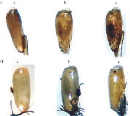
Embryological development- Changes in the appear-ance of developing embryos of P. humanus capitis and P. humanus humanus were assessed at different stages of egg development. The major changes were the visual ap-pearance and darkening of the ocular spots and the vi-sual appearance of the appendages. Pigmented eyes ap-peared as red spots and turned to black during embryo-genesis. The appendages were completely visible at the end of embryonic development.
Based on the colour of the ocular spots and the ap-pearance of the embryo appendages, the developing eggs of head and body lice were divided into three stages: early, medium, and late (Fig. 1). Early eggs were characterized by the absence of external markers, medium eggs showed reddish eyes and appendage outlines, and late eggs showed black eyes and clearly visible appendages (Fig. 1).
Laboratory egg incubation- The influence of tem-perature and humidity on the head louse embryo devel-opment was stated for early, medium, and late eggs. No eclosion was observed in any egg incubated at 18°C, so the effect of humidity at this temperature on development stage could not be assessed. Clearly, the continuous ex-posure of eggs to low temperature (18°C) induced 100% embryo death.
The average percentage of eclosion of early, medium, and late eggs incubated at 27°C and different RH, are shown in Fig. 2. For early embryos, the eclosion increased as humidity increased from 17 to 77%, and abruptly de-creased in eggs incubated at 99% RH (Fig. 2). For medium and late eggs, high percentage of hatched eggs was ob-served at 20, 26, 45, and 77% RH. Emergence was 80, 72, 87, and 86% for medium embryos, and 95, 86, 100, and 100% for late embryos. Low eclosion was observed at 17 and 99% RH in both stages of development (Fig. 2).
The average percentage of eclosion of early, medium, and late eggs incubated at 76-82 % RH and different tem-peratures are shown in Fig. 3. For all, the hatching in-creased as the temperature inin-creased, showing the high-est values from 27 to 31°C. Thus, the optimal laboratory conditions for rearing head louse eggs were 27-3°C and 45-75% RH. According to these ranges, the temperature and humidity finally chosen for the ovicidal test (28ºC, 75% RH), was based on practical criteria.
TABLE I
Relative humidity obtained with saturated aqueous solution of different salts
Salt % RH a ± SE % RH a ± SE
27ºC 18ºC
H2O 99 99
NaCl 76.8 ± 0.3 81.8 ± 0.3
K2CO3 45.4 ± 0.2 48.8 ± 0.2
CaCl2 25.9 ± 0.7
-LiCl 19.8 ± 0.6 29 ± 1.2
Silica gel b 17 ± 0
-a: obtained in closed plastic containers with saturated aqueous solution of different salts; b: obtained in closed plastic containers with granules of anhydrous silica gel.
Early, medium or late eggs were incubated at two tem-peratures (18 and 27 ± 1°C), each one evaluated with in-creasing percentages of humidity (Table I). Similarly, early, medium or late eggs were kept at 76-82% RH (closed con-tainers with saturated aqueous solutions of NaCl) and increasing temperatures (18, 23, 27, and 31 ± 1°C). The number of emerged nymphs was counted, and those with incomplete emergence were considered as dead. Three replicates of at least three groups of ten eggs were ex-posed to each one for each humidity/temperature condi-tion; the average percentage of eclosion was calculated and the fiducial limits at the temperature and humidity were estimated.
259 259 259 259 259 Mem Inst Oswaldo Cruz, Rio de Janeiro, Vol. 101(3), M ay 2006
Bioassay - The treatment of eggs of each develop-ment stage of body and head lice with spinosad, caused embryo death resulting in failure to hatch. In addition it was observed that the treatment with spinosad in early and medium stage did not interrupt embryo development but it failure to hatch.
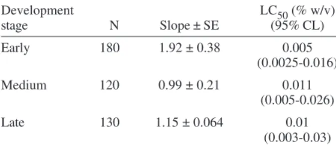
Toxicity values (LC50) to spinosad in early, medium, and late development stage, are shown in Table II. No significant difference was found between the three devel-opment stages according to the overlapped confidence limits.
Toxicity values (LC50) to spinosad and LCR between head and body lice eggs, are shown in Table III. There
was no significant difference in the susceptibility of spinosad between S-BL and GH-HL (LCR = 1).
DISCUSSION
Developing insect eggs represent a continuous chang-ing system that affects the toxicity of insecticides. Labo-ratory rearing of the developing eggs represents an im-portant tool to select similar aged embryos for the ovi-cidal tests. The present paper reports some changes in the morphology of human louse embryos observed through the transparent chorion at different stages of egg development. Based on these, external markers were de-fined for early, medium, and late eggs of head and body lice.
Fig. 1: external appearance of human louse eggs in three different development stages. I: Pediculus humanus capitis; II: P. humanus humanus. a: early development, b: medium development, c: late development. Ocular spots and appendages are visible in medium and late embryos.
Fig. 2: effect of the relative humidity (RH) on the eclosion of early,
260 260 260 260
260 Embryonic development of human lice • Gastón M ougabure Cueto et al.
Similar studies had been reported on the blood suck-ing bug Triatoma infestans (Klug) (Picollo de Villar et al. 1980). In this study, the changes in the external appear-ance of the eggs during the development allowed daily characterization of T. infestans embryogenesis, based mainly on ocular spots and appendages.
The development of the head louse embryo at differ-ent temperature and humidities, demonstrated that the optimal laboratory conditions for rearing P. humanus capi-tis eggs were 27-31°C and 45-75% RH. These intervals overlapped with those reported for P. humanus humanus
(29-31°C, 40-90% RH) (Buxton 1946, Hinton 1981, Kettle 1995). Narrow intervals of environmental conditions were also reported for other louse eggs. Embryos of Damalinia equi (Denny) developed at 31-39°C, and embryos of Li-nognathus pedalis (Osborne) hatched at 33-38°C (Hinton 1981). Considering humidity, embryos of D. ovis (Schr.) developed during water immersion with appreciable mor-tality after 7 day immersion, and exposure to 90% RH 24 h before hatching avoided emergence of first nymph (Hinton 1981). In P. humanus humanus, 70-90 % eclosion was found at 29-31°C and 40-90% RH (Buxton 1946, Hinton 1981, Kettle 1995).
Recent results from our laboratory demonstrated the high insecticide activity of spinosad against adult human lice (Mougabure Cueto et al. 2006 in press). Spinosad is an extract of fermentation of the actinomycete Sac-charopolyspora spinosa. The spinosad alters the func-tion of nicotinic receptors of acetylcholine and gamma
-aminobutyric acid (GABA)-gated chloride channels (Sparks et al. 2001). Based on the adult activity, it was an important goal to evaluate the effectiveness of spinosad against human louse eggs. To assess the ovicide action, we immersed eggs in acetone dilutions of spinosad. This method has not been previously reported for human louse eggs. The results showed similar susceptibility to spinosad in the 3 stages of the embryonic development of head lice. However, Picollo et al. (1976) reported that the early stage of T. infestans was more tolerant to organophos-phate insecticides than medium and late stages, and dem-onstrated that the degradative enzymes present in the early stage metabolized the insecticides before of the ap-pearance of the nervous system. On the contrary, spinosad is poorly or not readily metabolized in insect (Sparks et al. 2001). Consequently the insecticide applied to early eggs could be accumulate in the developing embryo and exert its toxic effect after the appearance of the target. This hypothesis is in accordance with the fact that head lice eggs treated in early stage almost completed its develop-ment before they died.
Toxicity values of head and body louse eggs showed similar susceptibility to spinosad, an insecticide that has not been used as pediculicide in Argentina. In agreement with these results, previous reports showed similar sus-ceptibility for postembryonic body and head lice to insecticides not used for the control of pediculosis (Mumcuoglu et al. 1990, Hemingway et al. 1999, Lee et al. 2000, Mougabure Cueto et al. 2006. in press).
Standardized laboratory development conditions for louse eggs, identification of external markers of different stages of development, and the immersion method de-scribed in this work represent a new and alternative ap-proach for evaluating insecticide toxicity in human louse eggs.
REFERENCES
Abbott WS 1925. A method of computing the effectiveness of an insecticide. J Econ Entomol 18: 265-267.
Berman EL, Firstenberg D 1979. The human body louse egg-correlative study of anatomy by SEM and light micros-copy. Scanning Elect Microsc 3: 197-202.
Burgess IF 1999. Dermatopharmacology of antiparasitics and insect repellents. In B Gabard, P Elsner, C Surber, P Treffel (eds), Dermatopharmacology of Topical Preparations, Springer-Verlag, Heidelberg, p. 157-178.
Burkhart CN, Arbogast J, Smythe P, Burkhart CG 1999a. His-tochemical analysis of the nit of Pediculus humanus capitis
(Anoplura: Pediculidae). J Med Entomol 36: 530-532.
Burkhart CN, Burkhart CG, Gunning WT, Arbogast J 1999b. Scanning electron microscopy of human head louse (Anoplura: Pediculidae) egg and its clinical ramifications. J Med Entomol 36: 454-456.
Burkhart CN, Stankiewicz BA, Pchalek I, Kruge MA Burkhart, CG 1999c. Molecular composition of the louse sheath. J Parasitol 85: 559-561.
Buxton PA 1946. The Louse: an Account of the One Which Infest Man, their Medical Importance and Control, 2nd ed., Edward Arnold & Co, London, 164 pp.
TABLE II
Comparative susceptible values (LC50) to spinosad in early, medium, and late embryos
Development LC50 (% w/v)
stage N Slope ± SE (95% CL)
Early 180 1.92 ± 0.38 0.005
(0.0025-0.016)
Medium 120 0.99 ± 0.21 0.011
(0.005-0.026)
Late 130 1.15 ± 0.064 0.01
(0.003-0.03)
LC50 expressed as percentage of insecticide (g) in 100 ml acetone solution.
TABLE III
Susceptibility values (LC50) to spinosad and lethal concentration ratio (LCR) in head and body louse eggs
Population N Slope ± SE LC50 (% w/v) LCR (95% CL) (95% CL)
S-BL 120 0.68 ± 0.046 0.01
-(0.004-0.045)
GH-HL 180 1.15 ± 0.064 0.01 1.0
(0.003-0.03) (0.76-1.42)
261 261 261 261 261 Mem Inst Oswaldo Cruz, Rio de Janeiro, Vol. 101(3), M ay 2006
Gilbert SF 1997. Development Biology, 5th ed., Sinauer Associ-ates Inc, Sunderland, Massachusetts, 918 pp.
Hatsushika R, Naramoto S, Miyoshi K 1983. Scanning electron microscope studies on head louse, Pediculus humanus capitis
(Anoplura: Pediculidae). Kawasaki Med J 9: 109-119.
Hemingway J, Miller J, Mumcuoglu KY 1999. Pyrethroid re-sistance mechanisms in the head louse Pediculus capitis
from Israel: implications for control. Med Vet Entomol 13: 89-96.
Hinton HE 1981. Phthiraptera. In HE Hinton, Biology of Insect Eggs, Pergamon Press, Oxford, p. 549-562.
Ibarra I 1993. Lice (Anoplura). In RP Lane, RP Crosskey (eds),
Medical Insects and Arachnids, Chapman & Hall, London, p. 517-529.
Kettle DS 1995. Phthiraptera. In DS Kettle, Medical and Vet-erinary Entomology, CAB International, Wallingford, p. 361-382.
Kim K, Ludwig HW 1978. The family classification of the Anoplura. Syst Entomol 3: 249-284.
Lee SH, Yoon K, Williamsom MS, Goodson SJ, Takano-Lee M, Edman JD, Devonshire AL, Clark M 2000. Molecular analy-sis of kdr-like reanaly-sistance in permethrin-reanaly-sistant strain of head lice, Pediculus capitis. Pestic Biochem Physiol 66: 130-143.
Lichfield JT, Wilcoxon FJ 1949. A simplifed method of evalu-ating dose-effect experiments. J Exp Therap 96:99-100.
Mougabure Cueto GA, Zerba EN, Picollo MI 2006. Permethrin resistant head lice in Argentine are susceptible to spinosad.
J Med Entomol (in press)
Mumcuoglu KY, Miller J, Galum R 1990. Susceptibility of the human head and body louse, Pediculus humanus (Anolplura: Pediculidae) to insecticides. Insect Sci Appl 11: 223-226.
Picollo de Villar MI, Zerba EN, Wood EJ, Licastro SM 1980. Neurogenesis and occurrence of cholinesterase in eggs of
Triatoma infestans. Comp Biochem Physiol 65: 65-70.
Picollo MI, Vassena C, Casadío A, Máximo J, Zerba EN 1998. Laboratory studies about susceptibility and resistance to insecticides in the head lice Pediculus capitis. J Med Entomol 35: 814-817.
Picollo MI, Wood EJ, Licastro SA, Zerba EN 1976. Acción ovicida de insecticidas organofosforados en Triatoma infestans. Acta Biochem Clin Latin 10: 309-319.
Robertson JL, Preisler HK 1992. Pesticide Bioassays with Arthropods, CRC, Boca Raton, FL, 127 pp.
Smallman BN, Mansingh A 1969. The cholinergic system in insect development. Ann Rev Entomol 14: 387-408.
Smith EH, Salkeld EH 1966. The use and action of ovicides.
Ann Rev Entomol 11: 331-368.
Smith EH, Wagenknecht AC 1959. The ovicidal action of orga-nophosphate insecticides. Can J Biochem Physiol 37: 1133-1144.
Sparks TC, Crouse GD, Durst G 2001. Naturals products as insecticides: the biology, biochemistry and quantitative structure-activity relationships of spinosyns and spinosoids. Pest Manag Sci 57: 896-905.
Tahmisian TN 1943. Enzimes in ontogenesis: choline-esterase in developing Melanoplus differentialis eggs. J Exp Zool 92: 199-213.
Vassena CV, Mougabure Cueto G, Gonzales Audino P, Alzogaray RA, Zerba EN, Picollo MI 2003. Prevalence and levels of permethrin resistance in Pediculus humanus capitis De Geer (Anoplura: Pediculidae) from Buenos Aires, Argentina.