Proliferating Cell Nuclear Antigen (PCNA) and p53
Protein Expression in Ameloblastoma and
Adenomatoid Odontogenic Tumor
Carlos Augusto Galvão BARBOZA1
Leão PEREIRA PINTO2
Roseana de Almeida FREITAS2
Antônio de Lisboa Lopes COSTA2
Lélia Batista de SOUZA2
1Department of Morphology, Faculty of Dentistry, Federal University of Rio Grande do Norte, Natal, RN, Brazil 2Department of Oral Pathology, Faculty of Dentistry, Federal University of Rio Grande do Norte, Natal, RN, Brazil
In this study, proliferating cell nuclear antigen (PCNA) and p53 protein expressions were analyzed in 16 cases of ameloblastoma and 8 cases of adenomatoid odontogenic tumor (AOT). The cases of ameloblastoma consisted of solid type tumors and histologic arrangements of different subtypes were observed. In some specimens, more than one histologic subtype was identified in the same lesion, and each tumor was categorized according to the predominant cell pattern. The odontogenic tumors were grouped as follows: follicular ameloblastoma (n=7), plexiform ameloblastoma (n=4), acanthomatous + follicular ameloblastoma (n=3), basal cell ameloblastoma (n=2), adenomatoid odontogenic tumor (n=8). PCNA immunohistochemical expression revealed stronger quantitative labeling index for the follicular ameloblastoma, while for p53 protein the strongest quantitative labeling index was detected in the plexiform type. Nevertheless, statistical analysis using ANOVA and Tukey’s test did not detect significant differences (p>0.05) among the histologic subtypes of ameloblastoma. The findings of this study suggest that the different histologic patterns of ameloblastoma did not show a direct correlation with their clinical behavior and consequently with the prognosis of the cases. The results also indicated that the ameloblastoma has greater proliferative potential than the AOT, which can contribute to explain its more aggressive and invasive characteristics.
Key Words: ameloblastoma, adenomatoid odontogenic tumor, PCNA, p53 protein.
Correspondence: Profa. Dra. Lélia Batista de Souza, Programa de Pós-Graduação em Patologia Oral, UFRN, Departamento de Odontologia, Av. Salgado Filho, 1787, Lagoa Nova, 59056-000 Natal, RN, Brasil. e-mail: [email protected]
INTRODUCTION
Odontogenic tumors are remarkable among oral lesions because of their clinic and histologic heteroge-neity. This diversity reflects in the complex develop-ment of dental structures because odontogenic tumors derive from aberrations in odontogenesis. The amelo-blastoma deserves special attention, not only because of its particular biologic behavior, exhibiting great infiltrative potential, high recurrence rate and capacity to metastasize, but also due to the relatively high fre-quency that it is diagnosed among odontogenic tumors. The adenomatoid odontogenic tumor (AOT), on the other hand, in spite of sharing a common origin
(epithelial tissue) with the ameloblastoma, causes less pain and can be treated with conservative surgery, with extremely few reports of recurrence or metastasis.
Three clinically distinct types of ameloblastoma are recognized: solid or multi-cystic, unicystic and peripheral, each of them presenting a specific biologic behavior and thus different prognosis and treatment (1). From a histologic standpoint, the ameloblastoma exhibits distinct microscopic characteristics and a vari-able histologic pattern, according to which they are classified as follicular, plexiform, granular, basal cell,
acanthomatousand desmoplastic (2). Despite this
Divergent opinions are found in the literature with respect to the possible relation between the histo-logic type and the clinical behavior of a lesion. It has been asserted (3) that the follicular ameloblastoma recurs more often than the plexiform type and that the unicystic ameloblastoma shows lower recurrence rate than the solid type.
Knowledge of the biologic behavior of patho-logic entities affecting the oral cavity, including the odontogenic tumors, is essential for rendering the most appropriate therapeutic approach and establishing a prognosis for each case. This has led several oral pa-thologists to investigate different aspects related to the molecular biology of cell populations in tumors, in an attempt to elucidate many points that still remain un-clear, resorting to a variety of methodologies. Among these are the tritiated thymidine ([3H]-thymidine) incor-poration followed by autoradiography; bromodeoxyuridine incorporation followed by anti-BrdU immunohistochemistry; flow cytometry analysis; and immunohistochemistry using specific monoclonal anti-bodies that recognize
antigens related to the dynamics of the cell cycle(4).
Several studies (5-8) have tried to explain the mechanisms of proliferative activity of various lesions with an oral origin. Regarding the ameloblastoma, vari-able results have been shown (9-13) with respect to its histologic subtypes.
The purpose of this immunohistochemical study was to investigate PCNA and p53 protein expressions in ameloblastoma and adenomatoid odontogenic tu-mor, as they are known to play important roles in cell proliferation and tumorigenesis.
MATERIAL AND METHODS
The material used in this study consisted of 16 cases of ameloblastoma and 8 cases of adenomatoid odontogenic tumor obtained from the files of the De-partment of Oral Pathology of the Federal University of Rio Grande do Norte, Natal, RN, Brazil.
The paraffin-embedded material was prepared in 5-µm-thick slices, which were stained with hema-toxylin and eosin, mounted on microscope slides and examined by optical microscopy to evaluate the mor-phologic aspects of the sample.
The cases of ameloblastoma consisted of solid type tumors and histologic arrangements of different subtypes were observed. In some specimens, more
than one histologic subtype was identified in the same lesion, and each tumor was categorized according to the predominant cell pattern. The odontogenic tumors were grouped as follows: follicular ameloblastoma (n=7), plexiform ameloblastoma (n=4), acanthomatous + fol-licular ameloblastoma (n=3), basal cell ameloblastoma (n=2), adenomatoid odontogenic tumor (n=8).
For the immunohistochemical reaction, 3-µ m-thick slices were obtained from the paraffin-embedded material and submitted to the following methodology: paraffin removal; hydration in a decreasing ethanol series; removal of formalin pigment with 10% ammonium hydroxide in 95% ethanol; treatment in a microwave oven (3 cycles of 5 min at maximum power of 700 watts); blocking of endogenous peroxidase using a hydrogen peroxide solution; incubation with primary anti PCNA antibody (PC-10, diluted 1:80; Biogenex, San Ramon, CA, USA) for 18 h and anti-p53 antibody (DO-7, diluted 1:50; DAKO, Carpinteria, CA, USA) for 18 h; incubation in secondary serum; streptoavidin-biotin complex (SABC) for 30 min at room temperature; development of the reaction with diaminobenzidine. Between stages, the material was immersed in a Tris phosphate buffer solution, pH 7.4 (Vetec, Rio de Janeiro, RJ, Brazil). After development of the reaction, the material was counterstained with Mayers hema-toxylin (Vetec) in Permount (Fisher Scientific, New Jersey, NY, USA).
The slices submitted to immunohistochemical reaction were observed by optical microscopy both for analysis of the presence of a reaction and for quantita-tive assessment of the areas corresponding to the histo-logic patterns of ameloblastoma and AOT.
The quantitative analysis for PCNA and p53 expressions was undertaken using the index of positiv-ity (IP), calculated by the relation between the number of PCNA and p53 positive cells per 1000 cells counted for each case studied, multiplied by 100 [(PCNA- and p53-positive cells/1000 cells) X 100]. Counting was undertaken using optical microscopy with X1000 mag-nification.
RESULTS
labeling.
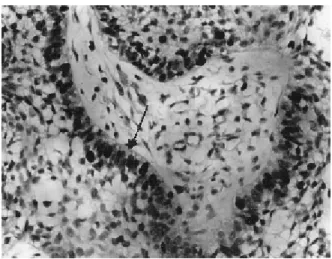
In the cases of ameloblastoma, the peripheral cylindrical epithelial cells of the islets presented, in general, strong PCNA labeling index (Fig. 1) and mod-erate to weak p53 protein labeling index (Fig. 2). Some cells in the central area of the follicles resembling the stellate reticulum of the enamel organ also presented positive PCNA labeling index. In the cases of AOT, the PCNA positive cells showed strong labeling (Fig. 3) while the cells labeled with anti-p53 antibody showed weak labeling (Fig. 4).
The results of the quantitative analysis of the PCNA and p53 positive cells are given in Table 1.
PCNA and p53 protein labeling indices were compared in the five groups of lesions using one-way ANOVA. The results showed no statistically signifi-cant difference (p>0.05) among the groups for PCNA, whereas significant difference (p<0.05) was detected for p53 protein. Multiples comparisons using Tukey’s test showed significant difference (p<0.05) between the follicular ameloblastoma and the AOT regarding the indices of positivity for the p53 protein.
Figure 1. Histologic section of the follicular ameloblastoma showing strong nuclear staining for PCNA (arrow). SABC. (original magnification X 200).
Figure 2. Histologic section of the follicular ameloblastoma showing p53 positive cells (arrow). SABC. (original magnification X 200).
Figure 3. Histologic section showing positive cells in AOT with moderate staining for PCNA (arrow). SABC. (original magnification X 200).
DISCUSSION
A possible correlation between the biologic be-havior of ameloblastomas and their histologic appear-ance has been investigated over the years in an attempt to establish histologic criteria that could be helpful not only in the treatment but also in the establishment of a
prognosis for these lesions. According to Gardner(1), it
is important to recognize the clinical types of ameloblastomas because the unicystic and peripheral tumors have better prognosis after conservative resec-tion than the solid ameloblastomas. The sample we examined in this study consisted of solid ameloblastomas and the following histologic subtypes were identified: follicular, plexiform, acanthomatous, basal cell and desmoplastic. In some cases, more than one histologic subtype was found in the same lesion, as reported in the literature. Histologic patterns of granular cells and clear cells were not observed.
Studies (1,14) have postulated that there is no correlation between the histologic types of ameloblastomas and the clinical behavior (and consequent prognosis) of these lesions because more than one cellular configuration can be seen in a single lesion. On the other hand, Ueno
et al.(15) stated that it is possible to establish a correlation
between both the patient’s age and the radiographic/
histologic aspects of a tumor and its clinical behavior. They also reported that the ameloblastoma with a follicu-lar histologic pattern presents higher recurrence rate, which is consistent
with the findings of Reichart et al.(3),
who reported that the follicular amelo-blastoma recurs more often than the plexiform ameloblastoma.
Studies using the AgNOR tech-nique (5,12) have found no differ-ences in the cellular activity of the ameloblastoma and the adenomatoid odontogenic tumor, but statistically significant differences were observed (5) between the follicular and plexi-form ameloblastoma subtypes. In the present study, there was statistically significant higher incidence of PCNA labeling in the cases of ameloblastoma (mean 76.0%) than in the cases of AOT (mean 56.6%). Higher PCNA labeling index may indicate higher cellular proliferation rate, which would explain the more aggressive biologic behavior of the ameloblastoma compared to the adenomatoid odontogenic tumor. However, PCNA expression probably results not only form cellular pro-liferation, but also from other sources, including DNA repair (16) and factors influencing the increase of autocrine and paracrine rates in messenger-RNA of the proliferating cell nuclear antigen (17). Because of these high levels, PCNA can be found in cells that are not in the cellular cycle. According to Scott et al. (18), another important issue that should be considered is that PCNA has a considerably longer half-life (approximately 20 h) compared to the rapid cell cycle time.
Taking into account the various histologic sub-types of ameloblastoma recognized in the sample, the 16 cases included in this study were categorized according to the predominant cell pattern. The follicular ameloblastoma presented the strongest PCNA labeling index (mean 78.4%), while the basal cell ameloblastoma showed the weakest labeling index (mean 69.7%). One-way analysis of variance did not detect statistically significant differences either among the histologic sub-types of ameloblastoma or between the cases of amelo-blastoma and AOT. These findings are consistent with those of Kim and Yook (6), who did not find differences
Table 1. Statistical results for PCNA and p53 indices of positivity in the odontogenic tumors, according to histologic subtype.
n Mean ± SD Minimum Maximum
PCNA index
Follicular ameloblastoma 7 78.4 ± 15.9 59.7 95.7
Plexiform ameloblastoma 4 74.3 ± 16.9 51.0 91.3
Follicular + acanthomatous 3 77.5 ± 8.2 68.9 85.3
ameloblastoma
Basal cell Ameloblastoma 2 69.7 ± 23.5 53.1 86.3
Adenomatoid odontogenic tumor 8 56.6 ± 18.4 28.9 82.0
p53 index
Follicular ameloblastoma 7 42.4 ± 14.1* 23.6 59.7
Plexiform ameloblastoma 4 46.0 ± 15.9 26.8 64.2
Follicular + acanthomatous 3 31.6 ± 27.8 3.8 59.3 ameloblastoma
Basal cell Ameloblastoma 2 26.1 ± 16.9 14.1 38.0
Adenomatoid odontogenic tumor 8 19.5 ± 11.1* 7.2 34.9
in the proliferative activity among the different histologic types of solid ameloblastoma and Carvalhais et al. (19) who did not find significant difference between the follicular and the plexiform ameloblastoma. Nevertheless, these findings diverge from those of Funaoka et al. (9), who reported higher PCNA labeling index for the follicular than for the plexiform ameloblastoma, and Kumamoto et al. (10), who stated that the basal cell ameloblastoma possesses more proliferative activity than other types of ameloblastoma. An alternative statistical analysis using the Student’s t-test, which is less conservative, was also done but no difference was found among the various histologic subtypes of ameloblastoma. Statistically significant difference was detected only between the follicular ameloblastoma and the AOT, the first presenting greater proliferative potential. According to Reichart et al. (3) and Ueno et al. (15), this could explain the higher recurrence rate of follicular ameloblastomas.
All lesions in this study presented p53 positive cell labeling, the mean labeling index being 37.2% among the 16 cases of ameloblastoma and 19.5% among the 8 cases of AOT. The cases of ameloblastoma pre-sented, in general, moderate to weak p53 labeling index, while the cases of AOT presented weak p53 labeling index. Considering the different histologic pattern of ameloblastoma, it was observed that the plexiform subtype had the strongest labeling index (mean 46.0%), followed by the follicular subtype (mean 42.4%). Comparing the different histologic patterns of ameloblastoma among each other and with the AOT, one-way ANOVA detected significant differences be-tween the groups of lesions. Tukey’s test for multiple comparisons identified significant difference between the follicular ameloblastoma and the AOT. An alterna-tive statistical analysis using the Student’s t-test, which is less conservative, detected significant difference be-tween the plexiform ameloblastoma and the AOT. The cases of plexiform ameloblastoma having the strongest p53 labeling index could be indicative of the higher level of cellular activity. However, we believe that comparison of our results for p53 labeling index to those of other studies may not be useful to assess the kinetic cellular events in cellular kinetic tumors be-cause variations in the methodology, such as undertak-ing or not antigenic recuperation in a microwave oven, can considerably alter the labeling index, as shown by Dowell and Ogden (20).
PCNA and p53 protein immunodetection results showed that the ameloblastoma has greater proliferative potential than the AOT, which can be helpful to explain the more aggressive biologic behavior of ameloblastomas. However, our results differ from those obtained using AgNOR technique (5,12), in which differences between the ameloblastoma and the AOT were established in terms of cellular activity.
In this study, the morphologic analysis of amelo-blastoma cases identified the presence of more than one histologic pattern in the same lesion. PCNA immuno-histochemical expression revealed stronger quantita-tive labeling index for the follicular ameloblastoma, while for p53 protein the strongest quantitative labeling index was detected in the plexiform type. Nevertheless, statistical analysis did not detect significant differences among the histologic subtypes of ameloblastoma. The findings of this study suggest that the different histo-logic patterns of ameloblastoma did not show a direct correlation with their clinical behavior and consequently with the prognosis of the cases. The results also indi-cated that the ameloblastoma has greater proliferative potential than the AOT, which can contribute to explain its more aggressive and invasive characteristics.
RESUMO
REFERENCES
1. Gardner DG. Some current concepts on the pathology of ameloblastomas. J Oral Surg Oral Med Oral Pathol Oral Radiol Endod 1996;82:660-669.
2. Waldron CA. Odontogenic cysts and tumors. In: Oral & Maxillofacial Pathology. Neville BW, Damm DD, Allen CM, Bouquot JE (Editors). Philadelphia: WB Saunders; 1995. p. 511-538.
3. Reichart PA, Philipsen HP, Sonner S. Ameloblastoma: biological profile of 367 cases. Eur J Cancer B Oral Oncol 1995;31B:86-99.
4. Garcia RL, Coltrera MD, Gown AM. Analysis of proliferative grade using anti-PCNA/cyclin monoclonal antibodies in fixed, embedded tissues: comparison with flow cytometric analysis. Am J Pathol 1989;134:134-139.
5. Do Carmo MA, Silva EC. Argyrophilic nucleolar organizer re-gions (AgNORs) in ameloblastomas and adenomatoid odontoge-nic tumours (AOTs). J Oral Pathol Med 1998;27:153-156. 6. Kim J, Yook JI. Immunohistochemical study on proliferating cell
nuclear antigen expression in ameloblastomas. Eur J Cancer B Oral Oncol 1994;30B:126-131.
7. Meer S, Galpin JS, Altini M, Coleman H, Ali H. Proliferating cell nuclear antigen and Ki67 immunoreactivity in ameloblastomas. Oral Surg Oral Med Oral Pathol Oral Radiol Endod 2003;95:213-221.
8. Jãaskelainen K, Jee KJ, Leivo I, Saloniemi I, Knuutila S, Heikinheimo K Cell proliferation and chromosomal changes in human ameloblastoma. Cancer Genet Cytogene 2002;136:31-37.
9. Funaoka K, Arisue M, Kobayashi S, Iizuka T, Kohgo T, Amemiya A, Totsuka Y. Immunohistochemical detection of proliferating cell nuclear antigen (PCNA) in 23 cases of ameloblastoma. Eur J Cancer B Oral Oncol 1996;32B:328-332.
10. Kumamoto H, Kimi K, Ooya K. Detection of cell cycle-related factors in ameloblastomas. J Oral Pathol Med 2001;30:309-315.
11. Sandra F, Mitsuyasu T, Nakamura N, Shiratsuchi Y, Ohishi M. Immunohistochemical evaluation of PCNA and Ki67 in ameloblastoma. Oral Oncol 2001;37:193-198.
12. Medeiros MCS, Freitas RA, Souza LB. Análise quantitativa das regiões organizadoras nucleolares nos diversos tipos histológicos do ameloblastoma e no tumor odontogênico adenomatóide. RGO 1997;45:67-70.
13. Souza LB, Sousa CV, Pereira Pinto L. Estudo morfológico e imuno-histoquímico da membrana basal em diversos padrões histológicos do ameloblastoma. RPG Rev Pós Grad 2001;8:40-45.
14. Thompson IO, van Rensburg LJ, Phillips VM. Desmoplastic ameloblastoma: correlative histopathology, radiology and CT-MR imaging. J Oral Pathol Med 1996;25:405-410.
15. Ueno S, Mushimoto K, Shirasu R. Prognostic evaluation of ameloblastoma based on histologic and radiographic typing. J Oral Maxillofac Surg 1989;47:11-15.
16. Shivji MKK, Kenny MK, Wood RD. Proliferating cell nuclear antigen is required for DNA excision repair. Cell 1992;69:367-374.
17. Hall PA, Levison DA, Woods AL. Yu CC, Kellock DB, Watkins JA, Barnes DM, Gillett CE, Camplejohn R, Dover R, Waseem NH, Lane DP. Proliferating cell nuclear antigen (PCNA) immu-nolocalization in paraffin sections: an index of cell proliferation with evidence of deregulated expression in some neoplasms. J Pathol 1990;162:285-294.
18. Scott RJ, Hall PA, Haldana JS, Van Noorden S, Price Y, Lane DP, Wrigh NA. A comparison of immunohistochemical markers of cell proliferation with experimentally determined growth frac-tion. J Pathol 1991;165:173-178.
19. Carvalhais JN, Aguiar MCF, Araújo VC, Araújo NS, Gomez RS. p53 and MDM2 expression in odontogenic cysts and tumours. Oral Dis 1999;5:218-222.
20. Dowell SP, Ogden GR. The use of antigen retrieval for immuno-histochemical detection of p53 over-expression in malignant and benign oral mucosa: a cautionary note. J Oral Pathol Med 1996;25:60-64.