1Institute of Neurology and Neuro s u rg e ry, Passo Fundo RS, Brazil;2D e p a rtment of Pathology, Hospital São Vicente de Paulo, Passo Fundo RS, Brazil
Received 1 February 2005, received in final form 29 April 2005. Accepted 31 May 2005.
D r. Cassiano Mateus Forcelini - Rua Paissandu 1644/302 - 99010-120 Passo Fundo RS - Brasil. E-mail: cassianoforc e l i n i @ b rt u r b o . c o m . b r, [email protected]
METASTATIC PROLACTINOMA
Case report with immunohistochemical
assessment for p53 and ki-67 antigens
Paulo S. Crusius
1, Cassiano M. Forcelini
1, Adroaldo B. Mallmann
1, Daniela A. Silveira
2,
Elder Lersch
2, Cláudio A. Seibert
1, Marcelo U. Crusius
1, Charles A. Carazzo
1,
Cassiano U. Crusius
1, Eduardo Goellner
1ABSTRACT - Pituitary carcinomas are rare neoplasms characterized by craniospinal and/or systemic metas-tases originated from the pituitary. Their histopathology is frequently indistinguishable from that of benign adenomas. The development of markers that better reflect their behavior is desirable. We present the case of a 47 year-old man with a pro l a c t i n - s e c reting macroadenoma who was submitted to surgeries, cranial rdiation therapy, and bromocriptine treatment, but evolved to a fatal outcome after the disclosure of int r a c r a-nial metastases. Tumor samples underwent p53 and Ki-67 immunohistochemical assessment. p53 was absent in all samples, a rare finding among pituitary carcinomas. Ki-67 proliferative index was 2.80% in the origi-nal tumor, 4.40% in the relapse, and 4.45% in the metastasis. The figure in the relapse is higher than the expected for a noninvasive adenoma. In conclusion, p53 staining is not positive in all pituitary carc i n o m a s . A high Ki-67 proliferative index in a pituitary adenoma might indicate a more aggressive behavior.
KEY WORDS: prolactinoma, pituitary carcinoma, metastasis, p53, Ki-67.
P rolactinoma metastático: relato de caso com avaliação imuno-histoquímica para os antígenos p53 e Ki-67
RESUMO - Carcinomas pituitários são neoplasias raras caracterizadas pela presença de metástases cranio-espinhais e/ou sistêmicas originadas da hipófise. Sua histopatologia é freqüentemente indistinguível daque-la dos adenomas benignos. O desenvolvimento de marc a d o res que melhor reflitam o seu comport a m e nto é desejável. Apresentamos o caso de um homem de 47 anos com um macroadenoma secretor de pro l a c t i n a que foi submetido a procedimentos cirúrgicos, radioterapia e tratamento com bromocriptina, mas que e v o l u i u para o óbito após o descobrimento de metástases intracranianas. Amostras do tumor foram submetidas à a n á-lise imuno-histoquímica para os antígenos p53 e Ki-67. A coloração para p53 foi negativa em todas as amos-tras, um achado raro entre os carcinomas pituitários. O índice proliferativo Ki-67 foi 2,80% no tumor origin a l , 4,40% na recidiva e 4,45% na metástase. O valor obtido na recidiva é maior que o esperado para um adeno-ma não-invasor. Concluindo, a coloração para p53 não é positiva em todos os carcinoadeno-mas pituitários. Um í n d i c e proliferativo Ki-67 alto em um adenoma pituitário poderia indicar um comportamento mais agressivo.
PALAVRAS-CHAVE: prolactinoma, carcinoma pituitário, metástase, p53, Ki-67.
The prevalence of clinically overt pituitary tu-mors is around 0.02% of the population1. Most are
m i c roadenomas (<10 mm), exhibit benign behavi-o r, and prbehavi-oduce behavi-one behavi-of the hbehavi-ormbehavi-ones secreted by the normal gland. Relatively few people develop m a c ro a d e n o m a s2, usually with a benign
phenoty-pe, but that can occasionally become aggressive, i n-filtrating adjacent stru c t u res and re c u rring re p e a
t-edly despite conventional treatment (invasive ade-n o m a s )3 , 4. Rare l y, adenomas present even more
a g g re s s i v e l y, developing intracranial and/or e x t r a c r a-nial metastases (pituitary carcinomas). The pre s e n-ce of tumor tissue not contiguous with sella turc i-ca is a condition re c q u i red for diagnosis of a pitui-t a ry carc i n o m a5. Its prevalence is estimated aro u n d
Few more than 140 cases of pituitary carc i n o m a s have been re p o rted in the literature7, 30% of them
represented by prolactin-secreting tumors8.
The etiology of pituitary carcinomas is poorly understood and their histology is generally indistin-guishable from that of benign adenomas9 - 1 1. Such
factors added to the poor response of such neo-plasms to cytotoxic chemotherapy and their fatal o u t c o m e7 , 1 1 - 1 3place a challenge towards more
pre-cocious diagnosis or suspicion of aggressive behavi-o r1 3. Survival after documentation of metastatic
disease is poor; 66% of patients die within 1 year7.
It has recently been proposed that markers of ag-g ressiveness other than standard histoloag-gy better reflect the behavior of metastatic pituitary adeno-m a s1 0 , 1 1 , 1 4. Immunohistochemical stainings for the
e x p ression of the protein produced by mutated p53 gene and for Ki-67 (MIB-1) index of cellular p roliferation have been suggested as the most pro-mising of those markers1 0 , 1 1. However, it re m a i n s
a theoretical issue whether such immunohistoche-mical analysis could identify a trend for local inva-sion or metastatic dissemination when applied to patients with macroadenomas.
CASE
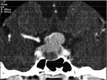
A 47-year-old white man initiated with re c u rrent e p i-sodes of dizziness and vomiting in 1994. Some months l a-ter a moderate sustained fronto-temporal headache, d ro-wsiness, and a defect on the visual fields have appeare d . F u rther health problems were smoking, obesity, hyperli-pidemia, and erectile dysfunction of recent onset. On M a rch 1995 a neuro s u rgeon ord e red a computed tomo-graphy (CT scan) of the head that exhibited a slight brain a t rophy and a space-occupying lesion in sella turc i c a with 2.5 cm on its largest diameter, invading suprasell a r c i s t e rn (Fig 1). Serum prolactin level was higher than 3 , 0 0 0 ng/ml (Table 1), and the rest of his endocrinal profile w a s not evaluated at that time. The patient was submitted to a surgical intervention through transsphenoidal ap-p roach on Aap-pril 1995. Histoap-pathological examination c o n f i rmed a pituitary adenoma. The prolactin dro p p e d after surg e ry (on the second day it was 634.4 ng/ml) ac-companied by the gradual abatement of the symptoms. On November 1995 a new CT scan demonstrated a tumoral relapse with 1.7 cm on its larger diameter, still invading suprasellar cistern. Further CT scans perf o rm e d 9 and 23 months later evidenced little additional gro w t h . At the end of 1997 the patient looked for assistance by our group. He was still complaining about visual diff i c u l-t y. Examinal-tion of l-the visual fields showed upper bil-tem- bitem-poral quadrantanoptic defect. The serum prolactin lev-el was 2,717 ng/ml (Table). A new surgical pro c e d u re w a s u n d e rtaken, this turn by transcranial approach (Novem-ber 1997). Even though the normalization of the
visu-al fields and a great fvisu-all in serum prolactin level have h a p-pened, the latter did not return to normal range.
On March 1998 a tumoral mass with a slight suprasel-lar prominence was again manifest on a CT scan, and se-rum prolactin level raised up to 1,025 ng/ml. The patient was then re f e rred to radiation theraphy, and received 50 Gy of external beam radiation in 35 fractions to sella thro-ughout 40 days. Some months later (August 1999) re m a r k-able decrease of tumoral dimensions on a new CT scan and of serum prolactin levels was evident (Table). A mag-netic resonance imaging (MRI) scan of brain and pituitary gland perf o rmed on June 2000 comproved the re d u c t i o n in tumor size: there was just a small intrasellar spot that had high signal on T2 and was less contrast-enhancing than the rest of the gland, a finding that was interpre t-ed as a surgical sequel or a small persistent tumor.
Since 1999 treatment with bromocriptine and hor-mone replacement (thyroxine, prednisone, testostero n e , and vasopressin) were carried out, the latter due to a h o r-monal pattern of panhypopituitarism. Adverse eff e c t s (nausea, stomacache) attributed to the increasing dai-ly doses of bromocriptine employed since 2000 made t h e patient unwilling to accept the therapy. From May to August 2000 he was submitted to a trial with carbego-line 1 mg a week, but serum prolactin level still rose ( f ro m 230.4 ng/ml to 413.0 ng/ml). On October 2000 he re f u s e d the re t u rn to high doses of bromocriptine proposed by medical staff (17.5 mg a day) and quit accompaniment. Even so, he kept the use of hormone replacement and a low daily dose of bromocriptine (5 mg).
On April 2001 the patient started to feel an intense pain on lumbar region with spread to the posterior as-pect on the right lower limb, what led him to consult an o rthopedist. A CT scan of lumbar spine showed an image suggestive of lumbar interv e rtebral disk prolapse on L5-S1 level. Surgical decompression was undertaken and t h e disk prolapse was confirmed and excised. However,
ing the pro c e d u rethe patient suff e red cardiac arrh y t h-mia followed by cardiac arrest that re q u i red more than 20 minutes to be reversed. After that, he exhibited an anoxic-ischemic encephalopathy: bilateral Babinski’s sign, limb withdrawal to pain, no verbal response, spont a-neous eye openning, no evident brain stem damage, a n d myoclonic jerks in limbs and face. Neurological accompa-niment by our staff was then solicited. A CT scan of the head disclosed a surprising finding: multiple hyperinten-se lesions with slight contrast enhancement scattered o n
left cerebral hemisphere convexity (Fig 2). No evident t u-mor in sella or parasellar region was detected.
In the following days a MRI scan of brain and pitui-t a ry gland confirmed pitui-the presence of pitui-the lesions and absence of a sellar mass. A biopsy of one of the menin-geal tumors revealed a pituitary adenoma. The contem-porary serum prolactin level was 2,373.0 ng/ml (Table). Evolution was unfavourable, with installation of pneu-monia and renal insuff i c i e n c y. Cardiovascular status remained critical, without evidence of myocardial
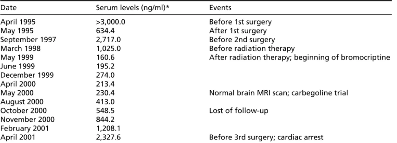
infarc-Table. Serum prolactin levels during the evolution of the present case.
Date Serum levels (ng/ml)* Events
April 1995 >3,000.0 Before 1st surgery
May 1995 634.4 After 1st surgery
September 1997 2,717.0 Before 2nd surgery
March 1998 1,025.0 Before radiation therapy
May 1999 160.6 After radiation therapy; beginning of bromocriptine
June 1999 195.2
December 1999 274.0
April 2000 213.4
May 2000 230.4 Normal brain MRI scan; carbegoline trial
August 2000 413.0
October 2000 548.5 Lost of follow-up
November 2000 844.2 February 2001 1,208.1
April 2001 2,327.6 Before 3rd surgery; cardiac arrest *Normal range: (Men) 2.0 to 14.5. MRI: magnetic ressonance imaging.
tion. The patient died 8 days after the surgical interv e n-tion. Autopsy was not performed.
Histopathological assessment – Tumor samples ob-tained from the three surgical interventions (transsphe-noidal approach of the original tumor in 1995; transcra-nial excision of tumor relapse in 1997; biopsy of menin-geal metastasis in 2001) were routinely analysed and c o m-p a red to control tissues. Immunostaining was m-perf o rm e d using the stre p t a v i d i n - b i o t i n - p e roxidase method. Primary antibodies were applied to against p53, Ki-67 (MIB-1), p rolactin and the growth (GH), thyroid-stimulating (TSH), a d re n o c o rt i c o t ropic (ACTH), follicle stimulation (FSH), and luteinizing (LH) hormones.
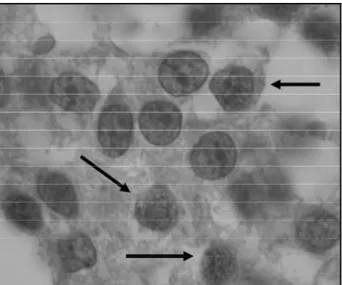
All tumor samples showed the appearance of a be-nign pituitary adenoma on standard histopathological examination, with a difuse (solid) pattern constituted by round cells with a predominantly basophilic cytoplasm. Nuclear pleomorphism, cytological atypia, and necro s i s w e re not detected, while mitotic figures were rare. Im-munostaining for pituitary hormones was positive only for the prolactin in the three samples, whereas p53 stain-ing was negative in all of them. Ki-67 stainstain-ing was detec-ted in all samples with the labeling indices bellow: 2.80% in original tumor (1995), 4.40% in relapse (1997), and 4.45% in metastasis (2001) (Fig 3).
DISCUSSION
P resent re p o rt shares many similarities with pre-vious published cases of pituitary carcinomas. Fro m the initial diagnosis of the macroadenoma to the d i s c l o s u re of metastatic disease a long interval of seven years has passed. In a series of 15 cases this l a-tency period ranged from 4 months to 18 years, with the mean of 6.6 years8. According to another study,
this period is 9.5 years for ACTH-producing lesions and 4.7 years for PRL-secreting tumors7. Vi rt u a l l y
all pituitary carcinomas arose from macro a d e n o-m a s8 , 1 1and most were submitted to one and, in m
a-ny times, two surgical approaches and radiation t h e-rapy years before appearance of metastases. In this setting, the process of origin seems like a ma-lignant transform a t i o n1 1. Radiation therapy and
surgical interventions are pointed by some authors as possible causes of such change in behavior1 2 , 1 5 , 1 6.
By the way, the patient with the longest latency period (18 years) in that series was the only who did not undego cranial surgery8.
A profile of resistance to bromocriptine action denoted by the increase in serum prolactin levels after some months or years of pharm a c o l o g i c a l t reatment is also a common characteristic among metastatic prolactinomas. This may re p resent a clue for suspition of an aggressive behavior, but is neither a specific nor an early manifestation. By the time of rise in prolactin levels in present case, no metastatic disease was evident on a MRI scan of brain (June 2000). Nevertheless, it became pa-tent on a CT scan ten months later. Immunostai-ning for pituitary hormones in the present case re i n-f o rced diagnosis on-f prolactinoma. The pro p o rt i o n of horm o n e - p roducing tumors among pituitary carcinomas is reported to be around 74%, repre-sented mostly by the prolactin- or ACTH-secre t i n g s u b t y p e s8 , 1 1. In fact, malignant prolactinomas
per-form 30% of pituitary carcinomas8.
Although histology of metastatic deposits in many cases tends to appear more aggressive than p r i m a ry tumor1 1, the aspect is commonly
indistin-guishable from that of benign adenomas1 0 , 1 1 , 1 7,
even on ultrastructural gro u n d s9. Traditional
histo-pathological features of malignancy (mitotic figu-res, nuclear pleomorphism, high cellularity, cytolo-gical atypias, presence of necrosis) are uncommon. When mitotic figures are present, they do pro v i d e some indication of the behavior and invasive po-tential of pituitary tumors, but for routine diagnos-tic purposes, however, discriminating power of this parameter is somewhat limited1 8. This discre p a
n-cy between clinical and pathological characterist i c s has led to a search for markers more informative of the behavior of pituitary carcinomas. For instan-ce, a myriad of genetic mutations has been re p o rt-ed in such tumors1 2 , 1 9 - 2 1, but their assessment has
shown little, if any, clinical usefulness1 2. Besides,
several biochemical and histopathological markers have recently been investigated in order to evalu-Fig 3. Positiveness for MIB-1 on immunostaining is re p re s e n t e d
ate the etiopathology of pituitary carcinomas, but still without conclusive results22-32.
In the last years an increasing amount of data about immunohistochemical assessment of the exp ression for mutant exprotein of exp53 gene and for K i -67 (MIB-1) antigen expressed during cellular pro l i-feration has suggested that such immunostainings may be promising markers of a malignant behav-ior in pituitary neoplasms. p53 is a nuclear pro t e i n encoded by a gene located on chro m o s s o m e 17p13.1, a tumor suppressor gene7. Its alteration
makes it act as a dominant oncogene. The gene is found mutated and/or deleted in a wide range of tumors, predominantly malignant1 4. In normal
tis-sues wild-type p53 protein cannot be detected by i m m u n o h i s t o c h e m i s t ry due to short half-life and rapid turn o v e r. However, once mutated, stabilized p rotein can be detected by antibodies1 0 , 1 4. Studies
about correlation between immunohistochemical data with actual mutations in p53 gene have re n-d e ren-d conflicting results, suggesting that immunos-taining may not re p resent the real genetic sta-t u s1 0 , 1 7 , 1 8. Despite the issue, the value of
positive-ness for p53 as a useful way for better represent-ing the phenotype of pituitary tumors has been e m p h a s i z e d1 0 , 1 4. An evaluation of 70 pituitary
ade-nomas re n d e red a pro p o rtion of p53 positive cas-es among noninvasive adenomas, invasive adeno-mas and carcinoadeno-mas of 0%, 15.2%, and 100% respectively10. Figures not so dramatic but also suggestive of an association of positiveness for p53 with an aggressive trend were pro d u c e d1 4. It is
dis-cussed whether technical diff e rences could be responsible for variable findings among studies1 0 , 3 3.
N e v e rtheless, absent p53 immunostaining in pitu-i t a ry carcpitu-inomas was recently re p o rt e d2 6 , 3 2 , 3 4, a
find-ing also encountered by our group. This suggests that this method is not so sensitive as first imagi n e d .
New histochemical methods of growth fraction determination were developed in last decades in o rder to provide rapid and cheap means of gaug-ing pituitary tumor behavior. One of the most pmising of these is detection of Ki-67 antigen, a p ro-liferation marker that is selectively expressed dur-ing G1, S1, G2, and M phases of cell cycle8 , 1 8 , 3 1 , 3 5. It
can be assessed through the use of the MIB-1 im-munostaining, the antibody against the Ki-67 for the paraffin pre p a r a t i o n s8 , 1 1. Studies with MIB-1
have revealed that growth fraction of pituitary tu-mors is low, with indices generally lower than 5 %1 1.
Vi rtually all noninvasive adenomas have a gro w t h fraction of less than 3%1 1. On the other hand,
gro-wth fractions are significantly higher among i n v a s
i-ve adenomas, and the highest among pituitary c a r-cinomas, when compared to noninvasive adenomas (4.7%, 11.9%, and 1.4%, re s p e c t i v e l y )3 6. Indeed,
p53-positive pituitary tumors (carcinomas and invasive adenomas) had four times higher indices of MIB-1 staining than p53-negative neoplasms1 0. The
pre-sent case shows a MIB-1 index of 2.80% in original tumor (1995). However, the figure reached 4.40% in the relapse (1997) and 4.45% in the metastatic deposit (2001). That is, there was an unexpected rise between first and second surgeries.
Even though such immunohistochemical stain-ings could better reflect the behavior of pituitary tumors, it remains unknown wheter those mark-ers could lead to an early suspicion of a malignant transformation when applied to patients with ma-croadenomas. In case this hypothesis is true, posi-tiveness for p53 or a high MIB-1 index could serv e as signs indicating that a macroadenoma is likely to metastatize or become locally aggressive. For instance, the present case shows a higher than ex-pected MIB-1 index at the second intervention, w h e n neither evidence of local invasion nor of metasta-tic dissemination were present. Some authors con-sider the concept that primary tumors that exhib-it mexhib-itotic activexhib-ity, an increased (> 3%) MIB-1 label-ing index, and/or p53 immunoreactivity should be t e rmed “atypical adenomas” to denote their a g g re s-sive potential and the possibility of future malig-nant transform a t i o n3 5. Would it indicate the need
of a more aggressive tretment, like chemotherapy, to prevent local invasion or metastases? This ques-tion could be answered with aid of pro s p e c t i v e studies that assess p53 and MIB-1 stainings ro u-tinely in every surgical intervention for pituitary a d e-nomas, since all knowledge about the issue is ba-sed on re t rospective and, perhaps, selected cases.
In conclusion, pituitary carcinomas re p resent a r a re neoplasm with fatal outcome, for which p53 and MIB-1 immunostainings emerged as clues for a better assessment of tumor behavior. However, not all pituitary carcinomas show positiveness for p53. On the other hand, a higher than expected Ki-67 proliferative index in a pituitary adenoma might alert to the possibility of a more aggre s s i v e behavior.
REFERENCES
1. Faglia G. Epidemiology and pathogenesis of pituitary adenomas. A c t a Endocrionol (Copenh) 1993;129:1-5.
2. G o l l a rd R, Kosty M, Cheney C, Copeland B, Bordin G. Pro l a c t i n - s e c re t-ing pituitary carcinoma with implants in the cheek pouch and metasta-ses to the ovaries: a case report and literature re v i e w. Cancer 1995; 76:1814-1820.
3 . Price PA. Cytotoxic chemotheraphy for aggressive pituitary tumours. In G rosman A (ed). Clinical endocrinology. Oxford: Blackwell, 1992;235-237. 4. Shelman WR, Laws ER, Scheithauer BW, Carpenter SM. The occure n c e of dural invasion in pituitary adenomas. J Neuro s u rg 1986;64:402-407. 5. Scheithauer BW, Kovacs KT, Laws ER, Randall RV. Pathology of invasi-ve pituitary tumours with special re f e rence to functional classification. J Neurosurg 1986;65:733-744.
6. Beauchesne P, Trouillas J, Brral F, Brunon J. Gonadotropic pituitary car-cinoma: case report. Neurosurgery 1995;37:810-816.
7. Ragel BT, Couldwell WT. Pituitary carcinoma: a review of the litera-ture. Neurosurg Focus 2004;16:E7.
8. Pernicone PJ, Scheithauer BW, Sebo TJ, et al. Pituitary carcinoma: a clin-icopathological study of 15 cases. Cancer 1997;79:804-812.
9 . Scheithauer BW, Fereidooni F, Horvath E, et al. Pituitary carcinoma: an u l t r a s t ructural study of eleven cases. Ultrastruct Pathol 2001;25:227-242. 10. Thapar K, Scheithauer BW, Kovacs K, Pernicone PJ, Laws ER Jr. p53 e x p ression in pituitary adenomas and carcinomas: correlation with in-vasiveness and tumor growth fraction [experimental studies]. Neuro-surgery 1996;38:765-771.
11. Thapar K, Kovacs K. Neoplasms of the sellar region. In Bigner DD, Mc-Lendon RE, Bruner JM (eds). Russel and Rubinstein’s pathology of tu-mors of the nervous system, 6.Ed. London: Arnold, 1998:561-677. 12. H u rel SJ, Harris PE, McNicol AM, Foster S, Kelly WF, Baylis P.
Metas-tatic prolactinoma: effect of octreotide, carbegoline, carboplatin and etoposide; immunocytochemical analysis of proto-oncogene expre s-sion. J Clin Endocrinol Metab 1997;82:2962-2965.
13. Kaltsas GA, Mukherjee JJ, Plowman PN, Monson JP, Grossman A B , Besser GM. The role of cytotoxic chemotherapy in the management of a g g ressive and malignant pituitary tumors. J Clin Endocrinol Metabol 1998;83:4233-4238.
14. Buckley N, Bates AS, Broome JC, et al. p53 protein accumulates in Cu-shings adenomas and invasive non-functional adenomas. J Clin En-docrinol Metabol 1994;79:1513-1516.
15. Mountcastle RB, Roof BS, Mayfield RK, Morde DB, Sagel J, Bigg PJ. Ca-se report: pituitary adenocarcinoma in an acromegalic patient: re s p o n s e to bromocriptine and pituitary testing: a review of the literature on 36 cases of pituitary carcinoma. Am J Med Sci 1989;298:109-118. 16. Papotti M, Limone P, Riva C, Gatti G, Bussolati G. Malignant
evolu-tion of an A C T H - p roducing pituitary tumor treated with intrasellar implantation of 90Y: case report and review of the literature. Appl Pa-thol 1984;2:10-21.
17. Bayindir C, Balak N, Gazioglu N. Pro l a c t i n - s e c reting carcinoma of the pituitary: clinicopathological and immunohistochemical study of a case with intracranial and intraspinal dissemination. Br J Neurosurg 1997; 11:350-355.
18. Thapar K, Yamada Y, Scheithauer B, Kovacs K, Yamada S, Stefaneanu L. Assessment of mitotic activity in pituitary adenomas and carc i n o-mas. Endocr Pathol 1996;7:215-221.
19. Rickert CH, Scheithauer BW, Paulus W. Chromossomal aberrations in pituitary carcinoma metastases. Acta Neuropathol 2001;102:117-120. 20. Yu R, Melmed S. Oncogene activation in pituitary tumors. Brain Pathol
2001;11:328-341.
21. Rickert CH, Scheithauer BW, Paulus W. Chromosomal aberrations in pi-tuitary carcinoma metastases. Acta Neuropathol 2001;102:117-120. 22. Kong YG, Ren ZY, Su CB, Wang RZ, Ma WB, Lian W. Elevated soluble
epidermal growth factor receptor level in pituitary adenoma and car-cinoma. Chin Med Sci J 2004;19:199-202.
23. O n g u ru O, Scheithauer BW, Kovacs K, et al. Analysis of epidermal gro-wth factor receptor and activated epidermal grogro-wth factor re c e p t o r e x p ression in pituitary adenomas and carcinomas. Mod Pathol 2004; 17:772-780.
24. O n g u ru O, Scheithauer BW, Kovacs K, et al. Analysis of Cox-2 and t h romboxane synthase expression in pituitary adenomas and carc i n o-mas. Endocr Pathol 2004;15:17-27.
25. Mulla A, Christian HC, Solito E, Mendoza N, Morris JF, Buckingham JC. Expression, subcellular localization and phosphorylation status of annexins 1 and 5 in human pituitary adenomas and a growth hormone-secreting carcinoma. Clin Endocrinol (Oxf) 2004;60:107-119. 26. R o n c a roli F, Nose V, Scheithauer BW et al. Gonadotropic pituitary carc
i-noma: HER-2/neu expression and gene amplification: report of two cases. J Neurosurg 2003;99:402-408.
27. Vidal S, Horvath E, Kovacs K, Kuroki T, Lloyd RV, Scheithauer B W. E x p ression of hypoxia-inducible factor-1alpha (HIF-1alpha) in pituitary tumours. Histol Histopathol 2003;18:679-686.
28. Jin L, Zhang S, Bayliss J, et al. Chromogranin a processing in human pituitary adenomas and carcinomas: analysis with region-specific anti-bodies. Endocr Pathol 2003;14:37-48.
29. Trouillas J, Daniel L, Guigard MP, et al. Polysialylated neural cell adhe-sion molecules expressed in human pituitary tumors and related to extrasellar invasion. J Neurosurg 2003;98:1084-1093.
30. Riss D, Jin L, Qian X, et al. Diff e rential expression of galectin-3 in pitui-tary tumors. Cancer Res 2003;63:2251-2255.
31. Lubke D, Saeger W, Ludecke DK. Proliferation markers and EGF in A C T H - s e c reting adenomas and carcinomas of the pituitary. Endocr Pa-thol 1995;6:45-55.
32. Nose-Alberti V, Mesquita MI, Martin LC, Kayath MJ. A d re n o c o r t i c o-t ro p i n - p roducing pio-tuio-tary carcinoma wio-th expression of c-erbB-2 and high PCNA index: a comparative study with pituitary adenomas and normal pituitary tissues. Endocr Pathol 1998;9:53-62.
33. Levy A, Hall L, Yeudall Wa, Lightman SL. p53 gene mutations in pitui-tary adenomas: rare events. Clin Endocrinol (Oxf) 1994;41:809-814. 3 4 . Kumar K, Macaulay RJ, Kelly M, Pirlot T. Absent p53
immunohistoche-mical staining in a pituitary carcinoma. Can J Neurol Sci 2001;28:174-178. 35. G a ffey TA, Scheithauer BW, Lloyd RV, et al. Corticotroph carcinoma of the pituitary: a clinicopathological study. Report of four cases. J Neuro-surg 2002;96:352-360.