Aging and immunoglobulin
isotype patterns in oral tolerance
Departamento de Bioquímica e Imunologia, Instituto de Ciências Biológicas, Universidade Federal de Minas Gerais, Belo Horizonte, MG, Brasil
A.M.C. Faria, S.M. Ficker, E. Speziali, J.S. Menezes, B. Stransky, B.A. Verdolin, W.M. Lahmann, V.S. Rodrigues and N.M. Vaz
Abstract
In the present review we address oral tolerance as an important biological phenomenon and discuss how it is affected by aging. Other factors such as frequency of feeding and previous digestion of the antigen also seem to influence the establishment of oral tolerance. We also analyze immunoglobulin isotypes of specific antibodies formed by tolerant and immunized animals of different ages submitted to different conditions of oral antigen administration. Isotypic patterns were studied as a parameter for assessing the pathways of B and T cell interactions leading to antibody production.
Correspondence
A.M.C. Faria
Departamento de Bioquímica e Imunologia, ICB, UFMG Av. Antônio Carlos, 6627 31270-901 Belo Horizonte, MG Brasil
Presented at the International Meeting on Cytokines, Angra dos Reis, RJ, Brasil, November 24-28, 1996.
Research supported by FAPEMIG (No. 2501/96), PRPq and CNPq (No. 53.0349/93-0).
Received September 4, 1997 Accepted November 6, 1997
Key words
•Oral tolerance
•Aging
•Isotypes
•Gut
•Mucosa
Feeding and immunological activities
Feeding is the major and most frequent occasion on which the organism contacts foreign proteins and, potentially, the major source of triggering immunological activi-ties. Through idiotype-anti-idiotype connec-tions, events initiated in the gut may influ-ence all immunological phenomena, regard-less of their origin. For example, effective vaccination against trachoma has been re-cently achieved by oral exposure to an anti-idiotypic antibody (1), showing that the gut provides a direct and effective access to the immune system by a physiological route.
Two misconceptions cause this evidence to be pratically ignored. First, ingested anti-genic molecules are thought to be totally di-gested and thus devoid of immunological rel-evance. This is simply incorrect and is contra-dicted by many experimental observations (2). Second, and more importantly, the immune
system has been conceptually divided into two systems. Secretory IgA immunoglobulins (sIgA) present on mucosal surfaces react with germs and viruses and may hinder the absorp-tion of antigenic macromolecules. sIgA is mainly produced in local mucosal-associated lymphoid tissues but lymphocytes stimulated at a local mucosal site migrate to other sites of the same mucosa and also to other mucosae (3,4). This suggests the operation of a web of intermucosal cell traffic independent of a more systemic immune circuitry. Thus, the organ-ism is seen as doubly protected from antigens in the gut: by digestion and by a secretory immune system or common mucosal im-mune system (5).
Different outcomes of oral contacts with antigens
Although different immunological out-comes may result from oral exposure to anti-gens, a basic pattern may be understood. Depending on several factors, related to the antigen and/or the organism, oral contacts with antigens may result either in oral toler-ance or in its reverse: local and/or sys-temic immunization, i.e., circulating anti-body formation (9-14). Intermittent feeding of mature or older mice with ovalbumin (Ova), depending on the antigen/strain com-bination (11), tends to result in serum anti-body formation (12,15). However, natural feeding of mice has been shown to result preferentially in oral tolerance to dietary components, especially to proteins repeat-edly ingested on several consecutive occa-sions (10,16).
Oral tolerance is defined as a suppression of the specific immune response to parenteral injections of the antigen previously presented by the oral route (7,17-19). This phenome-non has been reported in a number of animal species, including mice (20), hamsters (21), rats (22), guinea pigs (23), rabbits (24), and humans (25) and can be measured as a de-crease in delayed type hypersensitivity (DTH) reaction to the antigen (26) as well as a suppression in specific IgE and IgG produc-tion (27).
Several reports indicate that antigen/strain combination, the nature and dose of antigen (28), the immunological status of the animal (29), genetic factors linked or not to MHC (11,14), age at first oral contact (15,30,31), frequency of exposures and interval between them (10,12) are all factors determining the outcome of oral contacts with antigens.
Herein, we review past evidence and pres-ent data on the influence of aging and the rate of antigen intake on oral tolerance. We also analyze the isotypic patterns associated with oral tolerance at different ages and under different experimental conditions.
Immunoglobulin isotypes, aging and oral tolerance
Maturation of mice of different inbred strains to young adulthood - a period arbi-trarily defined between 8 and 24 weeks of age - coincides with a marked decrease in their susceptibility to the induction of oral tolerance and a parallel (consequent?) in-crease in their susceptibility to systemic oral immunization following repeated (intermit-tent) oral exposures to antigens (12,15,32,33). Moreau and Gaboriau-Routhiau (34) re-cently reported induction of oral tolerance to Ova in 20-month-old C3H/He mice. How-ever, in other studies using the parenteral route for tolerance induction, a decrease in ease of tolerance induction in aged BALB/c, C57BL/6 and CBA/CaJ mice (35,36) and a relative inability to induce tolerance in adult NZB and (NZB X NZW) F1 mice (37) have been described. The impairment of tolerance induction seems to affect both B and T cell populations in 6-month-old BALB/c mice (38,39).
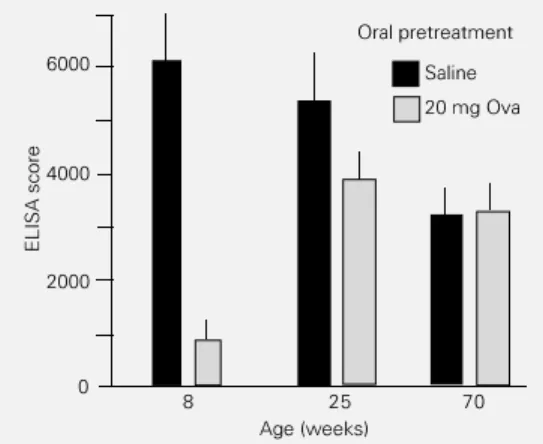
As shown in Figure 1, there is a marked decrease in the titers of anti-Ova antibody immune responsiveness to Ova (saline-fed groups) of 70-week-old mice when com-pared to 8- or 25-week-old mice.
titers of anti-Ova antibodies or to recall their previous tolerance status.
Several changes in immunological pa-rameters are observed during aging. B and T lymphocytes change in phenotype and rep-ertoire. The ratio of activated T cells (CD45RO) to resting T cells (CD45RA) in-creases. In mice and humans, the patterns of cytokines secreted by activated T cells change significantly, with a reduction of IL-2, IL-3, and GM-CSF and an increase of IL-4, IL-5, IFN-γ, IL-6 and IL-10 (40-43). Macrophage production of IL-1 and TNF also changes (44). There is a depression in early signal transduction as well as a reduction in cal-cium fluxes (45-48) in activated B and T lymphocytes in old animals. The levels of dimeric IgA produced in gut lamina propria as well as the number of CD4+ T cells in Peyers patches are significantly reduced in old animals (49,50).
All of these aging-associated changes can potentially interfere with the operation of the immune system as a whole and systemic phenomena such as tolerance are expected to be altered by senescence. We believe, however, that the discussion on the effect of aging on oral tolerance invokes the very mechanisms responsible for its induction and maintenance (33).
Suppression vs anergy in oral tolerance
The mechanisms underlying oral toler-ance induction and maintentoler-ance are still elu-sive but two proposals have been enthusias-tically discussed in the recent literature: clonal anergy and suppression by the differential production of cytokines by specific T cell clones.
Clonal anergy and clonal deletion are concepts derived from the notion that im-mune events are necessarily destructive and require mechanisms of contention to cir-cumvent immunological self-destruction. The original suggestion that self-tolerance relies
on the deletion of self-reactive (forbidden) lymphocyte clones was supported by the experiments of Brent, Billingham and Medawar on tolerance to allografts (51) and formed the backbone of the clonal selection theory of antibody formation created in the 1960s by Burnet (52). These initial studies showed that tolerance to allografts could only be achieved early during the neonatal period, suggesting that susceptibility to tol-erance is a characteristic of immunologi-cally immature hosts.
A whole array of findings in the last 30-35 years have shown these convictions to be basically incorrect. The original Brent, Billingham and Medawar experiments them-selves have been repeated (53) and have shown that tolerant animals, far from delet-ing alloreactive clones, maintain in their
bod-Figure 1 - Immune responses of normal and orally tolerant B6D2F1 mice of different ages. Mice were fed by gavage with either saline or 20 mg ovalbumin (Ova) 7 days be-fore primary ip immunization with 10 µg Ova + 1 mg Al(OH)3. All ani-mals were boosted ip with 10 µg Ova 14 days later and sera were collected 7 days after the booster. Anti-Ova antibodies were meas-ured by ELISA and the numbers represent the running sum of ab-sorbance ranging from 1/100 to 1/ 3200 in one group (N = 6-8). Signifi-cant differences between Ova-fed and saline-fed groups were calcu-lated by the two-tailed Student t -test. A significant difference (P<0.001) between groups was ob-served only in 8-week-old mice.
ELISA score
6000
2000
0 4000
Oral pretreatment
Saline
20 mg Ova
8 25 70
Age (weeks)
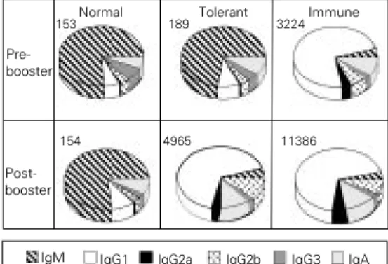
Table 1 - Maintenance of oral tolerance is not affected by aging.
B6D2F1 mice were fed either saline (normal and immune group) or 20 mg Ova (tolerant group) by gavage 7 days before the primary immunization and booster. The normal group was not immunized. The animals were classified as normal, tolerant and immune, and then, allowed to rest for 62 weeks before being boosted again with 10 µg Ova ip, 10 µg Ova + 1 mg Al(OH)3 and 10 µg Ova 7 days apart. Sera were collected 7 days after the last immunization and antibodies were measured by ELISA. ELISA scores and significant differences between tolerant and immune groups were calcu-lated as shown in the legend to Figure 1. *P<0.001 compared to the normal group.
Bleedings Normal Tolerant Immune
Pre-booster 153 ± 24 189 ± 62* 3224 ± 265
presenting cells (APC), renders mature thy-mic and peripheral T lymphocytes anergic (58). This has been shown in normal or cloned T cells stimulated with peptides pre-sented by chemically modified APC (59), purified MHC class II molecules incorpo-rated into planar lipid membranes (60), stim-ulation with concanavalin A in the absence of APC (61), or immobilized CD3 anti-body (62). In the anergic state, T cells are rendered refractory to restimulation with APC and Ag, and are unable to secrete IL-2 (58). It has been suggested that oral tolerance could also be a result of clonal anergy of specific T cell clones, mostly Th1 cells (63,64). According to these reports, high doses of orally administered antigen result in systemic antigen presentation after antigen passes through the gut and enters the sys-temic circulation either as intact protein or antigen fragments. Induction of clonal an-ergy would occur by defective presentation of the protein to the specific T lymphocytes. Evidence supporting the anergy model in-cludes the fact that spleen cells or lympho-cytes isolated from rats made tolerant with myelin basic protein (MBP) did not transfer tolerance to naive recipients. In vitro cell mixing studies have shown that the prolif-eration of lymphocytes from MBP-sensitized donors was not inhibited by the addition of lymphoid cells from tolerant donors (64). Other reports using Ova as antigen showed that lymphocytes from Ova-tolerant mice did not produce IL-2 or express IL-2R in response to Ova stimulation in vitro (65).
The present results, obtained using a high dose feeding protocol (20 mg Ova), again argue against any subtractive mechanism un-derlying oral tolerance establishment. Sus-ceptibility to oral tolerance may completely disappear in 70-week-old B6D2F1 mice which are among the most susceptible strains to ovalbumin tolerance induction when tested at 7-10 weeks of age. Furthermore, the adop-tive transfer of syngeneic spleen cells from young B6D2F1 donors to old recipient mice ies a large collection of these clones
acti-vated and coexisting with alloantigenic do-nor cells throughout life.
The phenomenon of oral tolerance repre-sents further strong evidence against these ideas since its induction is diametrically op-posite to allograft tolerance. Neonatal mice are not susceptible to oral tolerance (54) but may become susceptible if adoptively trans-ferred with syngeneic lymphocytes from adult donors (55). Thus, this is an active process requiring some degree of systemic organiza-tion.
Quite distantly from the neonatal period, the susceptibility to oral tolerance induction also wanes. Maturation to 20-25 weeks of age reduces and living one to one-and-a-half years (50-70 weeks) profoundly impairs the ability of mice to become orally tolerant (12,15). Some of these findings are discussed here (Figure 1). These protracted changes show that immunological activities comprise an ongoing epigenesis which extends far beyond the early phases of ontogenesis (56). The clonal selection theory tried to ex-plain tolerance based on three main prin-ciples: a) immune activity is clone specific; b) tolerance, as opposed to the immune re-sponse, represents the absence of such spe-cific immune activity, and c) tolerance, as a negative event, should take place in early life when immunocompetence is not yet com-pleted.
Several recent reports still insist that tol-erance must be achieved by the elimination of specific lymphocyte clones but the prob-lems raised by oral tolerance remain to be solved. Anergy is a convenient mechanism for the clonal theory since it provides an explanation for how tolerance, as a clonal negative event, may occur throughout life and not only early in ontogenesis.
antigen-may partially restore their susceptibility to oral tolerance and, reciprocally, adoptive transfer of cells from old donors to young recipients decreases their susceptibility to tolerance. On the other hand, transfer of syngeneic spleen cells from young or neo-nate (10 days old) BALB/c donors, which are refractory to tolerance induction, to old recipient mice fails to alter the lack of sus-ceptibility to oral tolerance (66).
Therefore, the induction of oral tolerance probably depends on a configuration of cells and molecules present in young but not in old mice (67,68). This configuration trans-ferred by spleen cells seems to provide the plastic connectivity between antibodies and the lymphocytes that animals need to incor-porate novel antigens into their immunologi-cal activities. Transfer of susceptibility to oral tolerance induction in our model rules out the possibility of any negative mech-anism such as anergy or clonal deletion as an operative explanation for oral tolerance.
Interestingly, old mice receiving cells from young mice and orally treated for toler-ance induction have an isotypic profile closer to that of young mice and different from that of old mice. This observation suggests that spleen cells were able to transfer not only the susceptibility to oral tolerance induction pres-ent in young mice but also its isotypic profile (69).
Suppression does not imply a differential activation of Th1 and Th2 clones
Another hypothesis indicates active sup-pression by specific T lymphocytes as a mechanism of oral tolerance establishment. Early studies have associated the induc-tion of oral tolerance with the activainduc-tion of suppressor T cells (20,29,70-72), but the nature and the properties of such cells are now disputed. Initially, CD8+ T cells were pointed out as the putative suppressor cells (72). Many recent articles still suggest that
oral tolerance is due to the activation of CD8+ suppressor T cells (73,74). These re-ports demonstrate that CD8+ lymphocytes can play a regulatory role in a number of immune activities either through classical cytotoxic effects on APC or via the produc-tion of cytokines such as IFN-γ and TGF-ß. However, other authors have shown that in vivo depletion of CD8+ cells at the time of feeding had no effect on the induction of oral tolerance, whereas depletion of CD4+ cells completely abolished the suppression of DTH and the antibody response (75,76). It has been suggested that cytokines secreted by special subsets of CD4+ T cells (Th1 and Th2) are involved. Cells belonging to the Th1 subset appear to be more susceptible to the induction of tolerance in vitro than Th2 cells (77) and Th1-mediated effector re-sponses in vivo are more easily suppressed than those requiring Th2 cells (78). Thus, tolerance induced by feeding low doses of antigen would reflect the preferential activa-tion of Th2 cells with subsequent down-regulation of Th1-dependent, cell-mediated immune responses by Th2-derived cytokines such as IL-4 and IL-10 (79).
induction result in the suppression of all isotypes.
In conclusion, the data presented here do not support the hypothesis of a differential activation of cells belonging to the Th2 sub-set as a mechanism to induce the suppres-sion described in oral tolerance experiments, nor do they support the idea of a subtractive process such as anergy or deletion. They indicate, on the contrary, that an active mech-anism must be involved in oral tolerance induction and, although an activation of Th2 cells cannot be regarded as a mechanism, they do not rule out the possibility of regula-tory cells being triggered by antigen feeding. The ways by which these regulatory cells would induce suppression might involve both idiotypic interaction and cytokine release. There are some reports on the secretion of cytokines such as GM-CSF and IFN-γ by mesenteric lymph node cells from mice fed ovalbumin 24 h before (82). It has also been shown that oral tolerance to myelin basic protein in rats is due to activation of CD8+ T cells which down-regulate Th-cell function through the release of TGF-ß (74,83). Many reports also indicate that idiotypic connec-tivity is involved in tolerance induction.
Mechanisms of tolerance/aging and idiotypic connectivity
There are a series of experiments dis-proving the idea that immunological toler-ance is a negative process. For insttoler-ance, in mice made tolerant to allografts during the neonatal period, the suppressed alloreactive lymphocytes belong to a population of large activated lymphocytes (53). This possibility also applies to self-tolerance (84,85), sug-gesting that tolerance depends on the addi-tion of certain types of activated lympho-cytes rather than on their subtraction.
If the development of oral tolerance is similar to tolerance to self components, it Figure 2 - Oral tolerance
drasti-cally reduces the magnitude of anti-ovalbumin (Ova) immune re-sponses, but preserves the isotypic profile of the antibodies formed. Mice were treated as described in the legend to Fig-ure 1. The numbers above the pies denote the global ELISA score for the group. Each sec-tion of the pie represents the percentage of each isotype in the overall antibody titer. Note that IgG1 and IgG2a antibodies are equally suppressed in the tolerant group.
IgM IgG1 IgG2a IgG2b IgG3 IgA
Immune Saline by gavage
Tolerant 20 mg Ova by gavage
7762 1617
decrease in eosinophilic infiltration in the lungs as well as in serum IgE and IgG1 antibody production.
might depend on the inclusion of specific activated lymphocyte precursors into self-maintained idiotypic lymphocyte circuits. In young animals, these endogenous circuits would be plastic enough to change upon contact with new elements in the diet. In this case, plasticity may depend on the use of V-gene families which allow a loose connec-tivity of the lymphocytes (68). In older ani-mals, tighter connections resulting from im-munological experience would be a hin-drance to this plasticity. Mouse strains which display abnormal patterns of idiotypic con-nectivity when young are susceptible to au-toimmune defects and tend to be refractory to oral tolerance induction (12,15,37,84). Aging is associated with a progressive de-cline in antibody responsiveness (see Figure 1) to foreign antigens, such as bacterial pro-teins, and SRBC, paralleled by a progressive rise in the production of self-reactive (auto) antibodies (67,86,87). According to Weksler et al. (67), most of the auto-antibodies pro-duced by old mice react with each other and their rise with aging results, at least in part, in an increased connectivity of the immune system. Studies on the auto-anti-idiotypic antibody response in young and old mice support this hypothesis (84,88,89), indicat-ing that agindicat-ing represents a cross-wirindicat-ing of the reactivities within the system and, conse-quently, an inevitable loss of ability to react with novel antigens.
Therefore, changes in immune activity with aging may be related to changes in the patterns of idiotypic connectivity among lym-phocytes. Our results are consistent with this hypothesis. Both oral tolerance and immuni-zation against a novel antigen are impaired in old mice (Figure 1) whereas the mainte-nance of these two kinds of immune activi-ties is preserved for antigens already pre-sented to the animal in early life (Table 1). Possibly, induction of oral tolerance requires incorporation of novelty into the dynamics of the immune system whereas its
mainte-nance represents the recursion of already established circuits.
Oral tolerance and the rate of antigen intake
A factor known to be important for the establishment of oral tolerance is the rate of antigen intake. The most efficient way to induce oral tolerance is the continuous ad-ministration of the antigen at small doses. Experiments in mice (10) and guinea pigs (23) demonstrated that ingestion of Ova for several consecutive days was able to sup-press the delayed hypersensitivity and serum antibody responses to a subsequent injection of the antigen in complete Freunds adju-vant. On the other hand, the animals were anaphylactically sensitized if the antigen was introduced in their diet all at once.
According to Stokes et al. (90), tolerance induction is related to a gradual and continu-ous absorption of the antigen. These authors showed that administration of 25 mg Ova to CBA and SWR/J mice by the intragastric route for 14 days did not induce tolerance but if the antigen was voluntarily ingested throughout the day in the same amounts, the animals could be rendered tolerant. Thus, continuous contact with the antigen appears to be necessary to trigger the tolerance cir-cuits.
We tested oral tolerance induction by two different protocols: a single gavage of 20 mg Ova or voluntary ingestion of 4 mg/ml
Figure 3 - Isotypic pattern of anti-Ova antibodies in 70-week-old mice orally treated when 8 weeks old and boosted after 52 weeks. Mice were treated as de-scribed in the legend to Table 1. The numbers above the pies de-note the global ELISA score of the groups. Note that the isotypic profile of tolerant mice is closer to that of normal mice before the booster and re-sembles the profile of the im-mune group after the booster.
Pre-booster
Post-booster
IgM IgG1 IgG2a IgG2b IgG3 IgA
Normal
153 189 Tolerant 3224Immune
11386 4965
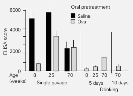
Ova solution for 5 or 10 days (the normal daily drinking volume of a mouse is mately 5 ml, so the animals ingest approxi-mately 20 mg Ova per day). As expected, 5-day voluntary ingestion of Ova was signifi-cantly more efficient for oral tolerance in-duction than a single feeding of 20 mg Ova (Figure 4). In 8-week-old B6D2F1 mice, both protocols were highly effective in in-ducing oral tolerance but the serum antibody titers after voluntary ingestion of Ova were even lower than after the single feeding pro-tocol. Voluntary ingestion of Ova for 5 days induced tolerance in 25-week-old B6D2F1 mice whereas a single feeding procedure did not. Furthermore, 70-week-old B6D2F1 mice, which are totally refractory to oral tolerance induction by a single gavage of Ova (Figure 1), can be rendered tolerant by voluntary ingestion of Ova. Ingestion for 5 days resulted in moderate suppression and ingestion for 10 days was highly effective.
Different antigen-presenting cells and cytokines may be triggered by differ-ent ways of antigen ingestion
The difference between the two proto-cols may be due to the way the antigen is processed and presented by mucosal anti-gen-presenting cells (APC). The first and obvious distinction is the antigen loading. When a high dose of antigen enters the mu-cosal lymphoid tissue at once, probably all APC available are recruited to process and present the protein, whereas when the same
amount of protein is gradually administered in small doses, only the most efficient APC (dendritic cells) are mobilized. This differ-ential presentation may trigger different path-ways of T cell activation resulting, for in-stance, in a distinct profile of cytokine re-lease.
Alternatively, the two protocols may lead to a distinct degree of antigen luminal diges-tion. In a voluntary ingestion regimen, anti-gen administered in continuous small a-mounts would be more exposed to digestive enzymes present in the intestinal lumen than a large amount of protein fed as a bolus. The result of a more extensive luminal process-ing of antigen may be a decrease in the requirements for processing by APC in the gut and differences in T cell responses, inas-much as different epitopes may be displayed. Several groups have recently focused on APC in the gut as potential regulators of mucosal immune responses. There are a num-ber of cell types that can process and present antigens in the intestinal lymphoid tissue: macrophages, dendritic cells, B cells present in Peyers patches and lamina propria as well as the intestinal epithelial cells (91,92). Antigen-presenting cells in the lamina pro-pria and Peyers patches use the classical class I or class II presentation pathway de-pending on whether the antigen gains access to the cytoplasm or to the endosomic com-partments of the cell (93). Intestinal epithe-lial cells, however, seem to take up soluble antigens and transport them by the endolyso-somal route but they do not use class II MHC as restriction elements for peptide presenta-tion. Instead, products of antigen processing within gut epithelial cells seem to associate with CD1b molecules and the antigen pres-entation process is very inefficient (94). The preferential subpopulation of T cells acti-vated by these APC are CD8+ (95) although it is not clear yet which other molecules are involved in cell-to-cell interactions. There-fore, several data support the idea that anti-gen presentation in gut lymphoid tissue may Figure 4 - Voluntary ingestion of
antigen on consecutive days is more effective than a single ga-vage for oral tolerance induction. B6D2F1 mice of different ages were treated either by a single gavage administration of 20 mg ovalbumin (Ova) or by voluntary ingestion of a solution of 4 mg/ ml Ova in drinking water for 5 to 10 days. The animals were im-munized 7 days after the last oral treatment and boosted 14 days thereafter. ELISA scores and sig-nificance were calculated as de-scribed in the legend to Figure 1. Only 8-week-old (P<0.001) and 25-week-old mice (P<0.025) could be made tolerant by a single gavage administration of Ova, whereas voluntary inges-tion was effective for all groups treated with Ova (P<0.001).
ELISA score
6000
4000
2000
0 Age (weeks)
8 25 70 8 25 70 70
Single gavage 5 days 10 days
Drinking Oral pretreatment
have unusual characteristics depending on the way the antigen is absorbed (95).
Our hypothesis that antigen uptake by voluntary ingestion would result in recruit-ment of APC distinct from that triggered by single gavage was tested in two experiments. First, we measured the isotypic profile of the anti-Ova antibodies induced in tolerant mice by the two protocols assuming that B cells become committed to producing different isotypes depending on the cytokines released by T helper cells. On the other hand, T-cell commitment to patterns of cytokine secre-tion could be altered by the way these cells are activated by different antigen-presenting cells. Second, we tested a protocol of toler-ance induction using intravenous (iv) injec-tion of Ova cleaved by cyanogen bromide (CNBr-Ova) and measured the isotypic pro-file of antibodies produced by this protocol. This alternative iv procedure was designed to investigate the role of previous digestion of antigen in tolerance induction.
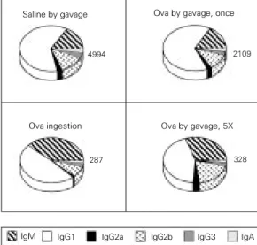
Figure 5 shows the isotypic pattern of anti-Ova antibodies in 8-week-old mice treated by the oral route with a single gavage of saline, a daily gavage of 20 mg Ova or voluntary ingestion of 4 mg/ml Ova in water for 5 days. All mice were challenged with Ova in adjuvant 7 days after the last oral exposure and sera for antibody testing were collected 7 days after the booster. As previ-ously shown in Figure 2, oral tolerance in-duction by a single gavage induced a sup-pression of all isotypes. However, the volun-tary ingestion protocol resulted in differen-tial suppression of IgG1 and a compensatory increase in the proportion of IgM antibodies. These results suggest not only that continu-ous feeding is a more efficient protocol for oral tolerance induction, but also that it trig-gers different pathways of T and B cell acti-vation.
Observing the low levels of anti-Ova an-tibodies induced in mice made tolerant by voluntary ingestion of Ova, one can argue that the antibody titers are so low that they
represent only natural IgM antibodies pres-ent in normal serum and that the isotypic profile simply shows the total absence of B cell activation by ovalbumin. In the follow-ing experiments, however, we confirm the present data under different conditions.
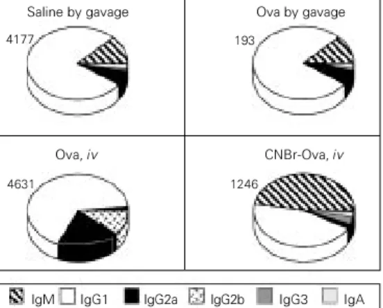
B6D2F1 mice injected iv with 500 µg CNBr-Ova 7 days before ip immunization with Ova in adjuvant showed a significant decrease in anti-Ova antibody titers as com-pared to a control group injected iv with saline (Figure 6). On the other hand, iv injec-tion of the same dose of native ovalbumin did not suppress the immunization to the protein. This result indicates that cleavage of the antigen, such as the degradation of pro-teins in the intestine, may have an important role in tolerance induction.
Curiously, the isotypic pattern of anti-Ova antibodies from mice rendered tolerant by iv injection of CNBr-Ova shown in Fig-ure 6 is closely similar to the voluntary-ingestion-tolerance pattern. There is a clear suppression of IgG1 antibodies and a rela-tive increase in IgM levels. Mice made toler-ant by a single gavage of 20 mg Ova again show suppression of all isotypes as com-pared to the control immune group. Injection of 500 µg Ova iv did not induce tolerance but triggered a change in the pattern of antibod-ies produced.
The levels of serum antibodies in mice
Figure 5 - Mice treated by ga-vage and voluntary ingestion of ovalbumin (Ova) show a dif-ferent pattern of suppression of their anti-Ova immune re-sponse. B6D2F1 mice at 8 weeks of age were treated with either salinene by ga-vage, one gavage of 20 mg Ova, voluntary ingestion of 4 mg/ml Ova for 5 days, or 5 consecutive daily gavages of 20 mg Ova. All mice were im-munized as described in the legend to Figure 1. All oral treatments induced oral toler-ance (P<0.001) while volun-tary ingestion and 5 gavages of Ova induced a preferential suppression of IgG.
Saline by gavage
2109 Ova by gavage, once
4994
Ova ingestion Ova by gavage, 5X
328 287
rendered tolerant by iv injection of CNBr-Ova shown in Figure 6 are not as low as those illustrated in Figure 4, although we observed an equivalent alteration in the isotype profile. This result suggests that an interference with the presentation of anti-gen, in this case by its cleavage, may result in a different activation of the B cells reactive to the antigen. It also reinforces the possibil-ity that the isotypic changes triggered by the voluntary ingestion protocol (Figure 5) do not represent a numerical artifact.
Therefore, our data indicate that the rate of antigenic intake could alter not only the efficiency but also the pathway of tolerance induction. In addition, the experiments re-ported here suggest that both the loading of APC and the partial digestion of the antigen are plausible hypotheses to explain the effi-ciency of the voluntary ingestion protocol.
Experiments performed with slow intra-venous antigen infusion indicate that the rate of antigenic entry into the circulation per se is neither relevant for the efficiency nor de-terminant for the occurrence of oral toler-ance. Using Harvard infusion pumps and Alzet osmotic pumps, which deliver antigen iv for 1 h and ip for 24 h, respectively, Stransky (96,97) demonstrated that slow parenteral infusion of Ova is not sufficient to induce tolerance in 8-week-old B6D2F1 mice. We were thus left with the hypothesis that the rate of antigen entry was a funda-mental requirement in the oral route.
Con-tact through mucosal tissues may involve other important factors such as previous di-gestion of the antigen, interaction with sev-eral lymphoid and non-lymphoid popula-tions only present there, typical cytokines, and a number of other unique molecules about which little information is available.
In order to test this hypothesis, we de-signed a protocol comparing voluntary in-gestion of ovalbumin and consecutive gavages as regimens for oral tolerance in-duction in susceptible B6D2F1 mice. Ani-mals were treated for 5 consecutive days either by a daily gavage of 20 mg Ova or by voluntary ingestion of 4 mg/ml Ova in their drinking water. We analyzed the efficiency of these treatments in inducing oral toler-ance and the isotypic profile of antibodies produced always comparing them with the single gavage protocol.
Figure 5 shows that, at least for young mice, 5 daily consecutive gavages are as efficient as voluntary ingestion of the same dose of ovalbumin for 5 days for oral toler-ance induction as compared to the single gavage protocol. Further experiments, using old mice, will help elucidate whether re-peated gavages would be able to replace continuous feeding as an efficient method to induce oral tolerance in these refractory ani-mals.
The isotypic profile of anti-Ova antibod-ies induced by a 5-gavage protocol is shown in Figure 5 along with the profiles of volun-tary ingestion, single gavage and saline pro-tocols. The pattern of isotypes induced by the 5-gavage protocol is similar to that in-duced by the voluntary ingestion regimen.
Results from these last experiments dem-onstrate that, in young mice, a multiple feed-ing protocol leads to a suppression compa-rable to that achieved by a continuous feed-ing of the same antigen.
We know from previous studies in adult and old mice (12,32) that intermittent expo-sures to an antigen by oral route do not induce tolerance but may lead to systemic Figure 6 - Previous cleavage of
the antigen influences tolerance induction. Eight-week-old B6D2 F1 mice were treated with 20 mg ovalbumin (Ova) by gavage or by intravenous injection of 500 µg native Ova or 500 µg of cyanogen bromide-treated Ova (CNBr-Ova) 7 days before ip munization. All mice were im-munized as described in the leg-end to Figure 1. The numbers above the pies indicate the glo-bal ELISA score of each group. Both oral treatment with Ova and iv injection of CNBr-Ova in-duced oral tolerance (P<0.001) but cleavage of the antigen led to a preferential suppression of IgG.
IgM IgG1 IgG2a IgG2b IgG3 IgA
Saline by gavage
193
Ova by gavage
4177
Ova, iv CNBr-Ova, iv
immunization. On the other hand, consecu-tive contacts with the same dose of antigen do not induce oral immunization. Therefore, episodic oral exposures appeared to be ideal for oral immunization whereas both repeti-tive and continuous oral contacts with a protein, such as in our daily meals, are suffi-cient to trigger the circuits responsible for oral tolerance induction.
The cellular and molecular components involved in the suppression observed as the oral tolerance phenomenon probably depend on reiterated stimulation, either because their action is of short duration (such as cytokine activities) or because they are quickly re-newed (such as surface molecules).
Conclusion
The formation of antibodies as well as the class of immunoglobulin produced is a process dependent on T cell stimulation of the B cells. A large amount of evidence indicate that the B-T cell interaction and cytokines released by T cells are determi-nant for the immunoglobulin class switch (98-100). Since we were using the suppres-sion of specific antibody production as a parameter for studying oral tolerance, we
decided to analyze the isotypic patterns un-der distinct experimental conditions as a way to assess the influence of age and rate of antigen intake on the interactions between B and T cells during oral tolerance establish-ment. We demonstrated here that oral toler-ance is an active phenomenon influenced by other natural events such as aging and condi-tions of antigen feeding. Senescence is asso-ciated with a decrease in susceptibility to oral tolerance induction but does not affect oral tolerance maintenance. On the other hand, both previous cleavage of the antigen and repeated oral exposures seem to favor the establishment of oral tolerance. These factors affect not only the susceptibility to oral tolerance induction but also the path-ways of B and T cell interactions that lead to antibody production.
Acknowledgments
We thank Mrs. Ilda Marsal de Souza for competent care of the animal colonies and Ms. Frankcinéia Aparecida de Assis and Ms. Ilma Marsal de Souza for technical assis-tance. We also thank Valéria Ruiz de Souza for help with editing this text.
References
1. Witthum-Hudson JA, An L-L, Saltzman
WM, Prendergast RA & MacDonald AB (1996). Oral immunization with an anti-idiotypic antibody to the exoglycolipid an-tigen protects against experimental Chla-mydia trachomatis infection. Nature Medi-cine, 2: 1116-1121.
2. Hemmings WA (1978). Antigen Absorp-tion by the Gut. MTP Press, Lancaster. 3. Husband AJ & Gowans JL (1978). The
origin and antigen-dependent distribution of IgA-containing cells in the intestine.
Journal of Experimental Medicine, 148: 1146-1151.
4. McDermont M & Bienenstock J (1979). Evidence for a common mucosal immu-nological system. Journal of Immunology, 122: 1892-1898.
5. Bienenstock J, McDermont M & Befus
AD (1979). A common mucosal immune system. In: Ogra PL & Dayton D (Editors),
Immunology of Breast Milk. Raven Press, New York.
6. Koster FT & Pierce NF (1983). Parenteral immunization causes antigen-specific cell-mediated suppression of an intestinal IgA response. Journal of Immunology, 131: 115-118.
7. Mowat AM (1987). The regulation of im-mune responses to dietary proteins. Im-munology Today, 8: 93-98.
8. Vaz NM & Carvalho CR (1994). Assimila-tion, tolerance and the end of innocence.
Ciência e Cultura, 46: 351-357.
9. Hanson DG (1980). Ontogeny of orally-induced tolerance to soluble proteins in mice. I. Priming and tolerance in newborns.
Journal of Immunology, 127: 1518-1524.
10. Saklayen MG, Pesce AJ, Pollak VE & Michael JG (1984). Kinetics of oral ance: study of variables affecting toler-ance induced by oral administration of an-tigen. International Archives of Allergy and Applied Immunology, 73: 5-9.
11. Vaz NM, Rios MJC, Lopes LM, Gontijo CM, Castanheira EB, Jacquemart F & Andrade LAB (1987). Genetics of suscep-tibility to oral tolerance to ovalbumin. Bra-zilian Journal of Medical and Biological Research, 20: 785-790.
im-munization. Immunology, 78: 147-151. 13. Peng H-J, Turner MW & Strobel S (1989).
The kinetics of oral hyposensitization to a protein antigen are determined by im-mune status and the timing, dose and frequency of antigen administration. Im-munology, 67: 425-430.
14. Mowat AM, Lamont AG & Bruce MG (1987). A genetically determined lack of oral tolerance to ovalbumin is due to fail-ure of the immune system to respond to intestinally-derived tolerogen. European Journal of Immunology, 17: 1673-1676. 15. Rios MJC, Pereira MAC, Lopes LM, Faria
AMC, Gontijo CM, Castanheira EB & Vaz NM (1988). Tolerance induction and im-munological priming initiated by mucosal contacts with protein antigens in inbred strains of mice. Brazilian Journal of Medi-cal and BiologiMedi-cal Research, 21: 825-831. 16. Faria AMC, Ficker SM, Rodrigues VS & Vaz NM (1995). Oral tolerance induction by gavage or by voluntary intake of anti-gen results in different patterns of isotype suppression. IV Latin American Congress of Immunology, ALAI, Mexico, M025. 17. Wells HG (1911). Studies on the
chemis-try of anaphylaxis. III. Experiments with isolated proteins, especially those of the hens egg. Journal of Infectious Diseases, 9: 147-156.
18. Thomas HC & Parrot DV (1974). The in-duction of tolerance to a soluble protein antigen by oral administration. Immunol-ogy, 27: 631-639.
19. Vaz NM, Maia LCS, Hanson DG & Lynch JM (1977). Inhibition of homocytotropic antibody responses in adult mice by pre-vious feeding with the specific antigen.
Journal of Allergy and Clinical Immunol-ogy, 60: 110-115.
20. Richman LK, Chiller JM, Brown WR, Hanson DG & Vaz NM (1978). Enterically-induced immunological tolerance. I. Induc-tion of suppressor T cells by intragastric administration of soluble proteins. Jour-nal of Immunology, 212: 2429-2435. 21. Dolezel J & Bienenstock J (1971).
Im-mune response of the hamster to oral and parenteral immunization. Cellular Immu-nology, 2: 54-60.
22. Stokes CR & Swarbrick ET (1977). Induc-tion of tolerance after oral feeding of soluble antigen. Biochemical Society Transactions, 5: 1573-1580.
23. Heppel JML & Kilshaw JP (1982). Immune responses of guinea pigs to dietary pro-tein. I. Induction of tolerance by feeding with ovalbumin. International Archives of Allergy and Applied Immunology, 68: 54-59.
24. Wicher V & Wicher K (1986). Immune responses of rabbits to intrarectal injec-tions of particulate and soluble antigens with and without enemas. Clinical Immu-nology and Immunopathology, 41: 443-452.
25. Husby S, Mestecky J, Moldoveanu Z, Hol-land S & Elson CO (1994). Oral tolerance in humans. Journal of Immunology, 152: 4663-4669.
26. Strobel S & Ferguson A (1987). Persis-tence of oral tolerance in mice fed ovalbu-min is different for humoral and cell-medi-ated immunity. Immunology, 60: 317-322. 27. Maia LCS, Vaz NM & Vaz EM (1974). Ef-fect of soluble antigen on IgE responses in the mouse. International Archives of Allergy and Applied Immunology, 46: 339-346.
28. Bruce MG & Ferguson A (1986). The influ-ence of intestinal processing on the im-munogenicity and molecular size of ab-sorbed, circulating ovalbumin in mice. Im-munology, 59: 295-300.
29. Hanson DG, Vaz NM, Rawlings LA & Lynch JL (1979). Inhibition of specific im-mune responses by feeding protein anti-gens. II. Effects of prior passive and ac-tive immunization. Journal of Immunol-ogy, 122: 2261-2266.
30. Strobel S, Mowat AM, Drummond HE & Ferguson A (1981). Age at first feed influ-ences the immune responses to fed anti-gens in mice. Pediatric Research, 15: 1193-1199.
31. Peng H-J, Turner MW & Strobel S (1989). Failure to induce oral tolerance to protein antigens in neonatal mice can be cor-rected by transfer of adult spleen cells.
Pediatric Research, 26: 486-490. 32. Verdolin BA, Faria AMC, Carvalho CR,
Lahmann WM & Vaz NM (1993). Systemic immunization of mature mice by the oral route. Brazilian Journal of Medical and Biological Research, 26: 725-734. 33. Vaz NM, Faria AMC, Verdolin BA &
Carvalho CR (1997). Immaturity, ageing and oral tolerance. Scandinavian Journal of Immunology, 46: 225-229.
34. Moreau M-C & Gaboriau-Routhiau V (1996). The absence of gut flora, the doses of antigen ingested and aging af-fect the long-term peripheral tolerance in-duced by ovalbumin feeding in mice. Re-search in Immunology, 147: 49-59. 35. Habicht GS (1980). Tolerance to human
IgG in aged C57BL/6 and Balb/c mice.
Federation Proceedings, 39: 2118-2124. 36. Weigle WO, Thoman ML & Goodman MG
(1988). The effect of aging on the induc-tion of tolerance in a subpopulainduc-tion of B
lymphocytes. Clinical Immunology, 111: 253-257.
37. Staples PJ & Talal N (1969). Relative in-ability to induce tolerance in adult NZB and (NZB X NZW) F1 mice. Journal of Experimental Medicine, 130: 123-130. 38. Doken J, Weksler ME & Siskind GW
(1980). Effect of age on ease of B-cell tolerance induction. Cellular Immunology, 55: 66-73.
39. Dekkruyff RH, Kan EAR, Weksler ME & Siskind GW (1980). Effect of aging on T cell tolerance induction. Cellular Immu-nology, 56: 58-67.
40. Wei J, Xu H, Davies JL & Hemmings GP (1992). Increase of plasma IL-6 concentra-tion with age in healthy subjects. Life Sci-ences, 51: 1953-1956.
41. Daynes RA & Araneo BA (1992). Preven-tion and reversal of age-associated changes in immunologic responses by supplemental dehydroepiandrosterone sulfate therapy. Immunology and Infec-tious Diseases, 3: 135-154.
42. Daynes RA, Araneo BA, Ershler WB, Maloney C & Li G-Z (1993). Altered regu-lation of IL-6 production with normal ag-ing. Journal of Immunology, 150: 5219-5230.
43. Araneo BA, Woods II ML & Daynes RA (1993). Reversal of the immunosenescent phenotype by dehydroepiandrosterone: hormone treatment provides an adjuvant effect on the immunization of aged mice with recombinant hepatitis B surface anti-gen. Journal of Infectious Diseases, 167: 830-840.
44. Inamizu T, Chang M-P & Makinodan T (1985). Influence of age on the production and regulation of interleukin 1 in mice.
Immunology, 55: 447-455.
45. Buckler AJ, Vie H, Sonenshein GE & Miller RA (1988). Defective T lymphocytes in old mice. Diminished production of ma-ture c-myc RNA after mitogen exposure not attributable to alterations in transcrip-tion or RNA stability. Journal of Immunol-ogy, 140: 2442-2446.
46. Thoman ML & Weigle WO (1989). The cellular and subcellular bases of immunosenescence. Advances in Immu-nology, 46: 221-261.
47. Ernest DN, Weigle WO, McQuitty DN, Rothermel AL & Hobbs MV (1989). Stimu-lation of murine T cell subsets with anti-CD3 antibody. Age-related defects in the expression of early activation molecules.
Journal of Immunology, 142: 1413-1421. 48. Philosophe B & Miller RA (1990).
in old mice. Journal of Gerontology, 45: B87-B93.
49. Senda S, Cheng E & Kawanishi H (1988). Aging-associated changes in murine in-testinal immunoglobulin A and M secre-tions. Scandinavian Journal of Immunol-ogy, 27: 157-164.
50. Kawanishi H, Senda S & Ajitsu S (1989). Aging-associated intrinsic defects in IgA production by murine Peyers patch B cells stimulated by autoreactive Peyers patch T cell hybridoma-derived B cell stimulatory factors (BSF). Mechanisms of Ageing and Development, 49: 61-78. 51. Owen RD (1945). Immunogenetic
conse-quences of vascular anastomosis be-tween bovine twins. Science, 102: 400-403.
52. Burnet MF (1959). The Clonal Selection Theory of Immunity. 1st edn. The Vanderbilt and Cambridge University Presses, Nashville/London.
53. Bandeira A, Carnaud C, Coutinho A, Jacquemart F & Forni L (1989). Transplan-tation tolerance correlates with high lev-els of lymphocyte activity. Proceedings of the National Academy of Sciences, USA, 86: 272-276.
54. Hanson DG & Morimoto T (1980). A role of digestion in orally induced tolerance to ovalbumin. Journal of Allergy and Clinical Immunology, 65: 227-228.
55. Hanson DG & Morimoto T (1987). De-layed recovery of orally-induced tolerance to proteins in irradiated and spleen-cell reconstituted mice. Advances in Experi-mental Medicine and Biology, 216A: 733-738.
56. Strobel S (1996). Neonatal oral tolerance.
Annals of the New York Academy of Sci-ences, 778: 88-102.
57. Ramsdell F & Fowlkes BJ (1990). Clonal deletion versus clonal anergy: the role of the thymus in inducing self tolerance. Sci-ence, 248: 1342-1344.
58. Schwartz RH (1990). A cell culture model for T lymphocyte clonal anergy. Science, 248: 1349-1352.
59. Jenkins MK & Schwartz RH (1987). Anti-gen presentation by chemically-modified splenocytes induces antigen-specific T cell unresponsiveness in vitro and in vivo.
Journal of Experimental Medicine, 165: 302-308.
60. Quill H & Schwartz RH (1987). Stimulation of normal inducer T cell clones with anti-gen presented by purified Ia molecules in planar lipid membranes: specific induc-tion of a long-lived state of proliferative nonresponsiveness. Journal of Immunol-ogy, 138: 3704-3708.
61. Mueller DL, Jenkins MK & Schwartz RH (1989). An accessory cell-derived costimu-latory signal acts independently of protein kinase C activation to allow T cell prolif-eration and prevent the induction of unre-sponsiveness. Journal of Immunology, 142: 2617-2622.
62. Jenkins MK, Chen C, Jung G, Mueller DL & Schwartz RH (1990). Inhibition of anti-gen-specific proliferation of type 1 murine T cell clones after stimulation with immo-bilized anti-CD3 monoclonal antibody.
Journal of Immunology, 144: 16-21. 63. Weiner HL, Friedman A, Miller A, Khoury
SJ, Al-Sabbagh A, Santos L, Sayegh M, Nussenblatt RB, Trentham DE & Hafler DA (1994). Oral tolerance: immunologic mechanisms and treatment of animal and human organ-specific autoimmune dis-eases by oral administration of autoanti-gens. Annual Review of Immunology, 12: 809-837.
64. Whitacre CC, Gienapp IE, Orosz CG & Bitar DM (1991). Oral tolerance in experi-mental autoimmune encephalomyelites. III. Evidence for clonal anergy. Journal of Immunology, 147: 2155-2163.
65. Melamed D & Friedman A (1994). In vivo
tolerization of Th1 lymphocytes following a single feeding with ovalbumin: anergy in the absence of suppression. European Journal of Immunology, 24: 1974-1981. 66. Lahmann W, Menezes JS, Verdolin BA &
Vaz NM (1992). Influence of age on the induction of oral tolerance in mice and its adoptive transfer by spleen cells. Brazilian Journal of Medical and Biological Re-search, 25: 813-821.
67. Weksler ME, Russo C & Siskind GW (1989). Peripheral T cells select the B-cell repertoire in old mice. Immunological Re-views, 110: 173-185.
68. Carlsson L & Holmberg D (1990). Genetic basis of the neonatal antibody network: high connectivity correlates with limited N-region diversity and germ-line V-gene expression. International Immunology, 2: 639-643.
69. Faria AMC, Ficker SM, Rodrigues VS, Lahman WM & Vaz NM (1995). Antigen feeding induces oral tolerance in young mice and changes the isotype of immune response in old mice. Clinical Immunol-ogy and ImmunopatholImmunol-ogy, 76: S110 (Ab-stract).
70. Mattingly JA & Waksman BH (1978). Im-munologic suppression after oral adminis-tration of antigen. I. Specific suppressor cells formed in rat Peyers patches after oral administration of sheep erythrocytes and their systemic migration. Journal of
Immunology, 121: 1878-1883.
71. Ngan J & Kind LS (1978). Suppressor T cells for IgE and IgG in Peyers patches of mice made tolerant by the oral adminis-tration of ovalbumin. Journal of Immunol-ogy, 120: 861-865.
72. Mowat AM (1985). The role of antigen recognition and suppressor cells in mice with oral tolerance to ovalbumin. Immu-nology, 56: 253-257.
73. McMenamin C & Holt PG (1993). The natural immune response to inhaled soluble protein antigens involves major histocompatibility complex (MHC) class-I-restricted CD8+ T cell-mediated but MHC class II-restricted CD4+ T cell dependent immune deviation resulting in selective suppression of immunoglobulin E produc-tion. Journal of Experimental Medicine, 178: 889-893.
74. Miller A, Lider O, Roberts AB, Sporn MB & Weiner HL (1992). Suppressor T cells generated by oral tolerization to myelin basic protein suppress both in vivo and in vitro immune response by the release of transforming growth factor beta after an-tigen-specific triggering. Proceedings of the National Academy of Sciences, USA, 89: 421-425.
75. Barone KS, Jain SL & Michael JG (1995). Effect of in vivo depletion of CD4+
and CD8+
cells on the induction and mainte-nance of oral tolerance. Cellular Immunol-ogy, 163: 19-29.
76. Garside P, Steel M, Liew FY & Mowat AM (1995). CD4+
but not CD8+
T cells are required for the induction of oral toler-ance. International Immunology, 7: 501-504.
77. Williams ME, Lichtman AH & Abbas AK (1990). Anti-CD3 antibody induces unre-sponsiveness to IL-2 in Th-1 but not in Th-2 clones. Journal of Immunology, 144: 1208-1212.
78. Burstein HJ, Shea CM & Abbas AK (1992). Aqueous antigens induce in vivo
tolerance selectively in IL-2 and IFN-γ producing (Th1) cells. Journal of Immu-nology, 148: 3687-3692.
79. Burstein HJ & Abbas AK (1993). In vivo
role of interleukin-4 in T cell tolerance induced by aqueous protein antigen.
Journal of Experimental Medicine, 177: 457-461.
81. Russo M, Jancar S, Siqueira ALP, Mello EAG, Mengel J, Ficker SM & Faria AMC (1996). Oral tolerance prevents the de-velopment of experimental asthma. Im-munology Letters (in press).
82. Hoyne GF, Callow MG, Kuhlman J & Tho-mas WR (1993). T-cell lymphokine re-sponse to orally administered proteins during priming and unresponsiveness.
Immunology, 78: 534-540.
83. Khoury SJ, Hancock WW & Weiner HL (1992). Oral tolerance to myelin basic pro-tein and natural recovery from experi-mental autoimmune encephalomyelites are associated with down-regulation of inflammatory cytokines and differential upregulation of transforming growth fac-tor ß, interleukin 4, and prostaglandin E expression in the brain. Journal of Exper-imental Medicine, 176: 1355-1364. 84. Dighiero G, Lim A, Poncet P, Daushik A,
Ge XR & Maxié J-C (1987). Age-related natural antibody specificities among hy-bridoma clones originating from NZB spleen. Immunology, 62: 341-347. 85. Bandeira A, Coutinho A, Martinez C &
Pereira P (1988). The origin of natural antibodies and the internal activity of the immune system. International Review of Immunology, 3: 47-58.
86. Paul JR & Bunnel WW (1932). Anti-SRBC agglutinin with age. American Journal of Medical Sciences, 183: 90-94.
87. Roberts-Thomson IC, Whittinghom S, Youngchaiyud U & Mackay IR (1968). Ag-ing, immune response and mortality.
Lancet, II: 24-28.
88. Goidl EA, Thorbecke GJ, Weksler ME & Siskind GW (1980). Production of auto-anti-idiotypic antibody during the normal immune response: changes in the auto-anti-idiotypic antibody response and idiotype repertoire associated with ag-ing. Proceedings of the National Acade-my of Sciences, USA, 77: 6788-6793. 89. Huetz F, Tai Kom Y & Coutinho A (1990).
Cellular basis for the age-associated in-crease in autoimmune reactions. Interna-tional Immunology, 2: 329-335. 90. Stokes CR, Swarbrick ET & Soothill JF
(1982). Genetic differences in immune exclusion and partial tolerance to in-gested antigens. Clinical and Experimen-tal Immunology, 53: 678-685.
91. Bland PW & Kambarage DM (1991). Anti-gen handling by the epithelium and lamina propria macrophages. Gastroen-terology Clinics of North America, 20: 3-10.
92. Panja A & Mayer L (1994). Diversity and function of antigen presenting cells in mucosal sites. In: Ogra MELPL, McGhee JR, Mestecky J, Strober W & Bienen-stock J (Editors), Handbook of Mucosal Immunology. Academic Press, San Di-ego, CA.
93. Mayer L, So LP, Yio XY & Small G (1996). Antigen trafficking in the intestine. In: Weiner WL & Mayer LF (Editors), Oral Tolerance: Mechanisms and Applica-tions. New York Academy of Sciences, New York.
94. Panja A, Blumberg RS, Balk SP & Mayer L (1993). CD1b is involved in T cell:epi-thelial cell interactions. Journal of Exper-imental Medicine, 178: 1115-1120. 95. Mayer L & Shlien R (1987). Evidence for
function of an Ia molecule on gut epithe-lial cells in man. Journal of Experimental Medicine, 166: 1471-1478.
96. Stransky BFA (1996). As fases iniciais da tolerância oral. Masters thesis, Departa-mento de Bioquímica e Imunologia, UFMG, Belo Horizonte.
97. Stransky B, Lahmann WM, Faria AMC & Vaz NM (1997). Concomitant parenteral exposure to antigen may block oral toler-ance induction. Brazilian Journal of Medi-cal and BiologiMedi-cal Research (in press). 98. Coffman RL, Seymour BWP, Lebman
DA, Hirak DD, Christiansen JA, Shrader B, Cherwinski HM, Savelkoul HFJ, Finkelman FD, Bond MW & Mosmann TR (1988). The role of helper T cell prod-ucts in mouse B cell differentiation and isotype regulation. Immunological Re-views, 102: 5-28.
99. Esser C & Radbruch A (1990). Immuno-globulin class switching: molecular and cellular analysis. Annual Review of Im-munology, 8: 717-735.
100. Finkelman FD, Holmes J, Katona IM, Urban JFJ, Beckman MP, Park L, Schooley KA, Coffman RL, Mosmann T & Paul W (1990). Lymphokine control of
in vivo immunoglobulin isotype selection.