Guanylin pe ptide s: cyclic GMP
signaling m e chanism s
1Harry S. Truman Veterans’ Hospital, Departments of
2Pharmacology, 3Physiologyand 4Pathology and Anatomical Sciences,
School of Medicine, Missouri University, Columbia, MO , USA L.R. Forte1,2,
R.H. Freeman3,
W.J. Krause4 and
R.M. London1,2
Abstract
Guanylate cyclases (GC) serve in two different signaling pathways involving cytosolic and membrane enzymes. Membrane GCs are receptors for guanylin and atriopeptin peptides, two families of cGMP-regulating peptides. Three subclasses of guanylin peptides contain one intramolecular disulfide (lymphoguanylin), two disulfides (guanylin and uroguanylin) and three disulfides (E. coli stable toxin, ST). The
peptides activate membrane receptor-GCs and regulate intestinal Cl
-and HCO3- secretion via cGMP in target enterocytes. Uroguanylin and
ST also elicit diuretic and natriuretic responses in the kidney. GC-C is an intestinal receptor-GC for guanylin and uroguanylin, but GC-C may not be involved in renal cGMP pathways. A novel receptor-GC expressed in the opossum kidney (OK-GC) has been identified by molecular cloning. OK-GC cDNAs encode receptor-GCs in renal tubules that are activated by guanylins. Lymphoguanylin is highly expressed in the kidney and heart where it may influence cGMP pathways. Guanylin and uroguanylin are highly expressed in intestinal mucosa to regulate intestinal salt and water transport via paracrine actions on GC-C. Uroguanylin and guanylin are also secreted from intestinal mucosa into plasma where uroguanylin serves as an intesti-nal natriuretic hormone to influence body Na+ homeostasis by
endo-crine mechanisms. Thus, guanylin peptides control salt and water transport in the kidney and intestine mediated by cGMP via membrane receptors with intrinsic guanylate cyclase activity.
Co rre spo nde nce
L.R. Forte
Department of Pharmacology School of Medicine Missouri University
M-515 Medical Sciences Building Columbia, MO 65212 USA
Fax: + 1-573-884-4558 E-mail: lrf@ missouri.edu
Presented at the Meeting “NO Brazil, Basic and Clinical Aspects of Nitric O xide”, Foz do Iguaçu, PR, Brazil, March 10-13, 1999.
Received May 28, 1999 Accepted June 22, 1999
Ke y wo rds
·Kidney
·Intestine
·Guanylate cyclase
·Chloride secretion
·Sodium excretion
Intro ductio n
Guanylin, uroguanylin and lymphogua-nylin are heat-stable peptides that regulate the enzymatic activity of cell-surface guany-late cyclase signaling molecules. Guanylin was the first endogenous peptide identified and was isolated as a 15 amino acid peptide from rat intestine that stimulates cGMP pro-duction in T84 intestinal cells (1). Urogua-nylin was next isolated from opossum urine
bond. A synthetic form of lymphoguanylin also activates guanylate cyclase (GC)-recep-tors in human T84 intestinal and opossum kidney (OK) cells. The family of guanylin regulatory peptides share similarities both in primary structures and biological activities with the heat-stable enterotoxin (stable toxin, ST) peptides secreted by strains of enteric microorganisms that cause a watery form of diarrhea similar to that seen in cholera (4). Thus, bacteria-derived ST peptides are mo-lecular mimics of guanylin peptides acting to stimulate fluid and electrolyte secretion into the lumen of the intestine by activation of native guanylin receptors located on the api-cal surfaces of enterocytes lining the intes-tine. Biologically active peptides in the guanylin family of proteins are found at the COOH-termini of longer precursor polypep-tides that are secreted as biologically inac-tive prohormones or protoxins (5-8). Pro-teolytic enzymes serve as converting en-zymes to activate the proguanylins or proSTs, but these important enzymes have not been identified thus far.
Physio lo gical actio ns o f guanylin pe ptide s
Physiological actions of the endogenous guanylin peptides include the regulation of intestinal fluid secretion during digestion, and neutralization of HCl in the duodenum and of organic acids derived from enteric bacteria in the large intestine (9,10). Guany-lins and STs stimulate the electrogenic se-cretion of both chloride and bicarbonate an-ions, which provides the physiological driv-ing force to accomplish fluid secretion into the intestinal lumen. Control of these intesti-nal functions by guanylin peptides is medi-ated via intracellular cGMP through activa-tion of cGMP-dependent protein kinase II (cG-kinase II) and/or cAMP-dependent pro-tein kinase II (cA-kinase II) with subsequent phosphorylation of the cystic fibrosis trans-membrane conductance regulator (CFTR)
protein (11,12). CFTR and GC-receptors are localized together in apical plasma mem-branes of target enterocytes. The first GC-receptor for guanylin family peptides was identified at the molecular level (i.e., GC-C) by molecular cloning of cDNAs encoding an intestinal membrane protein that binds 125
I-ST with high affinity and is activated by I-ST (13). Transgenic suckling mice with dis-abled GC-C genes have a marked reduction in the intestinal fluid secretion response to E. coli ST, indicating that GC-C is responsible
for a major component of the intestinal ac-tions of guanylin peptides to enhance fluid secretion (14,15). However, about 10% of specific 125I-ST binding to receptor sites on
intestinal membranes still remain functional in GC-C-knock out (GC-C-KO) animals (15). This suggests that an additional gene or mul-tiple genes encoding GC-receptors for guanylin agonists exist in the mouse ge-nome.
Bacterial STs were the first peptides shown to activate GC-receptor signaling molecules in the intestine, thus causing se-cretory diarrhea (4). Moreover, the intestinal GC targets for ST peptides in the intestine were considered unique and not existing outside the intestinal epithelium until we discovered that E. coli ST also stimulates
GC-receptors found on the surface of OK and potoroo kidney (PtK-2) cell lines (Fig-ure 1; Ref. 16). It can be seen from one of our original experiments that these two kidney cell lines have remarkable cGMP responses
to E. coli ST, but only small cGMP
receptors were also found in other epithelia of the opossum in addition to the receptors localized to brush border membranes (BBM) of epithelial cells lining the intestinal tract and within renal tubules (16-19). The exist-ence of renal, hepatic, airway and testicular ST receptors predicted that endogenous ST-like peptides exist to regulate the activity of the extra-intestinal as well as intestinal GC-receptors. These seminal experiments led directly to the subsequent isolation of guanylin and uroguanylin peptides from in-testine and urine, respectively (1,2). Both guanylin and uroguanylin are produced in the intestine, but uroguanylin is the major bioactive peptide found in urine, which con-tains either no guanylin or very small a-mounts of the peptide (2,20-22). Uroguany-lin and ST stimulate the enzymatic activity of renal tubular GC-receptors and increase the urinary excretion of sodium, potassium and water in both the perfused rat kidney ex vivo and the mouse in vivo (16-19,23-25).
Guanylin is less potent in the stimulation of urinary Na+ and water excretion compared
to either uroguanylin or ST, but guanylin does have marked kaliuretic activity in the perfused rat kidney (24). Thus, uroguanylin has biological activity consistent with a pep-tide hormone that influences renal function by regulating the urinary excretion of so-dium chloride as a physiological mechanism that contributes to the maintenance of Na+
balance in the body. A natriuretic peptide such as uroguanylin was predicted to exist in the digestive system for release into the blood-stream for the purpose of stimulating the urinary excretion of NaCl following a salty meal (26,27). Uroguanylin is a prime candi-date for this intestinal natriuretic hormone because it is produced at extraordinarily high concentrations in the upper small intestine and is released following a high salt meal (28-32). Uroguanylin mRNAs are most abun-dant in the small intestine compared to guanylin mRNA levels, which peak in the large intestine. Uroguanylin and
prourogua-nylin are also found in the circulation and it is likely that the gastrointestinal (GI) tract is a main source of the plasma peptides (7,33, 34). Secretion of uroguanylin from GI mu-cosa into the plasma in response to oral NaCl may explain the prolonged increase in uri-nary sodium excretion that occurs following a high salt meal (26,27).
Ide ntificatio n o f a kidne y GC-re ce pto r fo r uro guanylin
We sought to elucidate the primary struc-ture of a membrane GC expressed in cul-tured OK cells and in the opossum kidney because the prior discovery of this renal GC-receptor was a stimulus that ultimately led to the isolation of guanylin and uroguanylin (1,2,16-19). A PCR-based cloning strategy was used to isolate 3762-bp cDNAs from RNA/cDNAs expressed in OK cells and opossum kidney cortex (35,36). Transfec-tion and expression of the OK-guanylate cyclase (OK-GC) cDNA into COS and HEK293 cells produces a cell surface GC-receptor of ~160 kDa size that is activated by uroguanylin, guanylin and E. coli ST
pep-tides. OK-GC cDNA contains an open read-ing frame encodread-ing a 1049 residue mature
c
G
M
P
(
p
m
o
l/
1
0
6 c
e
lls
)
70000 13000
12000
11000
10000
9000
8000
7000
6000 500
250
0 60000
50000
40000
30000
20000 1000 750 500 250 0
Basal ST ANP-A Na-NP
c
G
M
P
(
p
m
o
l/
1
0
6 c
e
lls
)
OK PtK-2
Basal ST ANP-A Na-NP
protein belonging to the family of membrane GC-receptor signaling molecules. OK-GC is similar to other membrane GC proteins con-taining NH2-terminal agonist-binding do-mains, a single membrane span and intracel-lular kinase-like and GC catalytic domains. OK-GC is 72, 76 and 75% identical in its overall structure compared to the intestinal BBM-localized GC-C receptors for guanylin peptides found in rats, humans and pigs, respectively (13,37,38). The catalytic do-mains of OK-GC and GC-C receptors of rat, human and porcine intestine share 92, 94 and 95% identity, respectively. The most highly variable region of membrane GC-receptors occurs within the NH2-terminal ligand-binding domains of these proteins. OK-GC shares only 55-59% identity in this domain when compared to GC-C intestinal receptors for guanylin peptides. The GC-C receptors of the rat are more closely related to human and pig GC-C in the ligand-bind-ing domains with these proteins sharligand-bind-ing ~70% identity in this region. Thus, OK-GC is a distinctive renal GC-receptor that, along with the intestinal GC-C receptor, provides two distinctive molecular subtypes of GC-recep-tors for a growing family of membrane re-ceptors for the guanylin peptides.
OK-GC mRNA levels were measured in total RNA prepared from tissues of opos-sums by Northern and RT-PCR. A 3.8-kb mRNA was detected in kidney, OK cells, urinary bladder, adrenal gland, heart and intestine. Lower levels of OK-GC mRNA were detected in renal medulla compared to cortex. Tissues with lower levels of OK-GC mRNA are urinary bladder, adrenal gland and both the ventricles and atria of the heart. In the GI tract, high levels of OK-GC mRNA were measured in both the small and large intestine. Thus, OK-GC mRNA is highly expressed in the kidney cortex and intestinal mucosa with mRNA transcripts occurring at lower levels in a number of other organs.
OK-GC is a candidate for the renal tubu-lar receptor that is activated by uroguanylin
and/or guanylin peptides that signals via cGMP to regulate the urinary excretion of sodium, potassium and water (23-25). OK-GC is the first kidney receptor for guanylin family peptides to be fully defined at the molecular level. Prior to the identification of GC-receptor signaling molecules for E. coli
ST in the OK and PtK-2 cells and in the opossum kidney, it was thought that ST-stimulated GCs were restricted to intestinal mucosa (16-19). Thus, identification of OK-GC in OK cells and opossum kidney opened up a new field of inquiry that culminated recently with the discoveries of guanylin, uroguanylin and lymphoguanylin peptides (1-3). These peptides activate OK-GC and may serve as endogenous agonists for this membrane GC-receptor. The OK cell line has the differentiated properties of renal proximal tubules and receptor autoradiogra-phy experiments with 125I-ST have clearly
shown that specific binding sites for this uroguanylin-like radioligand are found in cells of both convoluted and straight por-tions of proximal tubules (16-19). High lev-els of 125I-ST-labeled receptors are also found
in BBMs isolated from the kidney and intes-tine, indicating that this GC-receptor is pref-erentially localized to apical plasma mem-branes of both kidney and intestinal target cells. It is likely that OK-GC serves as a physiological receptor for uroguanylin, which is the major bioactive guanylin family pep-tide in urine (2,20-22). However, guanylin was also isolated from opossum urine indi-cating that this peptide may influence renal function in vivo via cGMP (2). Active
Physi-ologically, uroguanylin may regulate kidney function via an endocrine axis linking the intestine to the kidney and/or through an intra-renal paracrine mechanism. Both pos-sibilities involve the activation of OK-GC as a key signaling molecule in renal tubular target cells that possess the guanylate cy-clase-cGMP signaling machinery.
Ide ntificatio n o f lympho guanylin
A third member of the guanylin family of peptides was sought since biologically ac-tive uroguanylin peptides were isolated from opossum urine (2). This inquiry culminated in the isolation of cDNAs encoding opossum preprolymphoguanylin, a third and unique member of the guanylin family of endoge-nous regulatory peptides (3). The identifica-tion of lymphoguanylin stems from recent experiments showing that mRNA transcripts for guanylin and/or uroguanylin and their GC-receptors are expressed broadly in tis-sues of the digestive, renal-urinary, cardio-vascular, reproductive, lymphoid-immune and central nervous organ systems (7,35). Moreover, hybridization assays revealed the existence of a novel mRNA transcript that hybridizes with both uroguanylin and guanylin cDNA probes, but is substantially longer than either the 1.2-kb uroguanylin or 0.8-kb guanylin mRNAs (3). We identified a third guanylin-related mRNA transcript by molecular cloning using a homology cloning strategy based on the PCR with cDNAs iso-lated from opossum tissues. The deduced polypeptide was named preprolymphogua-nylin because the first cDNAs isolated were derived from several different lymphoid or-gans and the encoded polypeptide is similar in primary structure to preprouroguanylin and preproguanylin (Figure 2). Sequence analyses of multiple independent cDNA clones reveals that the lymphoguanylin cDNA is 92.7% identical to the correspond-ing nucleotide sequences for preprourogua-nylin reported previously (7). An open
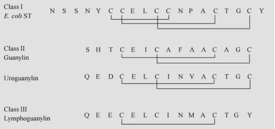
read-ing frame within the lymphoguanylin cDNAs encode a 109 amino acid polypeptide that is 84% identical to preprouroguanylin and 40% identical to preproguanylin (7,35). At the COOH-terminus of the 109 amino acid pre-prolymphoguanylin is a 15 amino acid pep-tide that is 80% identical to uroguanylin, but shares only 40% identity with guanylin (Fig-ure 3). Illustrated for comparison are the structures of opossum lymphoguanylin, uro-guanylin, guanylin and an E. coli ST peptide,
which form three different subclasses of guanylin peptides based on the number of intramolecular disulfides found within the active peptides. A major difference within lymphoguanylin is the tyrosine109 residue
because guanylins and uroguanylins have
CO2
-CO2
-CO2 -+
3HN
+ 3HN
+ 3HN
1
1
1
86
95
100
109
95 109
Guanylin
Uroguanylin
Lymphoguanylin
SHTCEICAFAACAGC
QEDCELCINVACTGC
QEECELCINM ACTGY
Figure 2 - Prepropeptide structures for guanylin, uroguanylin and lymphoguanylin. The amino acid sequences of the active peptides are show n using the single letter abbreviation of the amino acids w ithin the COOH-terminal regions of each opossum polypeptide.
Class I
E. coli ST
Class II Guanylin
Uroguanylin
Class III Lymphoguanylin
N S S N Y C C E L C C N P A C T G C Y
S H T C E I C A F A A C A G C
Q E D C E L C I N V A C T G C
Q E E C E L C I N M A C T G Y
cysteine residues at this position (1,2). The disulfide bonds formed between the first and third and second to fourth cysteines in the peptide chain were thought to be required for biological activity of guanylin and uro-guanylin. The replacement of cysteine109 with
the tyrosine109 residue is a novel molecular
change within the guanylin family of pep-tides. Lymphoguanylin is uroguanylin-like because it has two glutamate residues in its NH2-terminal domain. Opossum uroguany-lin has glutamate and aspartate residues and all other uroguanylin molecules in mamma-lian species have acidic residues at these positions (2,20,29,30,39). Shared between lymphoguanylin and uroguanylin is an inter-nal asparagine residue, which is also found in the bacterial ST peptides. Guanylin pep-tides have an aromatic amino acid at this position (1,2,6,7,35). The third difference is the methionine104 substitution in
lymphogua-nylin for the valine104 of uroguanylin.
A lymphoguanylin peptide was synthe-sized and oxidized to form an intramolecular disulfide bond between cysteine98 and
cys-teine106, with cysteine100 protected. Synthetic
lymphoguanylin containing a single disul-fide stimulates cGMP production in human T84 intestinal cells, but its potency is less than uroguanylin or guanylin (3). All three guanylin peptides are full agonists in the stimulation of intestinal GC-C expressed in human intestinal T84 cells (1-3). When the potencies of these peptides were examined in OK cells, we observed that the OK-GC receptors were also activated by lymphogua-nylin.
Lymphoguanylin mRNA transcripts of ~1.6 kb were detected in total RNA prepara-tions using Northern hybridization assays with a lymphoguanylin cDNA probe (3). Although lymphoguanylin cDNAs were iso-lated first from lymphoid tissues, we were surprised to discover that tissues with the most abundant lymphoguanylin mRNAs are the atria and ventricles of heart as well as kidney cortex. We also detected ~1.6 kb
mRNAs for lymphoguanylin using Northern assays in RNA from spleen, thymus and testis. Spleen appears to have the most abun-dant levels of lymphoguanylin mRNA within tissues of the lymphoid/immune system. RT-PCR was used to amplify the mRNA-cDNAs of spleen, thymus, lymph nodes, circulating white blood cells, bone marrow, cerebellum and testis. Lymphoguanylin cDNAs were cloned and sequenced to confirm that lym-phoguanylin mRNAs are expressed in thy-mus, lymph nodes, circulating white blood cells, bone marrow, spleen, cerebellum, kid-ney, OK cells, testis, ovary and heart of the opossum.
Co nclusio n
are learned concerning the cellular and mo-lecular mechanisms of action of these im-portant peptide hormones. Physiological roles for guanylin peptides in the regulation of target cell function via intracellular cGMP are likely to be documented in the future for the immune, reproductive, cardiovascular and
central nervous organ systems. This will complement the physiological roles of guanylin and uroguanylin that have been documented thus far in the regulation of fluid and electrolyte transport within the in-testine and kidney.
Re fe re nce s
1. Currie M G, Fok KF, Kato J, M oore RJ, Hamra FK, Duffin KL & Smith CE (1992). Guanylin: an endogenous activator of in-testinal guanylate cyclase. Proceedings of the National Academy of Sciences, USA, 89: 947-951.
2. Ham ra FK, Fort e LR, Eber SL, Pidhorodeckyj NV, Krause WJ, Freeman RH, Chin DT, Tompkins JA, Fok KF, Smith CE, Duffin KL, Siegel NR & Currie M G (1993). Uroguanylin: structure and activity of a second endogenous peptide that stimulates intestinal guanylate cyclase.
Proceedings of the National Academy of Sciences, USA, 90: 10464-10468. 3. Forte LR, Eber SL, Fan X, London RM ,
Wang Y, Row land LM , Chin DT, Freeman RH & Krause WJ (1999). Lymphoguany-lin: Cloning and characterization of a unique member of the guanylin peptide family. Endocrinology, 140: 1800-1806. 4. Hughes JM , M urad F, Chang B & Guerrant
RL (1978). Role of cyclic GM P in the ac-tion of heat-stable enterotoxin of Escheri-chia coli. Nature, 271: 755-756.
5. Wiegand RC, Kato J & Currie M G (1992). Rat guanylin cDNA: Characterization of the precursor of the endogenous activa-tor of intestinal guanylate cyclase. Bio-chemical and Biophysical Research Com-munications, 185: 812-817.
6. Wiegand RC, Kato J, Huang M D, Fok KF, Kachur JF & Currie M G (1992). Human guanylin: cDNA isolation, structure and activity. FEBS Letters, 311: 150-154. 7. Fan X, Hamra FK, Freeman RH, Eber SL,
Krause WJ, Lim RW, Pace VM , Currie M G & Forte LR (1996). Uroguanylin: cloning of preprouroguanylin cDNA, mRNA expres-sion in the intestine and heart and isola-tion of uroguanylin and prouroguanylin from plasma. Biochemical and Biophysi-cal Research Communications, 219: 457-462.
8. Hill O, Cetin Y, Cieslak A, M agert H-J & Forssmann W-G (1995). A new human
guanylate cyclase-activating peptide (uro-guanylin): precursor cDNA and colonic ex-pression. Biochimica et Biophysica Acta, 1253: 146-149.
9. Guba M , Kuhn M , Forssm ann W -G, Classen M , Gregor M & Seidler U (1996). Guanylin strongly stimulates rat duodenal HCO3- secretion: proposed mechanism and com parison w it h ot her secret a-gogues. Gastroenterology, 111: 1558-1568.
10. Joo NS, London RM , Kim HD, Forte LR & Clarke LL (1998). Regulation of intestinal Cl- and HCO3- secretion by uroguanylin.
American Journal of Physiology, 274: G633-G644.
11. Pfeifer A, Aszodi A, Seidler U, Ruth P, Hofmann F & Fassler R (1996). Intestinal secretory defects and dw arfism in mice lacking cGM P-dependent protein kinase II. Science, 274: 2082-2086.
12. Forte LR, Thorne PK, Eber SL, Krause WJ, Freeman RH, Francis SH & Corbin JD (1992). Stimulation of intestinal Cl- trans-port by heat-stable enterotoxin: activation of cAM P-dependent protein kinase by cGM P. American Journal of Physiology, 263: C607-C615.
13. Schulz S, Green CK, Yuen PST & Garbers DL (1990). Guanylyl cyclase is a heat-stable enterotoxin receptor. Cell, 63: 941-948.
14. Schulz S, Lopez M J, Kuhn M & Garbers DL (1997). Disruption of the guanylyl cy-clase-C gene leads to a paradoxical phe-notype of viable but heat-stable entero-toxin-resistant mice. Journal of Clinical In-vestigation, 100: 1590-1595.
15. M ann EA, Jump M L, Wu J, Yee E & Giannella RA (1997). M ice lacking the guanylyl cyclase C receptor are resistant to STa-induced intestinal secretion. Bio-chemical and Biophysical Research Com-munications, 239: 463-466.
16. Forte LR, Krause WJ & Freeman RH (1988). Receptors and cGM P signaling
mechanism for E. coli enterotoxin in opos-sum kidney. American Journal of Physiol-ogy, 255: F1040-F1046.
17. Forte LR, Krause WJ & Freeman RH (1989). Escherichia coli enterotoxin recep-tors: localization in opossum kidney, in-testine and testis. American Journal of Physiology, 257: F874-F881.
18. White AA, Krause WJ, Turner JT & Forte LR (1989). Opossum kidney contains a functional receptor for the Escherichia coli
heat-stable enterotoxin. Biochemical and Biophysical Research Communications, 159: 363-367.
19. Krause WJ, Freeman RH & Forte LR (1990). Autoradiographic demonstration of specific binding sites for E. coli entero-toxin in various epithelia of the North American opossum. Cell and Tissue Re-search, 260: 387-394.
20. Kita T, Smith CE, Fok KF, Duffin KL, M oore WM , Karabatsos PJ, Kachur JF, Hamra FK, Pidhorodeckyj NV, Forte LR & Currie M G (1994). Characterization of human u-roguanylin: member of the guanylin pep-tide family. American Journal of Physiolo-gy, 266: F342-F348.
21. Fan X, Hamra FK, London RM , Eber SL, Krause W J, Freem an RH, Sm ith CE, Currie M G & Forte LR (1997). Structure and activity of uroguanylin isolated from urine and intestine of rats. American Jour-nal of Physiology, 273: E957-E964. 22. Nakazato M , Yamaguchi H, Kinoshita H,
Kangaw a K, M at suo H, Chino N & M atsukura S (1996). Identification of bio-logically act ive and inact ive hum an uroguanylins in plasma and urine and their increases in renal insufficiency. Biochemi-cal and BiophysiBiochemi-cal Research Communi-cations, 220: 586-593.
23. Lima AAM , M onteiro HSA & Fonteles M C (1992). The effects of Escherichia coli
24. Fonteles M C, Greenberg RN, M onteiro HSA, Currie M G & Forte LR (1998). Natri-uretic and kaliNatri-uretic activities of guanylin and uroguanylin in the isolated perfused rat kidney. American Journal of Physiolo-gy, 275: F191-F197.
25. Greenberg RN, Hill M , Crytzer J, Krause WJ, Eber SL, Hamra FK & Forte LR (1997). Comparison of effects of uroguanylin, guanylin, Escherichia coli heat-stable en-terotoxin STa in mouse intestine and kid-ney: evidence that uroguanylin is an intes-tinal natriuretic hormone. Journal of In-vestigative M edicine, 45: 276-282. 26. Lennane RJ, Peart WS, Carey RM & Shaw
J (1975). Comparison of natriuresis after oral and intravenous sodium loading in sodium-depleted rabbits: evidence for a gastrointestinal or portal monitor of so-dium intake. Clinical Science and M olecu-lar M edicine, 49: 433-436.
27. Carey RM (1978). Evidence for a splanch-nic sodium input monitor regulating renal sodium excretion in man: lack of depend-ence upon aldosterone. Circulation Re-search, 43: 19-23.
28. Kinoshita H, Fujimoto S, Nakazato M , Yokota N, Date Y, Yamaguchi H, Hisanaga S & Eto T (1997). Urine and plasma levels of uroguanylin and its molecular forms in renal diseases. Kidney International, 52: 1028-1034.
29. Li Z, Perkins AG, Peters M F, Campa M J & Goy M F (1997). Purification, cDNA se-quence, and tissue distribution of rat uro-guanylin. Regulatory Peptides, 68: 45-56. 30. London RM , Krause WJ, Fan X, Eber SL & Forte LR (1997). Signal transduction path-w ays via guanylin and uroguanylin in the stomach and intestine. American Journal of Physiology, 273: G93-G105.
31. Nakazat o M , Yam aguchi H, Dat e Y, M iyazato M , Kangaw a K, Goy M F, Chino N & M atsukura S (1998). Tissue distribu-tion, cellular source and structural analy-sis of rat immunoreactive uroguanylin. En-docrinology, 139: 5247-5254.
32. M iyazato M , Nakazato M , M atsukura S, Kangaw a K & M atsuo H (1996). Urogua-nylin gene expression in the alimentary tract and extra-gastrointestinal tissues.
FEBSLetters, 398: 170-174.
33. Hess, R, Kuhn M , Schulz-Knappe P, Raida M , Fuchs M , Klodt J, Adermann K, Kaever V, Cetin Y & Forssmann W-G (1995). GCAP-II: isolation and characterization of the circulating form of human uroguany-lin. FEBS Letters, 374: 34-38.
34. Kinoshita H, Fujimoto S, Fukae H, Yokota N, Hisanaga S, Nakazato M & Eto T (1999). Plasma and urine levels of uroguanylin, a new natriuretic peptide, in nephrotic syn-drome. Nephron, 81: 160-164.
35. Fan X, Wang Y, London RM , Eber SL,
Krause WJ, Freeman RH & Forte LR (1997). Signaling pathw ays for guanylin and uroguanylin in the digestive, renal, central nervous, reproductive and lym-phoid systems. Endocrinology, 138: 4636-4648.
36. London RM , Eber SL, Visw esw ariah SS, Krause WJ & Forte LR (1999). Structure and activity of OK-GC: a kidney receptor-guanylate cyclase activated by guanylin peptides. American Journal of Physiolo-gy, 276: F882-F891.
37. de Sauvage FJ, Camerato TR & Goeddel DV (1991). Primary structure and func-tional expression of the human receptor for Escherichia coli heat-stable entero-toxin. Journal of Biological Chemistry, 266: 17912-17918.
38. Wada A, Hirayama T, Kitao S, Fujisaw a J, Hidaka Y & Shimonishi Y (1994). Pig intes-tinal membrane-bound receptor (guanylyl cyclase) for heat-stable enterotoxin: cDNA cloning functional expression, and charac-terization. M icrobiology and Immunology, 38: 535-541.
39. Whitaker TL, Witte DP, Scott M C & Cohen M B (1997). Uroguanylin and guanylin: dis-tinct but overlapping patterns of messen-ger RNA expression in mouse intestine.