Improved Structure and Function in
Autosomal Recessive Polycystic Rat Kidneys
with Renal Tubular Cell Therapy
K. J. Kelly1*, Jizhong Zhang1, Ling Han1, Malgorzata Kamocka1, Caroline Miller2, Vincent H. Gattone, II2†, Jesus H. Dominguez1,3
1Department of Medicine, Indiana University School of Medicine, Indianapolis, IN, United States of America, 2Department of Anatomy, Indiana University School of Medicine, Indianapolis, IN, United States of America, 3Department of Medicine, Veterans Affairs Medical Center, Indianapolis IN, United States of America
†Deceased. *[email protected]
Abstract
Autosomal recessive polycystic kidney disease is a truly catastrophic monogenetic disease, causing death and end stage renal disease in neonates and children. Using PCK female rats, an orthologous model of autosomal recessive polycystic kidney disease harboring mutantPkhd1, we tested the hypothesis that intravenous renal cell transplantation with nor-mal Sprague Dawley nor-male kidney cells would improve the polycystic kidney disease pheno-type. Cytotherapy with renal cells expressing wild typePkhd1and tubulogenic serum amyloid A1 had powerful and sustained beneficial effects on renal function and structure in the polycystic kidney disease model. Donor cell engraftment and both mutant and wild type
Pkhd1were found in treated but not control PCK kidneys 15 weeks after the final cell infu-sion. To examine the mechanisms of global protection with a small number of transplanted cells, we tested the hypothesis that exosomes derived from normal Sprague Dawley cells can limit the cystic phenotype of PCK recipient cells. We found that renal exosomes origi-nating from normal Sprague Dawley cells carried and transferred wild typePkhd1mRNA to PCK cellsin vivoandin vitroand restricted cyst formation by cultured PCK cells. The results indicate that transplantation with renal cells containing wild typePkhd1improves renal struc-ture and function in autosomal recessive polycystic kidney disease and may provide an intra-renal supply of normalPkhd1mRNA.
Introduction
Most patients with autosomal recessive polycystic kidney disease (ARPKD) who survive the neonatal period suffer from severe renal complications early in childhood [1,2]. ARPKD is a
monogenetic disease resulting from mutations inPKHD1, which encodes the cilial protein
fibrocystin [3]. Polycystic kidney disease (PKD) is currently incurable, although several
approaches have shown benefit [2,4–13]. The definitive treatment would be genetic, but, there
a11111
OPEN ACCESS
Citation:Kelly KJ, Zhang J, Han L, Kamocka M, Miller C, Gattone VH, II, et al. (2015) Improved Structure and Function in Autosomal Recessive Polycystic Rat Kidneys with Renal Tubular Cell Therapy. PLoS ONE 10(7): e0131677. doi:10.1371/ journal.pone.0131677
Editor:Benedetta Bussolati, Center for Molecular Biotechnology, ITALY
Received:December 19, 2014
Accepted:June 4, 2015
Published:July 2, 2015
Copyright:This is an open access article, free of all copyright, and may be freely reproduced, distributed, transmitted, modified, built upon, or otherwise used by anyone for any lawful purpose. The work is made available under theCreative Commons CC0public domain dedication.
Data Availability Statement:All relevant data are within the paper and its Supporting Information files.
Funding:Funding included National Institutes of Health DK082739 (KJK), Paul Teschan Research Fund of Dialysis Clinics, Inc. (KJK), US Department of Defense PR110473 (JHD) and US Department of Veterans' Affairs (JHD). The funders had no role in study design, data collection and analysis, decision to publish, or preparation of the manuscript.
are no current safe and effective clinical modes of gene transfer [14–16]. Hence, we examined a novel therapeutic strategy employing adult kidney cell transplantation, previously successful in
other renal failure models [17–20]. Our earlier data include successful long-term kidney cell
engraftment and improved renal structure and function in experimental diabetic nephropathy [19], as well as following cell auto-transplants in a chronic kidney disease model [20]. In the present study, we used adult primary kidney cells reprogrammed (via a non-viral vector) to express the tubulogenic protein serum amyloid A1 (SAA,[17]).
We now report the results of intravenous renal cell transplantation (IRCT) in the PCK rat, an orthologous model of ARPKD, derived from Sprague Dawley (SD) rats [21]. The overall aim was improved structure and function and we believe that goal was achieved. The transplanted
renal cells from SD rats expressed normalPkhd1and improved renal structure and function in
the PCK rats (as compared to rats that received no cells). Engrafted donor cells were identified in recipient kidneys 15 weeks after the last cell dose. We also report that SD renal exosomes
carry and transfer wild typePkhd1exo-mRNA and can limit cyst formation in matrigel
matri-ces. We propose that IRCT is a safe and very effective means to deliver the wild typePkhd1gene
and, more importantly, prevent progressive CKD in PKD. IRCT has the advantage that renal cells from one normal rat are sufficient to transplant multiple diseased animals, administration is non-invasive and the side effects of immunosuppression are obviated.
Materials and Methods
Primary Renal Tubular Cells
Primary renal cells from one age-matched male SD rat (Harlan, Indianapolis, IN) were equally distributed to four PCK female rats, one in each of the four cell treatment groups described below. After insuring adequate anesthesia, both kidneys were removed, the cortices minced in
S1 medium (Ham’s F-12/DMEM) with type 4 collagenase (Worthington, Lakewood, NJ), 6
mg/dl, at 37°C in 38% O2 and 5% CO2 for 50 minutes. Renal tubules were then separated by percoll gradient [20], divided into two sets, and transfected by electroporation. Control (SAA negative) tubules were co-transfected with empty vector pcDNA3.1 (30 ug), pAcGFP1-C1 (15 ug, GFP is the cytosolic label used to track cells in vivo, Clontech, Mountain View, CA). For SAA+ cells, pcDNA3.1 was replaced with pcDNA3.1-SAA1 plasmid, 30 ug, manufactured and sequenced in our laboratory as previously reported [17,20]. The isolated tubules were a mix of different tubule segments: approximately 1/4 proximal (positive for organic anion transporter 1), 20% thick ascending limb (positive for Tamm Horsfall protein), 15% collecting tubule (pos-itive for aquaporin-2) and 5% distal convoluted tubule (pos(pos-itive for thiazide-sens(pos-itive
co-trans-porter) [20]. Transfection efficiencies were>70% [20].
The co-transfected tubules were cultured in S1 medium with hepatocyte growth factor, 200 ng/ml; epidermal growth factor, 400 ng/ml (R&D Systems, Minneapolis, MN); hydrocortisone 100 ug/ml; insulin, 35 ug/ml; transferrin, 32 ug/ml; sodium selenite 42 ng/ml (Sigma, St. Louis MO); and 20% fetal calf serum. G418, 75 ug/ml, was added after 48 hours of culture for selec-tion. In preparation for transplantation, male renal tubular cells were lightly trypsinized after
7–8 days in culture, washed in PBS, and 106cells injected intravenously in the tail vein of PCK
female rats at 6 (2 days after surgery, below), 8 and 10 weeks of age.
Animal Protocols
isofluorane anesthesia and all efforts were made to minimize suffering. This included the administration of analgesia (buprenorphine) postoperatively. Although criteria (including minimal movement, not taking food, loss of more than 15% of body weight) for early euthana-sia were in place, early euthanaeuthana-sia was not necessary. The animals were monitored regularly: continuously while under anesthesia and then daily. The method of euthanasia, overdose of a barbituric acid derivative with subsequent exsanguination, is consistent with the American Veterinary Medical Association guidelines for the Euthanasia of Animals.
Experimental Design. Female PCK rats (Charles River, Wilmington, MA) were assigned to the 2 control and 4 experimental groups in a blinded manner. Rats underwent left renal ischemia or sham surgery at 6 weeks of age (between approximately 9 and 11 am) in laboratory space designated for rodent surgery. Anesthesia was accomplished with inhaled isofluorane
(0.5–1% to effect) prior to occlusion of the left renal pedicle for 50 minutes. Sham surgery
con-sisted of an identical procedure except the renal pedicle was not clamped [22]. Anesthesia was chosen to minimize recovery time and alterations in blood pressure that might cause further
renal injury. The rats were infused at 6, 8 and 10 weeks of age with donor cells (106
cells/infu-sion): one control (sham) group and one postischemia group received control (SAA-) cells. Additional control (sham) and postischemia groups received SAA+ cells. Control sham and postischemia rats received no cells. Weights, sera and urine were collected biweekly and chem-istries were measured by the Indianapolis VA clinical laboratory. Urine protein was measured
via ELISA according to the manufacturer’s protocol (Exocell, Philadelphia, PA). Cystic change
was quantified using point count stereology as described [23]. Dynamic contrast computed
tomography was performed on anesthetized animals 1–2 weeks before sacrifice using a high
speed CT scanner as described [24]. To examine the role of exosomes in the transfer of wild typePkhd1, one additional group of PCK rats underwent anesthesia as above with injection of
exosomes (20ug protein, exosomes from approximately 2 x 10⁶cells) into the renal pelvis and
sacrificed 24 hours later. Additional supporting information describing more details of animal use is in supplementary information (S1 File).
Histology and immunohistochemistry
Kidney sections were fixed in 3.8% paraformaldehyde, paraffin embedded and 5μM sections
obtained for Masson’s trichrome to stain collagen blue and periodic acid Schiff (PAS) to
evalu-ate morphology. The areas of glomerular and peritubular fibrosis were quantified in blinded sections and expressed as fractional areas, covering all available sections. Additional kidney sections were immunostained with anti-CD31 (PECAM) antibody (Santa Cruz Biotechnology, Santa Cruz, CA) and Texas-Red conjugated secondary antibody (Jackson Immunoresearch, West Grove, PA), to visualize the microvasculature. Kidney sections were also stained with anti-pan-keratin and anti-vimentin (both Cell Signaling, Danvers, MA) and Texas-Red conju-gated secondary antibodies. Anti-organic anion transporter 1 (OAT1, Alpha Diagnostics Inter-national, San Antonio, TX), anti-Tamm Horsfall protein (THP, Millipore, Temecula, CA), anti-aquaporin-2 (AQP, Millipore) and anti-thiazide-senstiive co-transporter (TSC, Alpha Diagnostics International) were used in combination with Texas Red conjugated secondary
antibodies as specific tubule segment markers. Paraformaldehyde fixed 100μm kidney sections
Fluorescent
in situ
hybridization (FISH) of the Y chromosome
FISH was used to localize the Y chromosome in female kidneys 15 weeks after IRCT with male renal cells as previously reported [20] employing the fluorescent labeled rat Y chromosome probe (Rat Idetet Chr Y Paint probe red, ID556, ID Labs Biotechnology Inc. London ON, Can-ada). Sections were counterstained with DAPI prior to imaging with Leica DMI 3000B fluores-cence microscope.
Renal SAA1 mRNA
The murine SAA1 mRNA was amplified from renal RNA (isolated using Trizol, Invitrogen,
Grand Island, NY via the supplier’s protocol) using PCR System 2400 (Perkin Elmer, San Jose,
CA) with the following primers [17]:
Forward 1:5’-CGCCACCATGGAGGGTTTTTTTCATTTGTTCAC-3’
Forward 2:5’-TACAGGCTAGCGCCACCATGGAGGGTTT-3’
Reverse 1/2:5’TCAGGTGGATCCCTCAGTATTTGTCAG-3’
Identification of DNA encoding the male sex-determining region on
chromosome Y (SRY
)
in female kidneys [
19
]
DNA was extracted from the recipient kidneys with the Wizard Genomic DNA Purification Kit as indicated by the manufacturer (Promega, Madison, WI). The specific SRY DNA was then amplified from extracted kidney DNA using PCR System 2400 with the following primers [25]:
Forward:5’-AAGCGCCCCATGAATGC-3’
Reverse:5’-AGCCAACTTGCGCCTCTCT-3’
Genotyping
Identification of wild type and mutatedPkhd1genes in the PCK rats was performed via PCR
(as above) using the primers specified by Charles River:
Mut-Forward:5’-AAG CCA AAT CTT TCT CTT TTC CT-3’
Mut-Reverse:5’- CTT GCT GTC CGA ATA CCA C -3’
Wild type-Forward:5’-ACT GCC TTT TAC TGA AGC ATT TAA C-3’
Wild type-Reverse:5’- TGG AAG GAA AAG TTG CCC T -3’
Exosome studies
Primary renal tubule cells from normal Sprague Dawley rats (Harlan, Indianapolis, IN) were isolated as above. After 2 days in culture, S1 medium with exosome-free fetal calf serum was used. Two days later, the cell culture supernatant was centrifuged at 300g for 10 minutes to remove cells, 2000g x 10 minutes to remove dead cells, 10,000g x 30 minutes to remove cells debris. The resultant supernatant was centrifuged at 100,000g x 70 minutes, washed and centri-fuged again at 100,000g x 70 minutes to obtain exosomes. After fixation in 2%
paraformalde-hyde/2% glutaraldehyde/0.1M phosphate buffer, the sample was adsorbed to a 200–400 mesh
carbon/formvar coated grid and the negative stain (Nanovan, Nanoprobes, Yaphank, NY) added. Exosome isolation was then verified by electron microscopy (Tecnai G2 12 Bio Twin microscope [FEI, Hillsboro, OR] equipped with an AMT CCD camera [Advanced Microscopy Techniques, Danvers, MA]). Prior to their addition to cultured PCK cells, SD exosome RNA was labeled with red fluorescent dye and exosome protein with green fluorescent dye via
Exo-Glow (SBI, Mountain View, CA) according to the supplier’s protocol. PCK tubular cells were
isolated by collagenase digestion and cultured as for Sprague Dawley cells (above). When the
fetal calf serum and fluorescently labeled exosomes (10μg protein/106cells) added to the cells and imaging performed approximately 16 hours later. Uptake of exosomes was documented by PCR genotyping (above). For these studies, prior to incubation with exosomes, some PCK cells were treated with cytochalasin D and chloropromazine (each 10ug/ml) to block exosome uptake (actin polymerization and endocytosis, respectively). In separate studies, exosome treated cells were cultured for 2 days prior to resuspension in matrigel (BD Biosciences, Bed-ford, MA) at a concentration of 100,000 cells/ml and incubated in glass bottom dishes. In some studies, PCK and SD cells were cultured together in the following proportions (Table 1)
Immunoblotting
Exosome and cell lystate samples and fibrocystin protein control (Santa Cruz Biotechnology, Santa Cruz, CA) were fractionated by electrophoresis through 16.5% polyacrylamide Tris-tri-cine gels. After transfer and blocking, blots were incubated with anti-fibrocystin or anti-CD63 (Santa Cruz).
Statistics
The experimental unit was one culture dish or one kidney (as right and left kidneys were treated differently). For albuminuria and BUN, the experimental unit was a single animal. Data are expressed as means ± 1 standard error. Analysis of variance was used to determine if
differ-ences among mean values reached statistical significance. Tukey’s test was used to correct for
multiple comparisons. Student's t test (2 tailed, 2 sample, unequal variance) was used for com-parisons between groups (GraphPad Prism, LaJolla, CA). The null hypothesis was rejected at p<0.05.
Results
Female PCK rats received either no cells, control cells or SAA+ cells intravenously when 6, 8 and 10 weeks old and were sacrificed at 25 weeks of age. Renal tubular cells from normal male SD rats were either transfected with empty vector (control cells) or SAA1. Both control and SAA+ cells were also transfected with green fluorescent protein (GFP) for tracking purposes.
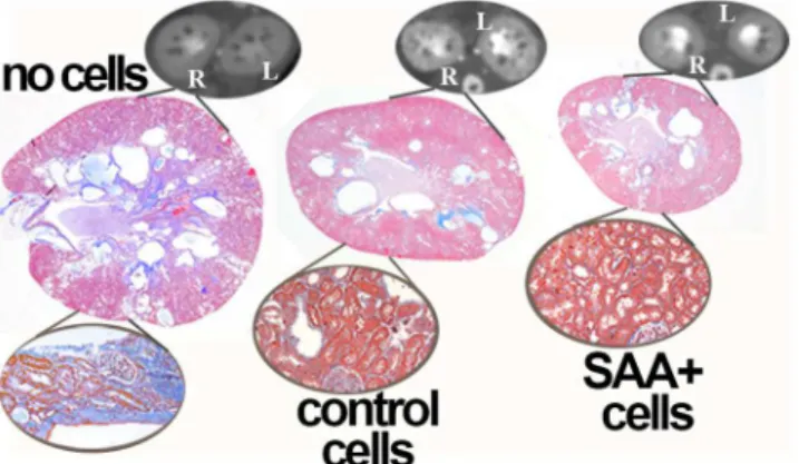
InFig 1is shown the marked improvement in cyst burden and renal histology in PCK rats
that were transplanted with control A renal cells (containing wild typePkhd1) and an even
greater positive effect in groups that received B renal cells (containing both wild typePkhd1
and SAA1) when compared to PCK rats that did not receive cells. In addition to decreased total cyst volume and kidney weights, better renal function was observed in the cell transplant
groups compared to the“no cell”rats as shown by decreased albuminuria and serum blood
Table 1.
PCK 100% 98% 90% 80% 0%
SD 0% 2% 10% 20% 100%
Cyst number was quantified in blinded images at 4 days. At 7 days, the cells werefixed in 4% paraformaldehyde, permeabilized in 0.1% Triton and incubated with rhodamine phalloidin (1:200) and Hoescht 3342 (both Life Technologies, Carlsbad, CA). Two photon images were acquired with an Olympus Fluoview FV-1000 MPE system (Olympus America, Central Valley, PA) using 839 nm excitation
wavelength and an Olympus XLPLN 25x, NA 1.05 water immersion objective. Z-stacks were collected to evaluate cyst formation in 3D. Volume rendering was performed using Amira software (FEI, Burlington, MA).
urea nitrogen (BUN) (Fig 2). Mean BUN was lower in the group that received SAA+ cells than in the control (SAA-) cell group.
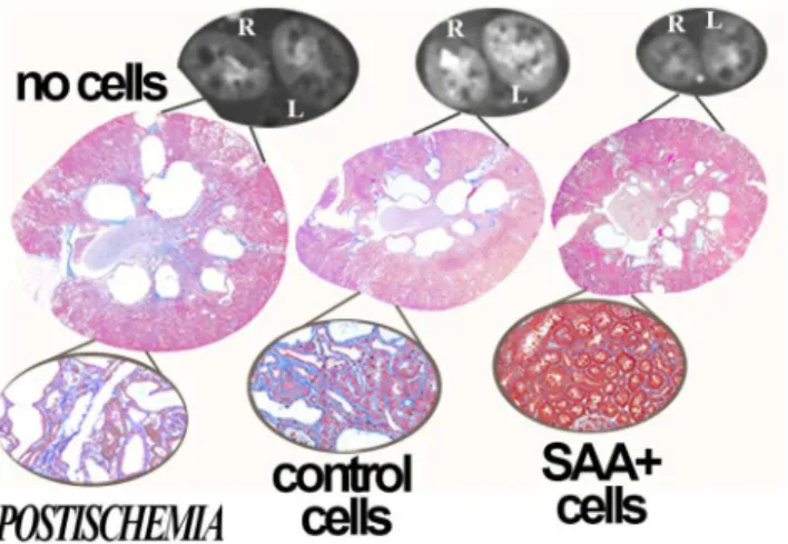
We hypothesized that renal ischemia, common in PKD, would reduce the number of mutant cells through mutant cell death and facilitate engraftment of transplanted cells on the denuded basement membrane. Thus, an additional three PCK groups were subjected to left (unilateral) renal ischemia at 6 weeks of age and transplanted with either no cells, control cells or SAA+ cells. One of the control ischemia rats died when 23 weeks of age. In the postischemia groups, decreased total cyst volume, kidney weights, albuminuria and BUN were also observed
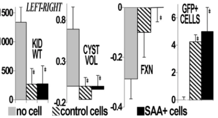
in the rats that received cytotherapy (Figs3and4). In addition, comparisons between the
post-ischemic left and sham right kidneys showed improvement in kidney weight, total cyst volume, split renal function by dynamic contrast computed tomography and increased numbers of GFP+ cells/hpf in the postischemic kidney (Fig 5). Despite this, function and structure were more impaired in the postischemia groups. Interestingly, mean heart/body weight was lower in the cytotherapy groups (4.46, 3.66 and 3.69 mg/g body weight in no cell, control and SAA+ cell
Fig 1. Cytotherapy decreases cyst burden.When compared to no cell transplant groups, treatment of PCK rats wiSAA+ or control cells improves cyst volume and structure at 25 weeks of age. The termination point was 15 weeks after the final cell transplant. Representative dynamic contrast CT images and PAS stained and trichrome stained sections (insets) are presented.
doi:10.1371/journal.pone.0131677.g001
Fig 2. Improvement in structure and function in PCK rat kidneys with cell transplant.When compared to no cell transplant groups, treatment of PCK rats with SAA+ or control (A) cells improves albuminuria (ALB), total cyst volume (CYST VOL), blood urea nitrogen (BUN), and kidney weight (KID WT) at 25 weeks of age, 15 weeks after the final cell transplant. Albuminuria is presented as g/g creatinine, cyst volume as ml/g body weight, BUN as mg/dl, and kidney weight as mg/g body weight.*p<0.05 vs no cell group, §p<0.05 vs control cells
groups, respectively, p<0.01 vs no cell). There were no significant differences in animal weights
(mean 302 ± 2.6 g) or final liver weights during the course of the study.
Fibrosis is a significant contributor to decreased function in PKD [26]. In addition to improvements in function and total cyst volume, decreases in both peritubular fibrosis and glo-merulosclerosis were also observed in treated kidneys. SAA+ cells results in larger improve-ments in fibrosis than SAA- cells (Fig 6).
In contrast to stem cell transplant protocols, we have clearly documented engraftment of
donor cells months after transplantation in other renal failure models [18–20]. Multiple
inde-pendent techniques were used to verify this critical mechanistic point (Fig 7): (1) fluorescence in situ hybridization (FISH) showed the Y chromosome in female recipient kidneys trans-planted with male cells, but not in normal females; (2) PCR genotyping demonstrated both
mutated and wild typePkhd1in the kidneys of transplanted rats, but not in those that did not
receive cells; PCR detected both (3) DNA encoding the male determining SRY gene in female
Fig 3. Protection in postischemia kidneys.When compared to no cell transplant groups, treatment of PCK rats with SAA+ or control cells also improves cyst volume and structure in postischemia kidneys at 25 weeks of age, 15 weeks after the final cell infusion. Representative dynamic contrast CT images and PAS stained and trichrome stained sections (insets) are presented.
doi:10.1371/journal.pone.0131677.g003
Fig 4. Improvement in structure and function in postischemia PCK rat kidneys with cell transplant. Treatment with SAA+ or control (A) cells improves albuminuria (ALB), total cyst volume (CYST VOL), blood urea nitrogen (BUN), and kidney weight (KID WT) in postischemia PCK rats. Albuminuria is presented as g/g creatinine, cyst volume as ml/kidney/g body weight, BUN as mg/dl, and kidney weight as mg/g body weight. ‡p<0.05 vs no cell/ischemia group, #p<0.05 vs control cell/ischemia group
kidneys transplanted with male cells but not in control females and (4) SAA mRNA in kidneys that received SAA+ but not control (SAA-) cells or in rats not given cells; (5) fluorescence microscopy showed GFP+ cells in kidneys of rats that received GFP+ control or SAA+ cells and (6) co-localization of immunoreactive SAA with GFP in kidneys from rats that received
SAA+ cells. In contrast to the kidneys, GFP+ donor cells were very rarely (<1 cell/hpf) seen in
lungs, spleen or liver in any of the groups. The majority of GFP+ cells were tubular with rare renal interstitial GFP+ cells (Fig 7). In summary, multiple independent tests showed that cell
transplantation can deliver normal genes to cystic kidneys, which is the goal of“gene therapy.”
We have postulated that the broad benefit seen with cell transplants [18–20] points to a
gen-eral action, potentially explained by improved vasculature with better delivery of oxygen and nutrients. It is known that major renal microvascular abnormalities aggravate human PKD, promoting renal dysfunction and cyst enlargement [27]. Thus, the renal microvasculature was
labeled with an anti-CD31 antibody to evaluate the role of cell transplantation,Fig 8.
Represen-tative images illustrate severe glomerular microvascular attenuation in control PCK rats and in those transplanted with SAA- cells. In contrast, glomerular vessels were much better preserved in the groups that received SAA+ cells. Pericystic hypervascularity, thought to contribute to cyst growth in human PKD [28] was markedly attenuated in cell treated rats.
Epithelial mesenchymal transition has also been implicated in the pathogenesis of polycystic kidney disease [29] and the mesenchymal marker vimentin has been found in cystic epithelia in the PCK rat [30]. Thus, we examined the epithelial marker pan-keratin and the mesenchy-mal marker vimentin in donor cells and PCK kidneys. The donor cells were of epithelial origin as indicated by uniform labeling with anti-pankeratin (not shown). The majority were also pos-itive for either OAT1 (24±3%), THP (18±3%) or AQP2 (16±2%) consistent with proximal tubule, thick ascending limb and collecting tubule phenotype, respectively [20]. Only a small
proportion (<10%) stained positive for the distal convoluted tubule marker TSC. Vimentin
expression was prominent in cyst lining epithelia in control PCK kidneys at study termination. This was markedly decreased in the kidneys from treated rats. GFP+ donor cells did not stain with anti-vimentin. Conversely, pan-keratin staining was significantly higher in kidneys from cell treated than from untreated rats (Fig 9).
Given that donor cells are only a small proportion of the host PCK kidney, and yet they altered the PCK phenotype, we hypothesized that engrafted cell exosomes influence neighbor-ing PCK cells [31]. This postulate is based on the fact that exosomes contain vast mRNA
Fig 5. The effect of ischemia.Comparison of left (postischemic) and right (contralateral, sham) kidneys in postischemia groups shows that the difference in kidney weight, total cyst volume, split renal function by dynamic contrast computed tomography between left and right kidneys is attenuated in the groups that received cell transplantation. In addition, more GFP+ cells were found in the postischemic (vs sham) kidneys. ‡p<0.05 vs no cell/ischemia group.
libraries [32] and might carry and transfer wild typePkhd1mRNA to PCK renal cells. To test
this hypothesis, we verified that SD cells produce nanovesicles that express CD63 and are of a
size consistent with exosomes (Fig 10) [33–35]. The SD exosomes also expressed the protein
product ofPkhd1, fibrocystin. Intra-exosome RNA (exoRNA) and protein were labeled with
Exo-Red and Exo-Green dyes (Exo-Glow, System Biosciences, Mountain View, CA), respec-tively. Labeled exosome cargo was taken up by cultured renal tubular cells from PCK rats,
resulting in expression of wild typePkhd1RNA in cells incubated with exosomes from SD cells
but not in untreated PCK cells (Fig 10). When PCK cells were grown in extracellular matrix
(matrigel), abundant 3D cystic structures were formed, for exampleFig 11G. These cystic
structures expanded for 7 days when they were imaged by 2-photon microscopy, confirming their cystic nature. SD renal cells, in contrast, did not form cysts under the same culture
Fig 6. Histology.Representative trichrome stained sections (resulting in blue labeling of fibrous tissue) of glomeruli (glom), cortex and medulla in each of the 6 groups are presented. Quantification of glomerulosclerosis (in a total of 5602 glomeruli) and peritubular fibrosis (in 4249 microscope fields) is presented in the graphs. GLOM, glomeruli; NO, no cells; CTRL, control;*p<0.05 vs no cell group;‡p<0.05 vs no cell/ischemia group; §p<0.05 vs control cell group; #p<0.05 control cell/ischemia group
conditions. When PCK cells were co-cultured with renal cells derived from SD rats, the number of cysts formed decreased as the proportion of SD cells increased. When PCK cells were cul-tured with SD exosomes prior to incubation in matrigel, the cells remained non-cystic and
formed“tubular”structures (Fig 11E). This result supports the hypothesis that exosomes
Fig 7. Engraftment of transplanted cells.Multiple methods were employed to demonstrate the persistence of infused cells in transplanted kidneys. (A) Fluorescence in situ hybridization (FISH) showed Y chromosome + (red) nuclei in kidneys of female rats that received male cells. The insets show green fluorescence of areas at arrows, demonstrating GFP and the Y chromosome in the same cells. Nuclei in this panel and panels E and F are stained with DAPI (blue). (B) Genotyping by PCR demonstrated the presence of both wild type (WT) and mutated (MUT)Pkhd1transcripts in kidneys from PCK rats that received SAA+ or control cells but not in PCK rats that received no cells. Only the wild type transcript is present in normal Sprague Dawley (SD) rats. (C) PCR for the SRY male determining gene showed similar results: SRY was present in female PCK rats transplanted with male cells and male (♂) SD but not female rats that received no cells. (D) mRNA for SAA is present only in groups that received SAA+ cells demonstrating transcriptional activity of the SAA gene from donor SAA+ cells. pcDNA3.3-SAA1 was used as the positive (+) control for SAA PCR. (E) GFP positive cells are also found in transplanted kidneys. (F) Immunostaining for SAA demonstrates co-localization (orange) with GFP in SAA+ groups. The insets in F show higher power confocal images for SAA in cyst epithelium. M = normal male; F = normal female; MW = molecular weight markers.
derived from normal cells transfer genetic material and the presence of wild type Pkhd1 results in decreased cystogenesis in PCK cells. In addition, co-culture of PCK cells with SD cells resulted in decreased cyst formation (Fig 11). These results demonstrate that normal renal tubular cells and exosomes derived from these cells contain wild type genetic material and can improve the phenotype in polycystic kidney disease. The results are consistent with the hypoth-esis that improved phenotype in the presence of normal SD cells results from transfer of genetic material from the SD cells via exosomes. Injection of SD exosomes into PCK rats also resulted
in the transfer of wild typePkhd1mRNA into PCK kidneys (Fig 12).
Discussion
The number of PKD end stage renal disease (ESRD) patients is increasing: US Renal Data Sys-tem (USRDS) 2013 [36] reports 28.2 (per million population) PKD patients on dialysis in 1985 and 92.5 in 2011. These data illustrate that PKD is a significant clinical issue. We hypothesized
Fig 8. Protection of microvasculature with cytotherapy.Representative images stained for CD31 (platelet endothelial cell adhesion molecule [PECAM], red) show better preserved glomerular vasculature in SAA+ cell groups. In addition, less vasculature surrounding abnormal cystic epithelium is seen in the groups that received SAA+ cells. Quantification of red pixel density representing CD31 staining in 266 images is shown in the graphs.*p<0.05 vs no cell group;‡p<0.05 vs no cell/ischemia group
Fig 9. Decreased vimentin in kidneys after cell treatment.Vimentin expression, consistent with epithelial to mesenchymal transition, has been documented in PKD and was found by immunohistochemistry (red) in the control (no cell) group in cyst lining cells. Immunoreactive vimentin was decreased in treated (control and SAA+ cells) kidneys with most tubule lining cells positive for the epithelial marker pan-keratin (not shown). GFP+ donor cells did not stain with anti-vimentin. Kidney sections were counterstained with DAPI to stain nuclei blue. The graphs show pixel density of vimentin (x10 to facilitate visualization on one axis) and pan-keratin. #p<0.05 vs no cells,‡p<0.05 vs control cells
doi:10.1371/journal.pone.0131677.g009
Fig 10. Exosome Uptake by PCK cells.Isolation of exosomes (exo) was confirmed by electron microscopy demonstrating vesicles of appropriate size after negative stain (EM, A) and enrichment of CD63 on
immunoblot (B). Exosomes from normal Sprague Dawley rats express fibrocystin (FPC, B). Internal exosome protein was fluorescently labeled green and RNA red prior to incubation of PCK cells with the exosomes for 16 hours. In panel C are representative fluorescence microscopy images demonstrating uptake of the exosomes by the primary PCK renal cells. The“no exo”control was incubated with exo-glow dye but no exosomes. PCR genotyping shows expression of both wild type and mutantPkhd1mRNA in the PCK cells exposed to exosomes (D). Wild typePkhd1is found in SD exosomes and SD cells (SD) and only mutant Pkhd1in control PCK cells. After the addition of the exosome uptake inhibitor chlorpromazine and the protein synthesis inhibitor cytochalasin D prior to exosomes, no wild typePkhd1was transferred to the mutant PCK cells. Graphs demonstrate quantification of band densities representing CD63 (corrected for actin) or mutant and wild typePkhd1. MW, molecular weight markers, + positive control (panel B)
Fig 11. The effect of exosomes on PCK cysts in culture.Representative confocal images of cells stained with rhodamine phalloidin to label actin red and Hoescht to label nuclei blue demonstrate cysts in tubule cells from PCK rats grown in matrigel without exosome treatment (A, F, G). Neither PCK cells cultured with exosomes (B, C, E) from Sprague Dawley (SD) rats nor SD tubular cells (D, H) form cysts in matrigel. The higher power images (A-D, scale bar 20um) show orthogonal projections to demonstrate the clear presence or absence of cysts. Panel F, showing a 3D reconstruction of multiple images, also demonstrates the cystic nature of the structures formed by PCK cells (scale bar 20um). The lower power images (E, G, H, scale bar 50um) demonstrate tubular structures in the presence of exosomes, multiple cysts in one field of PCK cells and the absence of cysts in SD cells, respectively. In PCK cells treated with exosomes (E),“tube-like”structures are seen. Quantification of cyst number in a total of 168 fields is presented in the graph (I). B exosomes are from SAA+ cells, A are from SAA- control cells. Both A and B exosomes contain wild typePkhd1(above).
doi:10.1371/journal.pone.0131677.g011
Fig 12. Wild type Pkhd1 in kidneys from PCK rats treated with SD exosomes.PCR genotype demonstrates mutant Pkhd1 in kidneys from control and treated PCK rats. Wild type Pkhd1 was found in kidneys from PCK rats treated with SD exosomes (24h after renal pelvic injection) but not in untreated rats. -, negative PCR control; exo, exosomes; MW, molecular weight markers.
that cytotherapy with cells containing the wild typePkhd1gene would result in renal chimeras
and improve structure and decrease cystogenesis in PKD. The PCK rat was studied because the
mutated gene in the PCK rat (Pkhd1) is orthologous to the human gene; and the phenotype is
very similar to the human phenotype in both ARPKD and autosomal dominant PKD
(ADPKD) [3,37,38]. The pathophysiology of ADPKD and ARPKD, and other renal cystic dis-eases, is thought to result from similar abnormalities in convergent and/or integrated signaling
pathways [26,38–40]. The products of the most commonly mutated genes, polycystin (PC)1
and PC2 in ADPKD and fibrocystin (FPC) in ARPKD, all localize to primary cilia and are believed to modulate essential cellular functions [41]. We report that infusion of a relatively small number of SD rat adult renal cells significantly improved the otherwise predetermined cystic phenotype of the PCK rat. Accordingly, we suggest that IRCT can be applied to limit untreatable PKD. Furthermore, multiple patients can potentially receive cells from a single donor, a critical point since many ESRD patients never receive renal transplants due to short-age of organs for donation [42]. All models have limitations, although in prior studies, we reported that donor cells expressing the tubulogenic SAA protein improved other models of CKD. However, in the PCK model, we found that the major differences existed between PCK rats receiving either SAA+ or control SAA- cells and PCK rats not receiving any cells, although those receiving SAA+ cells had slightly better renal structure (significantly decreased fibrosis) and function (significantly decreased BUN). Thus, in this model where the mechanism of renal failure is the destruction of normal renal architecture by cyst expansion, the provision of
nor-mal cells with wild typePkhd1may be more critical than the role of tubulogenic SAA1.
The present results corroborate the great potential of primary renal cell transplants. In
addi-tion to containing the wild typePkhd1gene, anchored donor tubular cells may positively
influ-ence recipient renal cells [20]. Thus, we have also shown that exosomes from normal SD cells
contain wild typePkhd1and its protein product, fibrocystin, and that wild typePkhd1can be
transferred from renal exosomes to PCK renal cells. In contrast to our cell transplant protocols,
stem cells have not been shown to become functional renal cells [43–46] and, in some cases,
the transplanted stem cells acquire a totally undesirable and uncontrolled phenotype in recipi-ent CKD kidneys [47] or results in embolization in the lung [48]. Microvesicles from mesen-chymal stem cells or endothelial progenitor cells have been shown to protect from ischemic
renal injury or partial nephrectomy in rodent models [49–52]. In a model of renal failure due
to ischemia followed by gentamicin, improvement in renal function and structure has been found using adult renal cells enriched for erythropoietin producing cells [53]. In the present study, the long term benefit of cell transplants on both structure and function points to a
wide-spread impact which may be due to intra-renal delivery of the wild typePkhd1gene as well as
improved renal vasculature with better delivery of oxygen/nutrients. In conclusion, we suggest that, in PKD, adult renal epithelial cells and exosomes offer a novel, physiological and effective means to deliver normal genes. These potential therapies effect preservation of renal structure and function and limit cyst formation and expansion in experimental PKD.
Supporting Information
place, early euthanasia was not necessary. The animals were monitored regularly: continuously while under anesthesia and then daily. The method of euthanasia, overdose of a barbituric acid derivative with subsequent exsanguination, is consistent with the American Veterinary Medical Association guidelines for the Euthanasia of Animals.
(DOCX)
Acknowledgments
We acknowledge the contribution of Dr. Gattone to the design of the experiments and analysis of total cyst volume prior to his untimely death. We thank Dr. Barbara Kluve-Beckerman for anti-SAA antibody. Two photon and confocal imaging was performed at the Indiana Center for Biological Microscopy, Indianapolis, IN. The results were presented, in part, at the annual meeting of the American Society of Nephrology in 2013.
Author Contributions
Conceived and designed the experiments: KJK VHG JHD. Performed the experiments: KJK JZ LH MK CM JHD. Analyzed the data: KJK JZ JHD. Contributed reagents/materials/analysis tools: MK VHG. Wrote the paper: KJK JHD.
References
1. Dell KMR, Avner ED (1993) Autosomal Recessive Polycystic Kidney Disease,In: Pagon RA, Adam MP, Ardinger HH et al, ed, GeneReviews, Seattle (WA), University of Washington, Seattle. 2. Gattone VH 2nd, Wang X, Harris PC, Torres VE (2003) Inhibition of renal cystic disease development
and progression by a vasopressin V2 receptor antagonist. Nat Med 9: 1323–1326. PMID:14502283 3. Ward CJ, Hogan MC, Rossetti S, Walker D, Sneddon T, Wang X, et al. (2002) The gene mutated in
autosomal recessive polycystic kidney disease encodes a large, receptor-like protein. Nat Genet 30: 259–269. PMID:11919560
4. Torres VE, Harris PC (2014) Strategies targeting cAMP signaling in the treatment of polycystic kidney disease. J Am Soc Nephrol 25: 18–32. doi:10.1681/ASN.2013040398PMID:24335972
5. Torres VE, Chapman AB, Devuyst O, Gansevoort RT, Grantham JJ, Higashihara E, et al. (2012) Tol-vaptan in patients with autosomal dominant polycystic kidney disease. N Engl J Med 367: 2407–2418. doi:10.1056/NEJMoa1205511PMID:23121377
6. Torres VE, Sweeney WE Jr, Wang X, Qian Q, Harris PC, Frost P, et al. (2003) EGF receptor tyrosine kinase inhibition attenuates the development of PKD in Han:SPRD rats. Kidney Int 64: 1573–1579. PMID:14531789
7. Tao Y, Zafar I, Kim J, Schrier RW, Edelstein CL (2008) Caspase-3 gene deletion prolongs survival in polycystic kidney disease. J Am Soc Nephrol 19: 749–755. doi:10.1681/ASN.2006121378PMID: 18272845
8. Sweeney WE, Chen Y, Nakanishi K, Frost P, Avner ED (2000) Treatment of polycystic kidney disease with a novel tyrosine kinase inhibitor. Kidney Int 57: 33–40. PMID:10620185
9. Richards WG, Sweeney WE, Yoder BK, Wilkinson JE, Woychik RP, Avner ED (1998) Epidermal growth factor receptor activity mediates renal cyst formation in polycystic kidney disease. J Clin Invest 101: 935–939. PMID:9486961
10. Gattone VH 2nd, Cowley BD Jr., Barash BD, Nagao S, Takahashi H, Yamaguchi T, et al. (1995) Methyl-prednisolone retards the progression of inherited polycystic kidney disease in rodents. Am J Kidney Dis 25: 302–313. PMID:7847359
11. Davis ID, MacRae Dell K, Sweeney WE, Avner ED (2001) Can progression of autosomal dominant or autosomal recessive polycystic kidney disease be prevented? Semin Nephrol 21: 430–440. PMID: 11559884
13. Blazer-Yost BL, Haydon J, Eggleston-Gulyas T, Chen JH, Wang X, Gattone V, et al. (2010) Pioglita-zone Attenuates Cystic Burden in the PCK Rodent Model of Polycystic Kidney Disease. PPAR Res 2010: 274376. doi:10.1155/2010/274376PMID:21052534
14. Rossetti S, Kubly VJ, Consugar MB, Hopp K, Roy S, Horsley SW, et al. (2009) Incompletely penetrant PKD1 alleles suggest a role for gene dosage in cyst initiation in polycystic kidney disease. Kidney Int 75: 848–855. doi:10.1038/ki.2008.686PMID:19165178
15. Weber GF (2013) Gene therapy—why can it fail? Med Hypotheses 80: 613–616. doi:10.1016/j.mehy. 2013.01.037PMID:23484673
16. Wirth T, Parker N, Yla-Herttuala S (2013) History of gene therapy. Gene 525: 162–169. doi:10.1016/j. gene.2013.03.137PMID:23618815
17. Kelly KJ, Kluve-Beckerman B, Dominguez JH (2009) Acute-phase response protein serum amyloid A stimulates renal tubule formation: studies in vitro and in vivo. Am J Physiol Renal Physiol 296: F1355– 1363. doi:10.1152/ajprenal.90622.2008PMID:19321596
18. Kelly KJ, Kluve-Beckerman B, Zhang J, Dominguez JH (2010) Intravenous cell therapy for acute renal failure with serum amyloid A protein reprogrammed cells. Am J Physiol Renal Physiol 299: F453–464. doi:10.1152/ajprenal.00050.2010PMID:20534870
19. Kelly KJ, Zhang J, Han L, Wang M, Zhang S, Dominguez JH (2013) Intravenous Renal Cell Transplan-tation (IRCT) with SAA1 positive cells prevents progression of chronic renal failure in rats with ische-mic-diabetic nephropathy. Am J Physiol Renal Physiol 305: F1804–1812. doi:10.1152/ajprenal. 00097.2013PMID:24133118
20. Kelly KJ, Zhang J, Wang M, Zhang S, Dominguez JH (2012) Intravenous Renal Cell Transplantation (IRCT) for rats with acute and chronic renal failure. Am J Physiol Renal Physiol 303: F357–F365. doi: 10.1152/ajprenal.00680.2011PMID:22592640
21. O'Meara CC, Hoffman M, Sweeney WE Jr., Tsaih SW, Xiao B, Jacob HJ, et al. (2012) Role of genetic modifiers in an orthologous rat model of ARPKD. Physiol Genomics 44: 741–753. doi:10.1152/ physiolgenomics.00187.2011PMID:22669842
22. Kelly KJ, Williams WW, Colvin RB, Bonventre JV (1994) Antibody to intercellular adhesion molecule-1 protects the kidney against ischemic injury. Proc Natl Acad Sci USA 91: 812–816. PMID:7904759 23. Gattone VH 2nd, Sinders RM, Hornberger TA, Robling AG (2009) Late progression of renal pathology
and cyst enlargement is reduced by rapamycin in a mouse model of nephronophthisis. Kidney Int 76: 178–182. doi:10.1038/ki.2009.147PMID:19421190
24. Mason SB, Liang Y, Sinders RM, Miller CA, Eggleston-Gulyas T, Crisler-Roberts R, et al. (2010) Dis-ease stage characterization of hepatorenal fibrocystic pathology in the PCK rat model of ARPKD. Anat Rec (Hoboken) 293: 1279–1288.
25. Seckert CK, Renzaho A, Tervo HM, Krause C, Deegen P, Kuhnapfel B, et al. (2009) Liver sinusoidal endothelial cells are a site of murine cytomegalovirus latency and reactivation. J Virol 83: 8869–8884. doi:10.1128/JVI.00870-09PMID:19535440
26. Wilson PD (2004) Polycystic kidney disease. N Engl J Med 350: 151–164. PMID:14711914 27. Peterson KM, Franchi F, Loeffler DL, Psaltis PJ, Harris PC, Lerman LO, et al. (2013) Endothelial
dys-function occurs prior to clinical evidence of polycystic kidney disease. Am J Nephrol 38: 233–240. doi: 10.1159/000354236PMID:24008943
28. Bello-Reuss E, Holubec K, Rajaraman S (2001) Angiogenesis in autosomal-dominant polycystic kidney disease. Kidney Int 60: 37–45. PMID:11422734
29. Chea SW, Lee KB (2009) TGF-beta mediated epithelial-mesenchymal transition in autosomal dominant polycystic kidney disease. Yonsei Med J 50: 105–111. doi:10.3349/ymj.2009.50.1.105PMID:19259356 30. Togawa H, Nakanishi K, Mukaiyama H, Hama T, Shima Y, Sako M, et al. (2011)
Epithelial-to-mesen-chymal transition in cyst lining epithelial cells in an orthologous PCK rat model of autosomal-recessive polycystic kidney disease. Am J Physiol Renal Physiol 300: F511–520. doi:10.1152/ajprenal.00038. 2010PMID:21084407
31. Turturici G, Tinnirello R, Sconzo G, Geraci F (2014) Extracellular membrane vesicles as a mechanism of cell-to-cell communication: advantages and disadvantages. Am J Physiol Cell Physiol 306: C621– 633. doi:10.1152/ajpcell.00228.2013PMID:24452373
32. Mathivanan S, Fahner CJ, Reid GE, Simpson RJ (2012) ExoCarta 2012: database of exosomal pro-teins, RNA and lipids. Nucleic Acids Res 40: D1241-1244. doi:10.1093/nar/gkr828PMID:21989406 33. Chambers AE, Stanley PF, Randeva H, Banerjee S (2011) Microvesicle-mediated release of soluble
34. Chen L, Charrier A, Zhou Y, Chen R, Yu B, Agarwal K, et al. (2014) Epigenetic regulation of connective tissue growth factor by MicroRNA-214 delivery in exosomes from mouse or human hepatic stellate cells. Hepatology 59: 1118–1129. doi:10.1002/hep.26768PMID:24122827
35. Hogan MC, Johnson KL, Zenka RM, Charlesworth MC, Madden BJ, Mahoney DW, et al. (2014) Sub-fractionation, characterization, and in-depth proteomic analysis of glomerular membrane vesicles in human urine. Kidney Int 85: 1225–1237. doi:10.1038/ki.2013.422PMID:24196483
36. U.S. Renal Data System, USRDS 2013 Annual Data Report: Atlas of Chronic Kidney Disease and End-Stage Renal Disease in the United States, National Institutes of Health, National Institute of Diabetes and Digestive and Kidney Diseases, Bethesda, MD, 2013.
37. Lager DJ, Qian Q, Bengal RJ, Ishibashi M, Torres VE (2001) The pck rat: a new model that resembles human autosomal dominant polycystic kidney and liver disease. Kidney Int 59: 126–136. PMID: 11135065
38. Kaimori JY, Germino GG (2008) ARPKD and ADPKD: first cousins or more distant relatives? J Am Soc Nephrol 19: 416–418. doi:10.1681/ASN.2008010033PMID:18272839
39. Woo YM, Park JH (2013) microRNA biomarkers in cystic diseases. BMB Rep 46: 338–345. PMID: 23884099
40. Pei Y, Watnick T, He N, Wang K, Liang Y, Parfrey P, et al. (1999) Somatic PKD2 mutations in individual kidney and liver cysts support a "two-hit" model of cystogenesis in type 2 autosomal dominant polycys-tic kidney disease. J Am Soc Nephrol 10: 1524–1529. PMID:10405208
41. Sweeney WE Jr., Avner ED (2006) Molecular and cellular pathophysiology of autosomal recessive polycystic kidney disease (ARPKD). Cell Tissue Res 326: 671–685. PMID:16767405
42. Wolfe RA, Ashby VB, Milford EL, Ojo AO, Ettenger RE, Agodoa LY, et al. (1999) Comparison of mortal-ity in all patients on dialysis, patients on dialysis awaiting transplantation, and recipients of a first cadav-eric transplant. N Engl J Med 341: 1725–1730. PMID:10580071
43. Duffield JS, Park KM, Hsiao LL, Kelley VR, Scadden DT, Ichimura T, et al. (2005) Restoration of tubular epithelial cells during repair of the postischemic kidney occurs independently of bone marrow-derived stem cells. J Clin Invest 115: 1743–1755. PMID:16007251
44. Feng Z, Ting J, Alfonso Z, Strem BM, Fraser JK, Rutenberg J, et al. (2010) Fresh and cryopreserved, uncultured adipose tissue-derived stem and regenerative cells ameliorate ischemia-reperfusion-induced acute kidney injury. Nephrol Dial Transplant 25: 3874–3884. doi:10.1093/ndt/gfq603PMID: 20921297
45. Guo JK, Cantley LG (2010) Cellular maintenance and repair of the kidney. Annu Rev Physiol 72: 357– 376. doi:10.1146/annurev.physiol.010908.163245PMID:20148680
46. Togel F, Isaac J, Hu Z, Weiss K, Westenfelder C (2005) Renal SDF-1 signals mobilization and homing of CXCR4-positive cells to the kidney after ischemic injury. Kidney Int 67: 1772–1784. PMID:15840024 47. Kunter U, Rong S, Boor P, Eitner F, Muller-Newen G, Djuric Z, et al. (2007) Mesenchymal stem cells prevent progressive experimental renal failure but maldifferentiate into glomerular adipocytes. J Am Soc Nephrol 18: 1754–1764. PMID:17460140
48. Humphreys BD (2014) Kidney injury, stem cells and regeneration. Curr Opin Nephrol Hypertens 23: 25–31. doi:10.1097/01.mnh.0000437332.31418.e0PMID:24231311
49. Cantaluppi V, Gatti S, Medica D, Figliolini F, Bruno S, Deregibus MC, et al. (2012) Microvesicles derived from endothelial progenitor cells protect the kidney from ischemia-reperfusion injury by micro-RNA-dependent reprogramming of resident renal cells. Kidney Int 82: 412–427. doi:10.1038/ki.2012. 105PMID:22495296
50. Gatti S, Bruno S, Deregibus MC, Sordi A, Cantaluppi V, Tetta C, et al. (2011) Microvesicles derived from human adult mesenchymal stem cells protect against ischaemia-reperfusion-induced acute and chronic kidney injury. Nephrol Dial Transplant 26: 1474–1483. doi:10.1093/ndt/gfr015PMID: 21324974
51. He J, Wang Y, Sun S, Yu M, Wang C, Pei X, et al. (2012) Bone marrow stem cells-derived microvesicles protect against renal injury in the mouse remnant kidney model. Nephrology (Carlton) 17: 493–500. 52. van Koppen A, Joles JA, van Balkom BW, Lim SK, de Kleijn D, Giles RH, et al. (2012) Human
embry-onic mesenchymal stem cell-derived conditioned medium rescues kidney function in rats with estab-lished chronic kidney disease. PLoS One 7: e38746. doi:10.1371/journal.pone.0038746PMID: 22723882


![Fig 8. Protection of microvasculature with cytotherapy. Representative images stained for CD31 (platelet endothelial cell adhesion molecule [PECAM], red) show better preserved glomerular vasculature in SAA+ cell groups](https://thumb-eu.123doks.com/thumbv2/123dok_br/16378001.191662/11.918.64.688.115.692/protection-microvasculature-cytotherapy-representative-endothelial-preserved-glomerular-vasculature.webp)


