ANATOMICAL AND PHYSIOLOGICAL MODIFICATIONS
OF MICROPROPAGATED ‘CAIPIRA’ BANANA PLANTS
UNDER NATURAL LIGHT
Frederico Henrique da Silva Costa1; Moacir Pasqual2*; Jonny Everson Scherwinski Pereira3; Evaristo Mauro de Castro1
1
UFLA - Programa de Pós-Graduação em Fitotecnia.
2
UFLA/Depto. de Fitotecnia, C.P. 3037 - 37200-000 - Lavras, SP - Brasil.
3
Embrapa Recursos Genéticos e Biotecnologia, Parque Estação Biológica - PqEB, Av. W5 Norte (final), C.P. 02372 - 70770-000 - Brasília, DF - Brasil.
*Corresponding author <[email protected]>
ABSTRACT: Research about the use of natural light associated to changes in sucrose levels demonstrated potential in promoting in vitro hardiness of tropical climate species, as well as reducing production costs. However, little is known about physiological and structural changes that happen in the process. This study evaluated the physiological and anatomic performance, and ex vitro survival of micropropagated banana plants in response to cultivation conditions, in the stage of in vitro rooting. Shoots of the ‘Caipira’ cultivar were cultivated in MS medium, supplemented with 1 mg L–1 NAA and 6 g
L–1 agar, in which the following treatments were applied: two sucrose concentrations (15 g L–1 or 30 g L–1)
and two cultivation conditions (Natural light – greenhouse and Artificial light – growth chamber). At the end of 45 days, the contents of chlorophyll a, b and total, the relative water content in the tissues, anatomic characteristics and the ex vitro survival were evaluated. Effects of growth environment and sucrose concentration were observed on micropropagated ‘Caipira’ banana anatomy, physiology and survival. In vitro rooting of the shoots under natural light in the medium containing 15 g L–1 or 30 g L–1 sucrose promoted major alteration in the increase of palisade and spongy parenchyma, as well as reducing leaf water loss and plant death. The results obtained in the present study confirm the potential of the use of natural light as a substitute for artificial light for micropropagation of tropical species. Key words: Musa spp., solar light, carbohydrate, water loss, anatomy
MODIFICAÇÕES ANATÔMICAS E FISIOLÓGICAS DE BANANEIRAS
‘CAIPIRA’ MICROPROPAGADAS SOB LUZ NATURAL
RESUMO: Pesquisas acerca do uso da luz natural associado a alterações nos níveis de sacarose têm apresentado potencial para promover a rustificação in vitro de espécies de clima tropical bem como reduzir os custos de produção. Todavia, pouco se conhece sobre as modificações fisiológicas e estruturais ocasionadas. Avaliaram-se o comportamento fisiológico, anatômico e a sobrevivência ex vitro de bananeiras micropropagadas em resposta às condições de cultivo, na fase de enraizamento in vitro. Para isso, brotações axilares da cultivar Caipira foram cultivadas em meio MS, adicionado de ANA (1 mg L–1) e agar (6 g L–1), onde foram aplicados os seguintes tratamentos: duas concentrações
de sacarose (15 g L–1e 30 g L–1) e dois ambientes de cultivo (Luz natural – casa de vegetação e Luz artificial – sala de crescimento). Ao final de 45 dias, foram avaliados os teores de clorofila a, b e total, conteúdo relativo de água nos tecidos, características anatômicas e a sobrevivência ex vitro. Houve efeito do ambiente de cultivo e concentrações de sacarose sobre a anatomia, fisiologia e sobrevivência de plantas de bananeira ‘Caipira’ micropropagada. O enraizamento in vitro das brotações sob luz natural e meio contendo 15 g L–1 ou 30 g L–1 de sacarose promove como principais alterações o aumento na espessura dos parênquimas paliçádico e esponjoso, bem como reduz a perda de água foliar e morte de plantas. Os resultados alcançados no presente estudo confirmam o potencial da luz natural em substituir a iluminação artificial para a micropropagação de espécies tropicais.
Palavras-chave: Musa spp, luz solar, carboidrato, perda de água, anatomia
INTRODUCTION
In vitro propagation has been extensively used
fast diffusion and validation of new genotypes, con-sisting on the basis for the recovery of propagated material with high phytosanitary standard (Gübbük & Pekmezci, 2004).
Despite of its importance, micropropagation un-der heterotrophic conditions is responsible for the in-duction of plant’s physiological and structural modifi-cations, considered as different from that obtained in ex vitro plants (Yokota et al., 2007), such as low regula-tion mechanism of water loss, mainly due to inefficient stomata functionality and the formation of epicuticular wax (Lamhanedi et al., 2003), and reduced development of photosynthetic tissues (Amâncio et al., 1999).
Some strategies have been adopted, such as reduction of carbohydrate level and relative humidity inside the flasks (Mohammed & Vidaver, 1990), varia-tions on irradiancy levels (Navarro et al., 1994), use of natural light (Kodym & Zapata-Arias, 1999; Talavera et al., 2005) and the increase in gas exchange (Valero-Aracama et al., 2007). Light is considered to be one of the most important due to the decisive influence on plant development. Light enables the photostimulation of the biosynthesis of compounds necessary for growth (Larcher, 2000) or promotes structural modi-fications needed for better plant adaptation to the ex-ternal environment (Whatley & Whatley, 1982). Nev-ertheless, studies include only few species, mainly us-ing the natural light as an alternative to the illumina-tion with fluorescent bulbs, but without evaluating the effects on plant morphology and physiology. Thus, the objective in this study was to evaluate the physiologi-cal and anatomic performance, and ex vitro survival of micropropagated banana plants in response to the cultivation conditions, in the stage of in vitro rooting.
MATERIAL AND METHODS
This study was carried out in Lavras (21º14’S, 45º17’W and 918 m asl), MG, Brazil, from March to July 2006. The plant material consisted of Musa cv. Caipira (AAA) shoots (2-3 cm and two expanded leaves), obtained from the in vitro establishment and multiplication of shoot tips of elite plants.
MS medium (Murashige & Skoog, 1962), con-taining 5 mg L–1
BA (6-Benzylaminopurine) was used to obtain the explants, and subculturing was carried out at 35 days interval. Standard growth conditions were 25 ± 2°C and 16 hours irradiance at 35 µmol m–2 s–1. Subsequently, the explants were transferred to culture medium composed of salts and vitamins used in the MS medium, containing 1 mg L–1 NAA (α -naphtha-lene acetic acid), in which the treatments for in vitro
rooting/elongation were applied, during 45 days. The treatments were analyzed in a 2 × 2 factorial design,
and were composed of sucrose concentrations (15 g L–1 or 30 g L–1) and culturing environments (green-house – natural light, and growth chamber – artificial light). All the tested media were solidified with 6 g L–1 agar, and the pH adjusted to 5.8 ± 0.1 before the ad-dition of the solidifying agent. Cultivation was carried out in 250-mL flasks containing 40 mL of medium and closed with clear plastic film. At the end of 45 days, the following parameters were evaluated: survival per-centage, chlorophyll contents, relative water content and anatomical characteristics.
The artificial light environment consisted of a growth chamber, with lighting provided by two cool white fluorescent bulbs (Osram 20 W each) (16 h irra-diance at 35 µmol m–2
s–1
) at 25 ± 2°C, while the natu-ral light environment corresponded to a greenhouse, covered by a clear polyethylene film (150 microns) and 70% shading, having the following environmental pa-rameters: maximum, minimum and average temperature of 26°C/32°C; 16°C/16°C and 20°C/23°C, and maxi-mum, minimum and average levels of irradiance of 432.17 µmol m–2
s–1
/918.57 µmol m–2
s–1
; 51.20 µmol m–2 s–1/49.04 µmol m–2 s–1 and 227.15 µmol m–2 s–1/ 457.38 µmol m–2 s–1, corresponding to cloudy and clear days typical of the experimentation period. The data of diurnal solar irradiance, incident at the top of the flasks, were obtained by irradiance sensors (LI-200SA, Li-cor, Lincoln, Nebrasca, USA), connected to a recording sys-tem (LI 1400; Li-cor, Neb.), with readings occurring at 30-minute intervals for 11 hours (from 7:00 AM to 6:00 PM). At the artificial environment, irradiance was measured only during 6 h, because the environmental conditions were rigorously controlled. Data correspond-ing to the weekly temperature means were obtained with a thermohygrograph.
Sub-sequently, the dry mass of the leaves was ALSO ob-tained (50°C) and the RWC for each time was esti-mated as: RWC (%) = [(FWt-DW) / (FWS-DW)] x 100, where FWt is the fresh matter at time t, FWs is the initial fresh matter (time zero) and DW is the dry matter (Romano & Martins-Loução, 2003).
Anatomic evaluations were carried out on the central part of the second expanded leaf, obtained from different plants per treatment. Plant material was pre-viously fixed in 70 FAA (formaldehyde, acetic acid and ethanol) (Johansen, 1940), during 72 h and conserved in 70% ethanol (v/v). The transversal and paradermal sections were obtained in a microtome and manually, by using a stainless-steel blade. Subsequently, they were subjected to clarification with sodium hypochlo-rite (50% v/v), rinsed three times in distilled water, stained with astra-safranine blue (transversal) and sa-franine 1% (paradermal), and mounted in water-glyc-erol for microscope observation (Kraus & Arduin, 1997). Transversal sections were observed in a Ken-a-vision 2100 light microscope, where the width of the epidermis and hypodermis (upper and lower faces), and the spongy and palisade parenchyma, after the fourth lateral leaf bundle sheath, were measured. Additionally, the foliar limb thickness was measured. For stomata characterization, only stomata density (number of sto-mata mm–2) was obtained using an Olympus CBB light microscope (Labouriau et al., 1961).
The acclimatization was carried out in a green-house with constant misting. Plants were removed from the flasks, and had their roots washed in tap water and pruned, and were transferred to 0.3 L root plug con-taining a mixture of soil and a commercial substrate: car-bonized rice husks (1:1:1 v/v), amended with 50 g L–1
of humus and 20 g L–1 of single superphosphate. A completely randomized experimental design was used for the quantification of the chlorophyll con-tent and for the anatomical analysis, with five and six replications of tissues measurements and stomata counting. The assay of the RWC was carried out as subdivided plots in time in a completely randomized design, with six replications per treatment. The data obtained were initially submitted to individual analyses of variance (for each cultivation environment), follow-ing a test of homogeneity of variances and the com-bined analyses of the environments, using the software Sisvar 4.3 (Ferreira, 2000) and test F (p ≤ 0.05). Plant survival was determined by visual observation.
RESULTS AND DISCUSSION
Chlorophyll contents
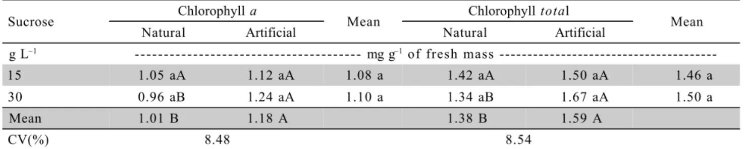
Differences for chlorophyll a and total, regard-ing the cultivation environment were found only for
the treatment containing 30 g L–1
sucrose. At this con-centration, high contents of both types of chlorophyll were obtained in conditions of artificial light, with 1.24 mg g–1 and 1.67 mg g–1 respectively. However, no dif-ferences were observed among the concentrations of carbohydrates in both environments (p ≤ 0.05) (Table 1). This result could be due to the lower irradiance observed in the growth chamber (artificial) environ-ment, because the processes of synthesis and degra-dation (photo-oxidegra-dation) of chlorophylls are directly correlated with irradiance, with pronounced reduction on chlorophyll content (per mass unit and, or area) under high levels (Lee et al., 2000; Atroch et al., 2001; Kitajima & Hogan, 2003; Deccetti, 2004).
No effects were observed for chlorophyll b
and the a/b ratio for all treatments, isolated or in as-sociation. This is consistent with the findings of Deccetti (2004) with plants of Amona glabra L., at the in vitro rooting stage, in which lower influence of the increase in irradiance (artificial) levels on a and b
chlorophylls was observed. Nevertheless, Lee et al. (2000) observed a reduction in chlorophyll concentra-tions and in the ratio of chlorophyll a/b by the expo-sure to a high PFD (Photon Flux Density) in two tree species native to Asia.
Relative water content (RWC)
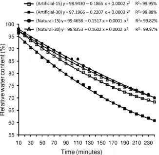
A significant interaction of the three factors (Environment × Sucrose × Time) evaluated was ob-tained for the RWC, where greater water loss (lower RWC) with exposition time occurred in plants grown in the artificial environment, and this loss was greater at 30 g L–1
sucrose (Figure 1). Lower RWC in plants
Figure 1 - Relative water contents of in micropropagated ‘Caipira’ (AAA) banana plants as a function of the cultivation environment and sucrose concentration.
55 60 65 70 75 80 85 90 95 100
10 30 50 70 90 110 130 150 170 190 210 230
Relative water content (%)
Time (minutes)
(Artificial-15) y = 98.9430 – 0.1865 x + 0.0002 x2 R2= 99.95%
(Artificial-30) y = 97.1966 – 0.2207 x + 0.0003 x2 R2= 99.88%
(Natural-15) y = 99.4658 – 0.1517 x + 0.0001 x2 R2= 99.82%
grown under artificial lighting and medium containing 30 g L–1
sucrose is observed only when the average of each growth environment and sucrose concentra-tion is evaluated, with 82.6% and 78.1% for the natu-ral and artificial conditions, and 81.8% and 78.9% for 15 and 30 g L–1
. Similar results have been observed reported by Decetti (2004) in leaf disks of Amona glabra L., who observed reduced water loss in leaf disks from plants grown under high in vitro irradiance (300 µmol m–2
s–1
).
The greatest RWC observed under natural light could have been favored by variations in relative hu-midity, irradiance and temperature inside the flasks, which would have favored a better response of sto-mata opening and closing mechanism after plant ex-posure to the external cultivation conditions, thus fa-voring the hardening of the plants. This hypothesis is supported by the observations of Silva (2006) with pineapple, who also conducted an experiment in the municipality of Lavras and under the same cultivation, irradiance and temperature conditions. According to Silva (2006), the relative humidity and temperature in-side the flasks, under natural light environment, var-ied from 58% to 95% and 10°C to 40°C; however, in the artificial environment they ranged from 96% to 98% and 23°C to 27°C. Hazarika (2003) stated that plants developed in vitro under conditions of low relative hu-midity showed lower transpiration and less ex vitro
translocation problems, and the persistent leaves are quite similar to the ones developed naturally.
Sciutti & Morini (1995) stated that the high relative humidity within the cultivation flasks was the main factor leading to modifications in tissue struc-ture and functioning. According to those authors, the increase in irradiance levels and or reduction in rela-tive humidity during in vitro cultivation have resulted in anatomical modifications similar to those observed during acclimatization in greenhouse, with the increase in deposition of epicuticular wax, reduction on stomata size and density, decreasing the loss of turgor and im-proving stomata functionality (Capellades et al., 1990; Sciutii & Morini, 1995; Decceti, 2004).
Moreover, Sciutti & Morini (1995) stated that
Prunus cerasifera plants rooted under 100% relative humidity lost more water (p ≤ 0.05) than plants rooted under relative humidity from 70% to 80%. In addi-tion, plants grown under relative humidity from 70% to 80% presented a visible thicker waxy layer on the leaves, the stomata were closed, not raised and with-out collapse, even after 30 minutes of exposure in growth chamber. In contrast, under 100% humidity the stomata were more numerous, raised on the leaf surface, with larger apertures and the epidermal cells collapsed after 30 minutes of exposure. In this as-pect, Capellades et al. (1990) reported that it is likely that the collapse of the epidermal cell could cause loss of integrity of the leaf surface, leading to more des-iccation.
These results are consistent with those of Khan et al. (1999), where greater water loss was ob-served in leaves of in vitroQuercus robur plants, with 90% and 80% loss in plants obtained in the multipli-cation and rooting stages, within 30 to 90 min. This was attributed to a high density of stomata and the heterogeneous functionality of the stomata developed in this environment. Similar results about greater wa-ter loss in in vitro leaf tissues was observed by Romano & Martins-Loução (2003), which occurred in leaves developed during the in vitro rooting, com-pared to the ones formed in the acclimatization stage, losing approximately 53% and 14%, respectively, in the first 30 minutes when exposed to 50% of rela-tive humidity at 21 ± 2°C. Additionally, the leaves formed in vitro showed opened stomata and collapse of guard-cells, which was not observed in acclimatized plants. High in vitro humidity, resulting from the low gas exchange in the flasks, lead the plants to open their stomata to keep the equilibrium with the atmosphere inside the flask, inducing an ab-normal development of stomata, thus remaining opened even when they are subjected to a drastic change in the environment (Shackel et al., 1990; Sutter et al., 1992).
Table 1 - Chlorophyll a and total contents of micropropagated ‘Caipira’ (AAA) banana plants as a function of the cultivation environment and sucrose concentration.
Sucrose Chlorophyll a Mean Chlorophyll total Mean
Natural Artificial Natural Artificial
g L–1 - - - mg g–1 of fresh mass
---15 1.05 aA 1.12 aA 1.08 a 1.42 aA 1.50 aA 1.46 a
30 0.96 aB 1.24 aA 1.10 a 1.34 aB 1.67 aA 1.50 a
Mean 1.01 B 1.18 A 1.38 B 1.59 A
CV(%) 8.48 8.54
Anatomical features
Differences related to sucrose concentration were observed only on the width of the upper epider-mis and on the foliar limb, with greater width in the medium amended with 15 g L–1
(p ≤ 0.05). For the cultivation environments, the plants grown under natu-ral light showed significant thickening in both the pali-sade and spongy parenchyma, in contrast to the up-per and lower epidermis, and lower hypodermis, which had greater thickening in the artificial environment (p
≤ 0.05) (Table 2). Sandoval et al. (1994) found that, despite in vitro ‘Grande Naine’ banana plants had epi-dermis and hypoepi-dermis with relatively thick cells, they showed thin walls, which could explain the greatest thickenings observed in the present study. As to the parenchyma, the increase in the width of these tissues due to increased irradiance was also observed by Hanba et al. (2002) in in vitroAcer species.
Greater thickening due to increase irradiance levels was also reported by Deccetti (2004). Accord-ing to that author, Annona glabra plants rooted in vitro
at 300 µmol m–2
s–1
had increase in the width of the palisade and spongy parenchyma, besides the pro-nounced differentiation of these tissues, responses that could be determinant to the photosynthetic process and
ex vitro survival. The occurrence of more elongated palisade cells consists of classic pattern of response to plant adaptation to greater light intensity, thus high-lighting the adaptable plasticity to the new environment (Lee et al., 2000). Moreover, structural modifications related to the development of the photosynthetic tis-sues could have an important role in increasing pho-tosynthetic activity, survival and acclimatization of plants (Amâncio et al., 1999; Serret & Trillas, 2000.
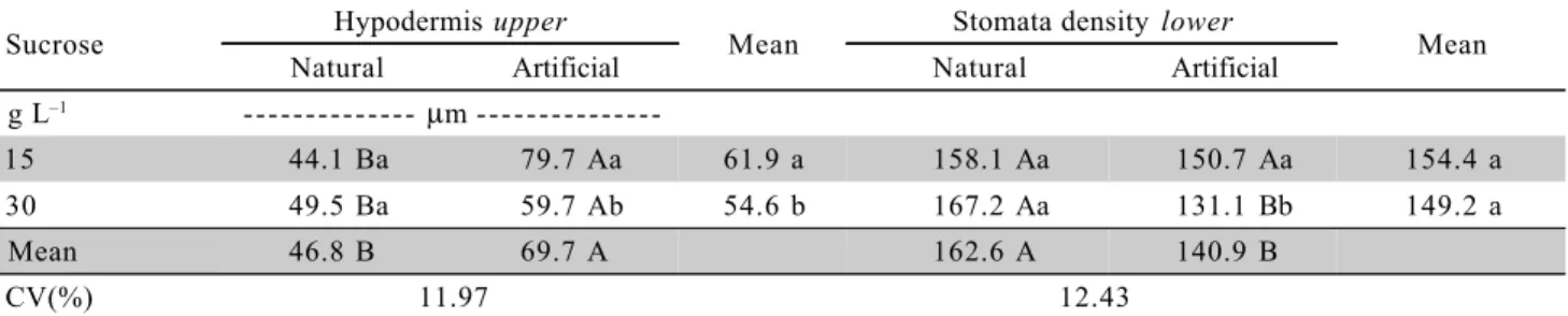
As to the upper hypodermis, greater thicken-ing (p ≤ 0.05) was observed in plants grown under artificial light environment, in both sucrose concentra-tions (Table 3). Greater width of the lower and upper hypodermis of ‘Prata-Anã’ banana plants rooted in a growth chamber (artificial light) was also observed by Rocha (2005). Regarding the characteristics of the sto-mata, the leaves of banana plants have stomata dis-tributed on both faces of the epidermis, however, with greater number on the lower face (Table 3), condition that classifies this species as amphistomatic, which agreed with Sandoval et al. (1994) and Rocha (2005).
Greater number of stomata per mm–2
was ob-served in the lower hypodermis in the natural environ-ment, only with 30 g L–1
sucrose (p ≤ 0.05). In con-trast, no effect for the factors studied was observed
Table 3 - Width of the upper hypodermis and stomata density of the lower hypodermis ‘Caipira’ (AAA) micropropagated banana plants as a function of the cultivation environment and sucrose concentration.
Means followed by distinct letters, small caption in the vertical and capital in the horizontal, within each variable, are different (F test at 5%).
Sucrose Hypodermis upper Mean Stomata density lower Mean
Natural Artificial Natural Artificial
g L–1 - - - mm
-15 44.1 Ba 79.7 Aa 61.9 a 158.1 Aa 150.7 Aa 154.4 a
30 49.5 Ba 59.7 Ab 54.6 b 167.2 Aa 131.1 Bb 149.2 a
Mean 46.8 B 69.7 A 162.6 A 140.9 B
CV(%) 11.97 12.43
Table 2 - Tissue width and stomata density in micropropagated ‘Caipira’ (AAA) banana plants as a function of cultivation environment and sucrose concentration.
Means followed by distinct letters in the vertical, within each variable, are different (F test at 5%). Sucrose Epidermis
upper
Palisade parenchyma
Spongy parenchyma
hypodermis lower
Epidermis
lower
Foliar limb
Stomate density upper
g L–1 - - - - mm
15 20.5 a 58.1 a 76.7 a 47.7 a 13.7 a 278.6 a 32.2 a
30 16.7 b 60.4 a 72.6 a 49.1 a 13.3 a 266.5 b 34.6 a
Environment
Natural 17.8 b 67.3 a 82.4 a 46.6 b 11.9 b 272.7 a 35.6 a
Artificial 19.4 a 51.1 b 67.0 b 50.2 a 15.1 a 272.4 a 31.3 a
on the upper surface (Table 3). Similar results were ob-tained by Rocha (2005) for the ‘Prata-Anã’ cultivar that showed greater stomata density on the lower surface in artificial environment with 15 g L–1 and natural with 15 g L–1
or 30 g L–1
sucrose. Similar effect of irradi-ance was also observed by Deccetti (2004) in Annona glabra plants subjected to in vitro rooting. The increase in stomata density can provide the plant with increased gas conductance, thus avoiding that photosynthesis to be limited under different environmental conditions (Kundu & Tigerstedt, 1998). Nevertheless, the occur-rence or not of diffeoccur-rences in the number of stomata depends on the species and the cultivation condition.
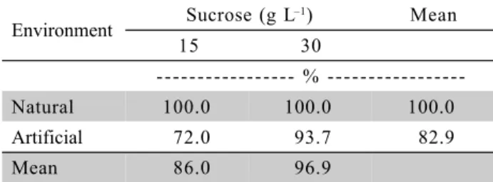
Ex vitro survival
Thirty days after transplanting, 100% survival was recorded in plants from the natural environment, in both sucrose concentrations. Conversely, in plants from the artificial environment, losses of 28.0% and 6.3% were observed with 15 g L–1 and 30 g L–1 of carbohydrate, respectively (Table 4).
Possibly, the exposure of the shoots to greater irradiance and temperature amplitudes promoted the hardening of the plants still in vitro, since the green-house conditions were more similar to that in which the plants were acclimatized, thus favoring lower stress in the transplanting and the ex vitro reestab-lishment.
Actually, the modifications observed to the RWC and anatomic parameters under natural light con-firm the better structural and physiological adaptation of the plants and justify the greater survival rates. Moreover, these results are consistent with those ob-tained by Kodym & Zapata-Arias (1999) in Musa cv. Grande Naine (AAA), and Talavera et al. (2005) in Co-cos nucifera plants grown under natural light. More-over, according to Kodym & Zapata-Arias (1999), plants grown under natural light were successfully hardened, with 100% survival after two months, and presented normal development.
The effect of sucrose on the success plant hardening is reported by Folliot & Marchal (1992), Skrebsky et al. (2004) and Pacheco et al. (2006) for
Musa spp., Pfaffia glomerata Spreng. Pedersen and
Arachis retusa, using between 15 g L–1 and 60 g L–1. In contrast, negative effects from the partial or total removal of sucrose on the ex vitro development of micropropagated plants were obtained by Skrebsky et al. (2004), Fuentes et al. (2005) and Pacheco et al. (2006). The low survival and the slow development of Cocos nucifera L. cultivated under the absence or low sucrose concentration is a result of the deficiency in the formation of carbon backbone and the alloca-tion of the stocks from the leaves formed in vitro to sustain the ex vitro reestablishment (Fuentes et al., 2005).
With a profound knowledge of the physiologi-cal and structural alterations induced in vitro, it is pos-sible to establish strategies for the most efficient man-ner of transferring micropropagated plants to ex vitro
conditions and, consequently, to maximize survival rates (Smith et al., 1997; Apóstolo et al., 2005). Thus, the results obtained in this study provide an explana-tion for the high survival rate observed in plants grown in natural light and contributes for the use of this kind of lighting for in vitro establishment. Similarly, the as-sociation of survival rate with morphological and ana-tomical features is important to demonstrate or not the hardening of plants subjected to modifications in the
in vitro environment, thus reducing the losses after the exposition to the ex vitro environment (Capellades et al., 1990).
CONCLUSIONS
In vitro rooting of ‘Caipira’ banana shoots, un-der natural lighting, with 15 g L–1
or 30 g L–1
sucrose promoted the thickening of the palisade and spongy parechyma, reduced leaf water loss, and increased ex vitro plant survival.
The study of structural and physiological char-acteristics provided a better understanding of the changes in banana plants subjected to environmental alterations in in vitro cultivation.
Substituting fluorescent lights for natural light-ing, during in vitro rooting confirms the potential of this lighting source in banana micropropagation pro-tocols.
ACKNOWLEDGMENTS
To Conselho Nacional de Desenvolvimento Científico e Tecnológico (CNPq) for the financial sup-port and the fellowship for the first author.
Table 4 - Survival of ‘Caipira’ (AAA) micropropagated banana plants as a function of the cultivation environment and sucrose concentrations, 30 days after the ex vitro transplanting.
Data obtained by the rate between the number of dead plants and or not developed and the total number of plants transferred to the ex vitro conditions, not being statistically analyzed.
Environment Sucrose (g L
–1) Mean
15 30
%
-Natural 100.0 100.0 100.0
Artificial 72.0 93.7 82.9
REFERENCES
AMÂNCIO, S.; REBORDÃO, J.P.; CHAVES, M.M. Improvement of acclimatization of micropropagated grapevine: photosynthetic competence and carbon allocation. Plant Cell, Tissue and Organ Culture, v.58, p.31-37, 1999.
APÓSTOLO, N.M.; BRUTTI, C.B.; LLORENTE, B.E. Leaf anatomy of Cynara scolymus L. in successive micropropagation stages. In Vitro Cellular Development Biology – Plant, v.41, p.307-313, 2005.
ARNON, D.I. Copper enzymes in isolated choroplasts. Polyphenoloxidase in Beta vulgaris. Plant Physiology, v.24, p.1–15, 1949.
ATROCH, E.M.A.C.; SOARES, A.M.; ALVARENGA, A.A.; CASTRO, E.M. Crescimento, teor de clorofilas, distribuição de biomassa e características anatômicas de plantas jovens de
Bauhinia forficata Link. submetidas a diferentes condições de sombreamento. Ciência e Agrotecnologia, v.25, p.853-862, 2001.
CAPELLADES, M.; FONTARNAU, R.; CARULLA, C.; DEBERGH, P. Environment influences anatomy of stomata and epidermal cells in tissue-cultured Rosa multiflora. Journal of the American Society for Horticultural Science, v.115, p.141-145, 1990.
CASTRO, E.M.; PINTO, J.E.B.P.; MELO, H.C.; SOARES, A.M.; ALVARENGA, A.A.; LIMA JÚNIOR, E.C. Aspectos anatômicos e fisiológicos de plantas de guaco submetidas a fotoperíodos. Horticultura Brasileira, v.23, p.846-850, 2005.
DECCETTI, S.F.C. Ambiente de cultivo e respostas morfofisiológicas durante o processo de micropropagação de
Annona glabra L.Lavras: Universidade Federal de Lavras, 2004. 93p. Tese (Doutorado).
FERREIRA, D.F. SISVAR 4.3: sistema de análise estatística.Lavras: UFLA/DEX, 2000.
FOLLIOT, M.; MARCHAL, J. Croissance in vitro des bananiers: influence de la concentration en saccharose du milieu de culture sur le développement des plants du cultivar Petite Naine. Fruits, v.47, p.649-655, 1992.
FUENTES, G.; TALAVERA, C.; OROPEZA, C.; DESJARDINS, Y.; SANTAMARÍA, J.M. Exogenous sucrose can decrease in vitro
photosynthesis but improve field survival and growth of coconut (Cocos nucifera L.) in vitro plantlets. In Vitro Cellular & Developmental Biology-Plant, v.41, p.69-76, 2005. GÜBBÜK, H.; PEKMEZCI, M. In vitro propagation of some new
banana types (Musa spp.). Turkish Journal of Agriculture and Forestry, v.28, p.355-361, 2004.
HANBA, Y.T.; KOGAMI, H.; TERASHIMA, L. The effects of growth irradiance on leaf anatomy and photosynthesis in Acer
species differing in light demand. Plant Cell and Enviroment, v.25, p.1021-1030, 2002.
HAZARIKA, B.N. Morpho-physiological disorders in in vitro culture of plants. Scientia Horticulturae, v.108, p.105-120, 2006.
HAZARIKA, B.N. Acclimatization of tissue-cultured plants. Current Science, v.85, p.1704-1712, 2003.
JOHANSEN, B.A. Plant microtechnique. New York: McGraw-Hill, 1940. 523p.
KHAN, P.S.S.V.; EVERS, D.; HAUSMAN, J.F. Stomatal characteristics and water relations of in vitro grown Quercus robur NL 100 in relation to acclimatization. Silvae Genetica, v.48, p.83-87, 1999.
KITAJIMA, K.; HOGAN, K.P. Increases of chlorophyll a/b ratios during acclimatization of tropical woody seedlings to nitrogen limitation and high light. Plant Cell and Environment, v.26, p.857-865, 2003.
KODYM, A.; ZAPATA-ARIAS, F.J. Natural light as an alternative light source for the in vitro culture of banana (Musa acuminata
cv. ‘Grande Naine’) Plant Cell, Tissue and Organ Culture, v.55, p.141–14, 1999.
KRAUS, J.E.; ARDUIM, M. Manual básico de métodos em morfologia vegetal. Rio de Janeiro: EDUR, 1997. 198p. KUNDU, S.K.; TIGERSTEDT, P.M.A. Variation in net
photosynthesis, stomate characteristics, leaf area and whole plant phytomass production among ten provenances of neem (Azadirachta indica). Tree Physiology, n.19, p.47-52, 1998.
LABOURIAU, L.G.; OLIVEIRA, J.G.; SALGADOLABOURIAU, M.L. Transpiração de Schizolobium parahyba (Vell) Toledo I. Comportamento na estação chuvosa, nas condições de Caeté, Minas Gerais. Anais da Academia Brasileira de Ciência, v.33, p.237-257, 1961.
LAMHANEDI, M.; CHAMBERLAND, H.; TREMBLAY, F.M. Epidermal transpiration, ultrastuctural characteristics and net photosynthesis of white spruce somatic seedlings in response to in vitro acclimatization. Physiologia Plantarum, v.118, p.554-561, 2003.
LARCHER, W. Ecofisiologia vegetal. São Carlos: Rima, 2000. 531p.
LEE, D.W.; OBERBAUER, S.F.; JOHNSON, P.; KRISHNAPILAY, B.; MANSOR, M.; MOHAMAD, H.; YAP, S.K. Effects of irradiance and spectral quality on leaf structure and function in seedlings of two Southeast Asian hopea (Dipterocarpaceae) species. American Journal of Botany, v.87, p.447-455, 2000.
MOHAMMED, G.H.; VIDAVER, W.E. The influence of acclimatization treatment and plantlet morphology on early greenhouse-performance of tissue-cultured Douglas fir
[Pseudotsuga menziesii (Mirb.) Franco]. Plant Cell, Tissue and Organ Culture, v.21, p.111–117, 1990.
MURASHIGE, T.; SKOOG, F.A. A revised medium for rapid growth and bioassays with tobacco tissue cultures. Physiologia Plantarum, v.15, p.473-497, 1962.
NAVARRO, C.; TEISSON, C.; CÔTE, F.; GANRY, J. Effects of light intensity and CO2 concentration on growth of banana plants (Musa AAA, cultivar ‘Petite Naine’) in vitro and subsequent growth following acclimatization. Scientia Horticulturae, v.60, p.41-54, 1994.
PACHECO, G.; GAGLIARDI, R.F.; COGLIATTI, M.B.; MANHÃES, H.B.; CARNEIRO, L.A.; VALLS, J.F.M.; MANSUR, E. Influence of substrates and in vitro preconditioning treatments on ex vitro acclimatization of Arachis retusa. Pesquisa Agropecuária Brasileira, v.41, p.165-169, 2006.
ROCHA, H.S. Luz e sacarose na micropropagação da bananeira “Prata Anã”: alterações morfoanatômicas. Lavras: Universidade Federal de Lavras, 2005. 98p. Dissertação (Mestrado). ROMANO, A.; MARTINS-LOUÇÃO, M.A. Water loss and
morphological modifications in leaves during acclimatization of cork oak micropropagated plantlets. Acta Horticulturae, v.616, p.439-442, 2003.
SANDOVAL, J.A.; MÜLLER, L.E.; WEBERLING, F. Foliar morphology and anatomy of Musa cv. Grande Naine (AAA) plants grown in vitro and during hardening as compared to field-grown plants. Fruits, v.49, p.37-46, 1994.
SCIUTTI, R.; MORINI, S. Water loss and photosynthesis of plum plantlets is influenced by relative humidity during rooting in vitro. Journal of Horticultural Science, v.10, p.221-228, 1995.
SERRET, M.D.; TRILLAS, M.I. Effects of light and sucrose levels on the anatomy, ultrastructure, and photosynthesis of Gardenia jasminoides Ellis leaflets cultured in vitro. International Journal of Plant Science, v.161, p.281-289, 2000. SHACKEL, K.A.; NOVELLO, V.; SUTTER, E.G. Stomatal function
and cuticular conductance in whole tissue cultured apple plants. Journal of American Society Horticultural, v.115, p.468-472, 1990.
SKREBSKY, E.C.; NICOLOSO, F.T; FERRÃO, G.E. Sacarose e período de cultivo in vitro na aclimatização ex vitro de ginseng brasileiro (Pfaffia glomerata Spreng. Pedersen). Ciência Rural, v.34, p.1471–1477, 2004.
SMITH, W.K.; VOGELMANN, T.C.; DELUCIA, E.H.; BELL, D.T.; SHEPHERD, K.A. Leaf form and photosynthesis. BioScience, v.47, p.785-793, 1997.
SUTTER, E.G.; SHACKEL, K.; DIAZ, J.C. Acclimatization of tissue cultured plants. Acta Hoticulturae, v.314, p.115–119, 1992. TALAVERA, C.; CONTRERAS, F.; ESPADAS, F.; FUENTES, G.; SANTAMARÍA, J.M. Cultivating in vitro coconut palms (Cocos nucifera) under glasshouse conditions with natural light, improves in vitro photosynthesis nursery survival and growth. Plant Cell, Tissue and Organ Culture, v.83, p.287-292, 2005.
VALERO-ARACAMA, C.; WILSON, S.B.; KANE, M.E.; PHILMAN, N.L. Influence of in vitro growth conditions on in vitro and ex vitro photosynthetic rates of easy- and difficult-to-acclimatize sea oats (Uniola paniculata L.) genotypes. In Vitro Cellular Development Biology – Plant, v.43, p.237-246, 2007.
YOKOTA, S.; KARIM, M. Z.; AZAD, M.A.K.; RAHMAN, M.M.; EIZAWA, J.; SAITO, Y.; ISHIGURI, F.; IIZUKA, K.; YAHARA, S.; YOSHIZAWA1, N. Histological observation of changes in leaf structure during successive micropropagation stages in Aralia elata and Phellodendron amurense. Plant Biotechnology, v.24, p.221-226, 2007.
WHATLEY, F.H.; WHATLEY, F.R. A Luz e a vida das plantas. São Paulo: EPU-EDUSP, 1982. 101 p. (Temas de Biologia, 30).