http://www.uem.br/acta ISSN printed: 1679-9275 ISSN on-line: 1807-8621
Doi: 10.4025/actasciagron.v34i1.12309
Protoplast production and isolation from
Etlingera elatior
Jessé Marques da Silva Júnior1, Renato Paiva1, Ana Carolina Atala Lombelo Campos1, Marcelo Rodrigues1*, Milene Alves de Figueiredo Carvalho1 and Wagner Campos Otoni2
1
Departamento de Biologia, Setor de Fisiologia Vegetal, Universidade Federal de Lavras, Cx. Postal 3037, 37200-000, Lavras, Minas Gerais, Brazil. 2
Departamento de Biologia Vegetal, Laboratório de Cultura de Tecidos, Universidade Federal de Viçosa, Viçosa, Minas Gerais, Brazil. *Author for correspondence. E-mail: [email protected]
ABSTRACT. The technique of hybridization using plant protoplasts is widely used in plant breeding programs. The purpose of our study is to further characterize the process of protoplast isolation from the ornamental species Etlingera elatior (Jack) R. M. Smith. Protoplasts were isolated from different tissues: in vitro leaves, in vitro pseudostem, and leaves from plants cultivated hydroponically. We tested six enzymatic combinations, four incubation time periods, the rotary system (40 rpm) or steady in the dark, and three concentrations of mannitol (0.5, 0.6 and 0.7 M). The diameter and viability of obtained protoplasts were evaluated. The best source of explants used for protoplast isolation was the in vitro leaves, which yielded 22x105 protoplasts g-1 of fresh matter. The optimal incubation period was 15 hours. The in vitro leaves
presented a greater viability (96%) and larger protoplasts (36.7 μm diameter). Greater yields were obtained using a rotatory system with protoplasts incubated in the dark. The best enzymatic combination was 3% Cellulase “Onozuca” R-10 + 2% Meicelase + 1% Driselase + 1% Dextran + 5 mM MES, followed by the addition of 0.6 M mannitol.
Keywords: FDA, ornamental plant, enzymatic combinations, incubation period.
Produção e isolamento de protoplasto de
Etlingera elatior
RESUMO.Com o objetivo de realizar hibridações que auxiliam em programas de melhoramento genético de flores ornamentais, protoplastos foram isolados a partir de diferentes tecidos (folhas in vitro, pseudocaules in vitro e folhas em sistema hidropônico) de Etlnigera elatior (Jack) R. M. Smith. Foram testados seis diferentes combinações enzimáticas, quatro períodos de incubação, sistema rotatório (40 rpm) ou estacionário no escuro, concentrações de manitol (0,5; 0,6 e 0,7 M), o diâmetro e a viabilidade dos protoplastos isolados. A melhor fonte de explante utilizado no isolamento de protoplastos foi folha in vitro, com rendimento de 22 x105 protoplastos g-1 MF. O melhor tempo de incubação foi 15 horas, pois períodos
superiores a este causavam diminuição no rendimento e viabilidade dos protoplastos. Protoplastos de folhas in vitro apresentaram viabilidade de 96% e diâmetro de 36,7 μm. Maiores rendimentos foram alcançados em sistema rotatório e no escuro. A melhor combinação enzimática utilizada no atual trabalho foi a 3% Cellulase “Onozuka” R-10 + 2% Meicelase + 1% Driselase + 1% Dextran + 5 mM MES. A melhor concentração de manitol foi de 0,6 M.
Palavras-chave: FDA, planta ornamental, combinações enzimáticas, período de incubação.
Introduction
Etlingera elatior (Jack) R. M. Smith is an ornamental plant extensively commercialized in the flower market. In Brazil, it is used as a cut flower and in landscapes of parks and stands (LAMAS, 2002). Its propagation is mainly made by in vitro cultivation, due to the occurrence of several diseases that affect this species. Plant breeding programs are, therefore, attempting to increase E. elatior pathogen resistance.
Somatic hybridization by protoplast fusion is a promising technique for breeding ornamental species and requires reliable in vitro protocols. Somatic hybridization can fuse two complete
genomes, which is an alternative to sexual reproduction (WU et al., 2009). This technique was successfully used to breed citrus, sunflower, brassica and wheat (DAVEY et al., 2005).
expression in Arabidopsis (WU et al., 2009) and the production of secondary metabolites used commercially (FONTES et al., 2010).
According to Wu et al. (2009), a protoplast is a transitory state of a cell lacking its cell wall and can be obtained using pectocelulolitics enzymes. Without cell walls, protoplasts can incorporate materials such as DNA and fuse. Somatic hybrids can be obtained when protoplasts of different species are fused.
For protoplast isolation, tissues should be pre-plasmolysed with enzymatic solutions followed by washing with CPW-Cell Washing Protoplasts and 13% mannitol. The most-used enzymes for protoplast isolation are Cellulase “Onozuka” R-10 (Yakult Honsha), Macerozyme R-10 (Yakult Honsha), Cellulase Cellulysin, hemicellulase Rhozyme and pectinase Pectoyase Y-23 (DORNELAS et al., 1995). In well-established cultures, protoplasts can maintain cell totipotency; rebuilding their walls, dividing, forming callus, and regenerating plants through embryogenesis or organogenesis (WU et al., 2009). Therefore, this study further characterizes the conditions used for isolating protoplasts from E. elatior because different tissues (invitro leaves, in vitro pseudostems and leaves from a hydroponic system), different combinations of enzymes, different incubation periods, using a rotating or stationary system in light or dark, and mannitol concentrations can affect the diameter and viability of isolated protoplasts.
Material and methods
Plant material
For protoplast isolation, three plant tissues were used based on their different cell wall structures: in vitro leaves, in vitro pseudostems, and leaves from plants cultivated hydroponically.
To isolate in vitro leaves and pseudostems, we grew seedlings in Ms medium (MURASHIGE; SKOOG, 1962) combined with the B5-Gamborg vitamin mixture. The culture medium was supplemented with 3.0 mg L-1 of BAP
(6-benzilaminopurine, 3% (w v-1) sucrose and 0.7%
(w v-1) of agar; Sigma Chemical Co., USA). The pH
was adjusted to 5.7 ± 0.1, before autoclaving. Plants were maintained in a growth chamber for 30 to 45 days under 36 µmol m-2 s-1 of photon irradiance
at 26 ± 2°C and 16 hours of light per day. Plants were grown hydroponically using a solution with 35% of its ionic strength under to isolate protoplasts from leaves.
The protoplasts’ diameters from the plant material used was determined based on digital
images (Canon PowerShot A710 7MP). The images were analyzed using Sigma Scan Pro 5® software.
For each plant material, 200 protoplasts were evaluated, and the percentage of the protoplasts belonging to different diameter categories was determined. For this experiment we used protoplasts incubated in 9 M CPW solution.
Protoplast isolation
Under aseptic conditions, the in vitro leaves were sectioned parallel to the middle vein, resulting in 1 - 1.5 mm wide pieces. The pseudostems were transversally sectioned into pieces of 50 mm. For hydroponically cultivated plants, leaves were sterilized with 70% ethanol for 2 minutes, followed by 40% sodium hypochlorite for 20 minutes, and rinsed with autoclaved demi-water (5 times). The epidermis was removed from the leaves using tweezers (peeling) to maximize infiltration of the enzymatic solution in the tissue.
The plant material obtained was transferred to 60 x 15 mm Petri dishes containing 10 mL of CPW solution. Three mannitol concentrations were tested: 0.5 M (9 g 100 mL-1 CPW), 0.6 M (11 g
100 mL-1 CPW), and 0.7 M (13 g 100 mL-1 CPW).
Approximately 0.5 g of plant material was pre-plasmolysed with this solution for one hour in the dark. Next, the 0.5, 0.6 and 0.7 M CPW solutions were discarded using Pasteur pipettes, followed by the addition of 10 mL of the enzymatic mixture.
Six enzyme combinations were used: A - 3% (p v-1) Cellulase “Onozuka” R-10 (Yakult Honsha)
+ 1% (w v-1) Pectolyase (Seishim Pharmaceutical.,
USA) + 0.5% (w v-1) Driselase; B - 3% (w v-1)
Cellulase “Onozuka” R-10 + 1% (w v-1) Pectolyase
+ 1% (w v-1) Driselase (Sigma London Chemical
Co Ltd); C – 3.75% (w v-1) Cellulase “RS” (Yakult
Honsha) + 1% (w v-1) Driselase; D - 2% (w v-1)
Rhozyme HP150 (Rohm & Haas Co., USA) + 1% (w v-1) Macerozyme R-10 + 0.5% (w v-1) Driselase;
E - 3% (w v-1) Cellulase Onozuka R-10 + 2%
(w v-1) Meicelase (Meiji Seika Haisha Ltd., Japan) +
1% (w v-1) Driselase; and F - 3% (w v-1) Cellulase
Aspergillus niger (Fluka Chemicals Ltd) + 1% (w v-1)
Pectinase Aspergillus niger (Fluka Chemicals Ltd.). The enzymatic solutions were buffered with 5 mM MES and 1% dextran followed by dilution in three mannitol concentrations (0.5, 0.6 and 0.7 M). The pH of the mannitol solutions was adjusted to 5.6.
evaluated. The experimental design was randomized, with a factorial of 6 x 3 (6 enzymatic solutions, 3 concentrations of mannitol); each replicate was constituted by a single Petri dish.
After the incubation phase, the suspension obtained (isolated protoplasts and tissues that were not digested) was filtered using a 64 μm nylon mesh (Wilson Sieves, Nottingham, UK) and centrifuged at 700 rpm for 5 minutes (3x). The precipitate was re-suspended and transferred to a new centrifuge tube, being the volume completed with the following CPW solutions with different sucrose gradients: 30, 25, 20, 21, 18, and 15S (5 mL for each solution sucrose). Finally, the suspension was centrifuged at 700 rpm for 5 minutes. The purified protoplasts, localized in the interface between the two media, were collected with a Pasteur pipette and transferred to new tubes.
The number of isolated protoplasts was determined using a Fuchs-Rosenthal-B.S. 74B hemacytometer (Weber Scientific Int. LTD., Sussex, U.K.) and an optic microscope (Hausser Scientific, USA). The viability of the protoplasts was determined based on staining with diacetate of fluorescein (FDA). For this test, a mixture of equal volumes of protoplast suspension and the FDA solution (0.01%) was incubated at room temperature for 3 to 5 minutes. The solution was observed using an inverted optic microscope 40 x (Olympus IMT 2) under UV light (with a blue filter). The viable protoplasts were indicated by a green fluorescence, and viability was defined by the percentage of observed fluorescent protoplasts (ADITYA; BAKER, 2003). The experimental design consisted of 2 replicates, each corresponding to the observation of 200 protoplasts. Data were analyzed by ANOVA and the separation of means test SNK (5%).
Results and discussion
The enzyme combinations B and E after 15 hours produced the best results for protoplast isolation and were significantly different according to the SNK test (5%) compared to other enzyme combinations. In vitro leaves of E. elatior incubated for 15 hours with shaking yielded 22.0 x 10⁵ and
12.30 x 10⁵ protoplasts g-1 of fresh tissue for the
enzyme combinations E and B, respectively (Table 1 and Figure 1).
Incubation periods longer than 15 hours resulted in a decrease in yield of isolated protoplasts caused by increased membrane instability and nonselectivity of the enzymatic solution. Similar results were obtained by Costa et al. (2002) using an enzymatic solution of 1% Cellulase “Onozuca” R-10
+ 0.2% Macerozyme R-10 and 0.1% Driselase, with a yield of 23.68 x 10⁶ protoplasts 500 g-1 of callus
from a variety of citrus. Previous studies with protoplast regeneration from leaf explants of Robinia pseudoacacia L. was obtained using an enzyme combination of 2% Celulose + 0.3% Macerozyme and incubated for 20 hours (KANWAR et al., 2009). Kanchanapooma et al. (2001) isolated protoplasts from Dendrobium pompadour with an enzyme mixture of 1% Cellulase “Onozuka” + 1% Macerozyme + 0.5% Driselase in a 0.4 M mannitol, which yielded 22.0 x 10⁵ (light) and 21.7 x 10⁵ (dark) of protoplasts g-1
of leaf tissue with a diameter of 50 to 80 μm.
Table 1. Efficiency of different enzyme combinations and incubation periods for E. elatior protoplast isolation from in vitro
leaves (average ± standard deviation).
Incubation period (h) Protoplasts income
a (x105 g-1 MF) B* E ** 5 0.38 ± 0.18 b 1.87 ± 0.73 a 10 3.23 ± 0.61 b 13.76 ± 1.22 a 15 12.30 ± 1.26 b 22.00 ± 2.37a 20 9.25 ± 2.45 b 19.5 ± 1.51 a
*3% (w v-1) Cellulase “Onozuka” R-10 + 1% (w v-1) Pectolyase + 1% (w v-1) Driselase
(Sigma London Chemical Co Ltd) + 0.6 M (mannitol) + 1% Dextran + 5 mM MES.
**3% (w v-1) Cellulase Onozuka R-10 + 2% (w v-1) Meicelase (Meiji Seika Haisha Ltd.,
Japan) + 1% (w v-1) Driselase + 0.6 M (mannitol) + 1% Dextran + 5 mM MES. aNumber of protoplasts obtained by enzymatic digestion of 0.5 g of tissue. Average of
200 protoplasts (2 replicates). Same letters in a row indicate values that do not differ for the SNK test (5%).
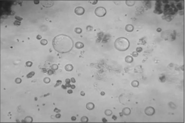
Figure 1. E. elatior protoplasts isolated after 15 hours of incubation in enzyme solution E (40x).
Castelblanque et al. (2010) isolated protoplasts from leaf explants of the ornamental species Kalanchoe blossfeldiana with the enzyme mixture of 0.4% of Cellulase “Onozuka” R-10 + 0.2% of Driselase, which yielded 6.0 x 10⁵ protoplasts per
gram of fresh tissue.
The isolation of protoplasts was described in several studies with the gender Passiflora (DORNELAS et al., 1995). Protoplast yields varied according to the species genotype and explant used.
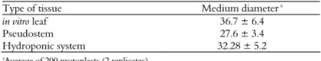
average diameters (36.7 μm) followed by leaves from plants grown hydroponically (32.28 μm) and in vitro pseudostems (27.6 μm) (Table 2).
Table 2. Average diameter of protoplasts isolated from 3 different explants of E. elatior. Enzyme solution E: 3% (w v-1) Cellulase “Onozuka” R-10 + 2% (w v-1) Meicelase (Meiji Seika Haisha Ltd., Japan) + 1% (w v-1
) Driselase + 0.6 M (mannitol) + 1% Dextran + 5 mM MES. (average ± standard deviation).
Type of tissue Medium diameter a
in vitro leaf 36.7 ± 6.4
Pseudostem 27.6 ± 3.4 Hydroponic system 32.28 ± 5.2
a
Average of 200 protoplasts (2 replicates).
According to Rodríguez and Dallos (2004), using protoplasts isolated from leaf mesophyll from Passiflora edulis var. flavicarpa, different diameters were observed depending on the tissue. Fully expanded leaves had protoplasts of a greater average diameter (19.45 ± 0.50 μm) when compared to cotyledons (28.90 ± 0.62 μm). Oliveira et al. (1995), using two citrus species, observed a variation of 4.8 to 16.8 μm in the diameter of isolated protoplasts. Protoplast diameter is information that can be used in hybridization studies using electriofusion so that an inverse relationship exists between protoplast diameter and the voltage necessary to promote protoplast fusion.
According to Dornelas et al. (1995), the average size of protoplasts depends on the species analyzed and the explant used and varies from 19 to 47 mm, when isolated from leaf tissues, or from 30 to 60 mm, when derived from cotyledon tissues. Protoplasts obtained from the mesophyll of dicotyledons tend to be smaller than those isolated from callus or cell suspensions (OCHATT, 1993). In monocotyledons, the protoplasts size is, in general, less than 30 μm, independent of tissue source.
Two systems were used for the protoplast incubation: stationary and shaking (40 rpm) in the dark. For this experiment, in vitro leaves of E. elatior were incubated in the enzymatic solution E. A higher yield of protoplasts was obtained for leaves incubated in the shaking system for 15 hours (22.0 x 10⁵ protoplasts g-1 of fresh tissue) (Table 3).
Expressive results were obtained by Monteiro et al. (2003) when they isolated protoplasts from the alfalfa Medicago sativa using the system of continuous shaking (35 rpm) in the dark.
From the different mannitol concentrations used in this study, a greater yield (19.95 x 10⁵ protoplasts
g-1 of fresh matter of protoplasts) was obtained with
0.6 M (11 g 100 mL-1) mannitol in combination
with enzyme solution E incubated for 15 hours;
however, no differences resulted as determined by the SNK test (5%) for protoplast isolation with 20 hours of incubation (19, 21 x 105 protoplasts g-1 of
fresh matter of protoplasts). In addition, these protoplasts had the highest viability percentage (96.7%; Table 5).
Table 3. The protoplast isolation efficiency from E. elatior for tissues incubated in the shaking (40 rpm) and stationary systems for different periods of time (average ± standard deviation). Enzymatic solution E: 3% (w v-1) Cellulase Onozuka R-10 + 2% (w v-1
) Meicelase (Meiji Seika Haisha Ltd., Japan) + 1% (w v-1) Driselase + 0,6 M (mannitol) + 1% Dextran + 5 mM MES.
Incubation period (h)
Protoplasts yield a (x105 g-1 MF) ST* SH** 5 4.53 ± 0.32 a 4.87 ±0.73 a 10 15.20 ± 0.48 a 15.75 ± 1.22 a 15 20.30 ± 0.26 b 22.00 ± 1.37 a 20 19.25 ± 0.45 a 19.74 ± 0.51 a *Stationary (dark) **Shaking (40 rpm in the dark). aNumber of protoplasts obtained
from the enzymatic digestion of 1 g of tissue. Average of 200 protoplasts (2 replicates). Same letters in a row indicate values that do not differ for the SNK test (5%).
Table 4. Protoplast isolation efficiency from E. elatior for tissues incubated in enzyme solution E with different concentrations of mannitol for different incubation periods (average ± standard deviation).
Incubation period (h) Protoplasts yield
a (x105 g-1 MF) 0.5* 0.6** 0.7*** 5 1.38 ± 0.18 b 4.29 ± 1.73 a 4.24 ± 2.75 a 10 5.21 ± 0.61b 13.76 ± 3.22 a 12.2 ± 3.12 a 15 12.30 ± 1.26c 19.95 ± 3.37a 16.45 ± 3.75b 20 15.65 ± 2.45c 19.21 ± 2.51a 17.33 ± 1.75 b *Mannitol 0.5 M (9 g 100 mL-1 CPW). **Mannitol 0.6 M (11 g 100 mL-1 CPW). ***Mannitol
0.7 M (13 g 100 mL-1 CPW). aNumber of protoplasts obtained from the enzymatic digestion of
1 g of tissue. Average of 2 replicates. E: 3% (w v-1) Cellulase “Onozuka” R-10 + 2% (w v-1)
Meicelase (Meiji Seika Haisha Ltd., Japan) + 1% (w v-1) Driselase + 0.6 M (mannitol) + 1%
Dextran + 5 mM MES. Average of 200 protoplasts (2 replicates). Same letters in a row indicate values that do not differ for the SNK test (5%).
The CPW 13 solution is commonly used for pre-plasmolysis, dissolution of enzymes and protoplast washing of tissues from Passiflora. This solution is composed of CPW medium salts and 13% mannitol (DORNELAS et al., 1995).
The highest viability percentages were obtained for protoplasts incubated with the enzyme solution E and purified with 0.6 or 0.5 M mannitol, with 96.7 and 81.8% of viable protoplasts, respectively (Table 5 and Figure 2).
Table 5. Viability of E elatior protoplasts isolated from in vitro
leaves using three concentrations of mannitol after 15 hours of isolation in enzyme solution E: 3% (w v-1
) Cellulase “Onozuka” R-10 + 2% (w v-1) Meicelase (Meiji Seika Haisha Ltd., Japan) + 1% (w v-1
) Driselase + 0.6 M (mannitol) + 1% Dextran + 5 mM MES. (average ± standard deviation).
Mannitol Viabilityb
0.5 M 81.8 ± 2.7 b
0.6 M 96.7 ± 3.9 a
0.7 M 30.2 ± 1.2 c
bPercentage of protoplasts with green fluorescence compared to total protoplasts.
The SNK test (5%) showed that these values differed from the average value for the highest concentration of mannitol (0.7 M) with (30.2%) viability of protoplasts.
Previous studies with protoplasts isolated from Dendrobiun pompadour using three concentrations of mannitol (0.4, 0.5 and 0.6 M) revealed that 0.4 M yielded 19.89 x 10⁵ protoplasts, which was greater
than from the other concentrations tested, 13.59 x 10⁵ and 6.95 x 10⁵, respectively (3-hour incubation).
Greater values for protoplasts viability (89%) were observed when 0.4 M mannitol solution was used (KANCHANAPOOMA et al., 2001).
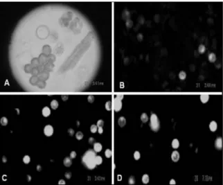
Figure 2. (A) Protoplasts recently isolated. (B) Viability test using 0.7 M mannitol. (C) Viability test using 0.6 M mannitol. (D) Viability test using 0.5 M mannitol. Enzyme solution E: 3% (w v-1) Cellulase “Onozuka” R-10 + 2% (w v-1) Meicelase (Meiji Seika Haisha Ltd., Japan) + 1% (w v-1) Driselase + 0.6 M (mannitol) + 1% Dextran + 5 mM MES.
Evaluating viability after isolation is important for determining the plating density to use for protoplast cultivation, which influences cell division and differentiation.
Conclusion
The optimal condition to isolate E. elatior protoplasts is enzyme solution E composed of 3% Cellulase “Onozuca” R-10, 2% Meicelase, 1% Driselase, 1% Dextran and 5 mM MES combined with 0.6 M mannitol and incubated for 15 hours with a shaking system (40 rpm) in the dark.
Acknowledgements
This work was financially supported by the following Brazilian agencies: Fundação de Amparo à Pesquisa do Estado de Minas Gerais (Fapemig), Coordenação de Aperfeiçoamento de
Pessoal de Nível Superior (Capes) and Conselho Nacional de Desenvolvimento Científico e Tecnológico (CNPq).
References
ADITYA, T. L.; BAKER, D. A. Optimization of protoplast isolation from NaCl stressed primary, secondary and tertiary calli derived from mature seeds of Bangladeshi indica rice cultivar Binnatoa. Plant Growth Regulation, v. 41, n. 1, p. 49-56, 2003.
CASTELBLANQUE, L.; GARCÍA-SOGO, B.; PINEDA, B.; MORENO, V. Efficient plant regeneration from protoplasts of Kalanchoe blossfeldiana via organogenesis. Plant Cell Tissue and Organ Culture, v. 100, n. 1, p. 107-112, 2010.
COSTA, M. A. P. C.; MOURÃO FILHO, F. A. A.; MENDES, B. M. J. Isolamento e eficiência de plaqueamento de protoplastos de citros. Revista Brasileira de Fruticultura, v. 24, n. 2, p. 472-476, 2002.
DAVEY, M. R.; ANTHONY, P.; POWER, J. B.; LOWE, K. C. Plant protoplasts: status and biothecnological perspectives. Biotechnology Advances, v. 23, n. 2, p. 131-171, 2005. DORNELAS, M. C.; TAVARES, F. C. A.; OLIVEIRA, J. C.; VIEIRA, M. L. C. Plant regeneration from protoplast fusion in Passiflora ssp. Plant Cell Reports, v. 15, n. 1, p. 106-110, 1995.
FONTES, N.; SILVA, R.; VIGNAULT, C.; LECOURIEUX, F.; GERÓS, H.; DELROT, S. Purification and functional characterization of protoplasts and intact vacuoles from grape cells. Biomedcentral Research Notes, v. 3, n. 19, p. 1-7, 2010.
KANCHANAPOOMA, K.; JANTAROB, S.; RAKCHADB, D. Isolation and fusion of protoplasts from Mesophyll Cells of Dendrobium Pompadour. Science Asia, n. 27, n. 1, p. 29-34, 2001.
KANWAR, K.; BHARDWAJ, A.; DEEPIKA, R. Efficient regeneration of plantlets from callus and mesophyll derived protoplasts of Robinia pseudoacacia L. Plant Cell Tissue and Organ Culture, v. 96, n. 1, p. 95-103, 2009.
LAMAS, A. M. Floricultura tropical: técnicas de cultivo. Recife: SEBRAE/PE, 2002. (Série Empreendedor, v. 5). MONTEIRO, M.; APPEZZATO-DA-GLÓRIA, B.; VALARINI, M. J.; OLIVEIRA, C. A.; VIEIRA, M. L. C. Plant regeneration from protoplasts of alfalfa (Medicago sativa) via somatic embryogenesis. Scientia Agricola, v. 60, n. 4, p. 683-689, 2003.
MURASHIGE, T.; SKOOG, F. A revised medium for rapid growth and bioassays with tabacco tissue culture. Physiologia Plantarum, v. 15, n. 3, p. 473-497, 1962. OCHATT, S. J. An efficient protoplast-to-plant system for the hybrid ornamental shrub, weigela x florida cv bristol ruby (Caprifoliaceae). Plant Cell Tissue and Organ Culture, v. 33, n. 3, p. 315-320, 1993.
protoplastos de porta-enxertos de citros. Scientia Agricola, v. 52, n. 2, p. 244-248, 1995.
RODRÍGUEZ, R. R.; DALLOS, M. P. Aislamiento y cultivo de protoplastos en maracuyá isolation and cultive of protoplast in passion fruit. Acta Biológica Colombiana, v. 9, n. 2, p. 35-46, 2004.
WU, F. H.; SHEN, S. C.; LEE, L. Y.; LEE, S. H.; CHAN, M. T.; LIN, C. S. Tape-Arabidopsis Sandwich - a simpler
Arabidopsis protoplast isolation method. Plant Methods, v. 5, n. 16, p. 1-10, 2009.
Received on January 20, 2011. Accepted on April 29, 2011.