Contents lists available atScienceDirect
Toxicology Letters
journal homepage:www.elsevier.com/locate/toxlet
Marinobufagin, a molecule from poisonous frogs, causes biochemical,
morphological and cell cycle changes in human neoplasms and vegetal cells
Kátia da Conceição Machado
a, Lívia Queiroz de Sousa
a, Daisy Jereissati Barbosa Lima
b,
Bruno Marques Soares
b, Bruno Coêlho Cavalcanti
b, Sarah Sant'Anna Maranhão
b,
Janaina da Costa de Noronha
c, Domingos de Jesus Rodrigues
c,
Gardenia Carmen Gadelha Militão
d, Mariana Helena Chaves
e, Gerardo Magela Vieira-Júnior
e,
Cláudia Pessoa
b, Manoel Odorico de Moraes
b, João Marcelo de Castro e Sousa
a,f,
Ana Amélia de Carvalho Melo-Cavalcante
a, Paulo Michel Pinheiro Ferreira
a,g,⁎aPostgraduate Program in Pharmaceutical Sciences, Federal University of Piauí, Teresina, Brazil bDepartment of Physiology and Pharmacology, Federal University of Ceará, Fortaleza, Brazil cInstitute of Natural, Humanities and Social Sciences, Federal University of Mato Grosso, Sinop, Brazil dDepartment of Physiology and Pharmacology, Federal University of Pernambuco, Recife, Brazil eDepartment of Chemistry, Federal University of Piauí, Teresina, Brazil
fDepartment of Biological Sciences, Federal University of Piauí, Picos, Brazil
gDepartment of Biophysics and Physiology, Laboratory of Experimental Cancerology, Federal University of Piauí, Teresina, Brazil
G R A P H I C A L A B S T R A C T
A R T I C L E I N F O
Keywords:
Anticancer action Apoptosis Murine lines
Chromosomal alterations
Allium cepa
A B S T R A C T
Skin toad secretion present physiologically active molecules to protect them against microorganisms, predators and infections. This work detailed the antiproliferative action of marinobufagin on tumor and normal lines, investigate its mechanism on HL-60 leukemia cells and its toxic effects on Allium cepa meristematic cells. Initially, cytotoxic action was assessed by colorimetric assays. Next, HL-60 cells were analyzed by morphological andflow cytometry techniques and growingA. ceparoots were examined after 72 h exposure. Marinobufagin
https://doi.org/10.1016/j.toxlet.2017.12.018
Received 27 June 2017; Received in revised form 7 November 2017; Accepted 21 December 2017
⁎Corresponding author at: Department of Biophysics and Physiology, Laboratory of Experimental Cancerology, Federal University of Piauí, Teresina, Brazil.
E-mail address:pmpf@ufpi.edu.br(P.M.P. Ferreira).
Available online 26 December 2017
0378-4274/ © 2017 Elsevier B.V. All rights reserved.
presented high antiproliferative action against all human tumor lines [IC50values ranging from 0.15 (leukemia) to 7.35 (larynx)μM] and it failed against human erythrocytes and murine lines. Human normal peripheral blood mononuclear cells (PBMC) were up to 72.5-fold less sensitive [IC50:10.88μM] to marinobufagin than HL-60 line, but DNA strand breaks were no detected. Leukemia treaded cells exhibited cell viability reduction, DNA frag-mentation, phosphatidylserine externalization, binucleation, nuclear condensation and cytoplasmic vacuoles. Marinobufagin also reduced the growth ofA. ceparoots (EC50: 7.5μM) and mitotic index, caused cell cycle arrest and chromosomal alterations (micronuclei, delays and C-metaphases) in meristematic cells. So, tofind out partially targeted natural molecules on human leukemia cells, like marinobufagin, is an amazing and stimulating way to continue the battle against cancer.
1. Introduction
Skin of amphibians has several important functions, such as breathing, transport of water and solutes, body temperature control and defense against microorganisms and predators, which is considered one of the main factors that ensured survival and animal permanence in terrestrial environments. The main adaptive physiological mechanisms are related to desiccation and blood pressure control, and production of compounds with antibiotic activity to protect animals against bacterial, viral and fungal infections and as defense against predators. Interestingly, some compounds are also responsible for Na+excretion in amphibians and their levels and secretion compositions depend on environmental salinity and climate (Lichtstein et al., 1991;Daly, 1995; Toledo and Jared, 1995;Clarke, 1997;Hickman-Júnior et al., 2009).
Since toad secretion present pharmacologically active aliphatic, aromatic and heterocyclic molecules, toxin-producing animals are also part of the traditional medicine in several countries around the world, especially in Egyptian and Asian communities. By the way, Asian ci-vilizations (Chinese were thefirst) used different toad skin poisons to prepare medicines (Duellman and Trueb, 1996;Krenn and Kopp, 1998; Costa-Neto, 2005). Venoms from frogs have demonstrated trypanocidal, leishmanicide, antibacterial, antifungal (Riera et al., 2003;Cunha-Flho et al., 2005;Tempone et al., 2008), insecticide (Supratman et al., 2000), antiviral (Wang et al., 2011), antiprotozoal (Schmeda-Hirschmann et al., 2017), and cardiotonic (Imai et al., 1965;Mijatovic et al., 2012) properties.Chan'Su, the toad dry poison of Bufo bufo gargarizansand Bufo melanostictus, is extensively used due to its anaesthetic, anti-in-flammatory, cardiotonic, diuretic, tonsillitis, sore throat, palpitations and hemostatic properties. Bufalin and cinobufagin are two important bufadienolides ofChan'Suthat have been widely used in cancer clinical therapy in China (Steyn and Heerden, 1998;Ye et al., 2004;Su et al., 2009;Qi et al., 2011;Wang and Bi, 2014;Li et al., 2015).
Therapeutic and toxicological activities of toad secretions from Bufonidae specimens are mostly attributed to the bufadienolides, a class of about 250 members also identified with a long time of biological reports (Toledo and Jared, 1995;Steyn and Heerden, 1998;Nogawa et al., 2001;Bick et al., 2002; Dmitrieva et al., 2000;Xu-Tao et al., 2009;Ferreira et al., 2013;Córdova et al., 2016).
In Brazil, Bufonidae family is represented by seven genera, and Rhinellais the most representative one, with approximately 40 species. They are commonly known as “sapo-cururu” (kuru'ru from Tupi, meaning big toad). Other specimens found in the Amazon basin are Leptodactylus labyrinthicus (Leptodactylidae), Rhaebo guttatus (Bufonidae) andPhyllomedusa camba(Hylidae) (Duellman and Trueb, 1996;Clarke, 1997;Sousa et al., 2017).
Recently, we have showed promising cytotoxic activity ofRhinella marina and Rhaebo guttatus venom extracts from Amazon Forest and displayed that marinobufagin, a bufadienolide mainly synthetized through the mevalonate-independent pathway, is the main active component into the extracts (Ferreira et al., 2013; Kerkhoff et al., 2016). However, its cytotoxic properties are unclear. So, this work detailed the antiproliferative action of marinobufagin on tumor and normal lines, and investigated, for thefirst time, its mechanism on HL-60 leukemia cells and toxicity upon meristematic cells of Allium cepa
roots.
2. Material and methods
2.1. Collection of the biological sample, chemical analysis and isolation
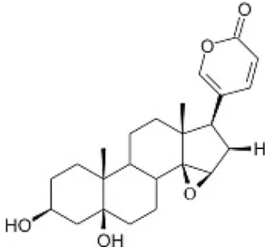
Toad venom collection following Brazilian guidelines (IBAMA, SISBIO: number 30034-1) was obtained fromR. marinasecretions in Mato Grosso State, in the southern Brazilian Amazon. The animals were correctly identified by biologists (Janaina da Costa de Noronha and Domingos de Jesus Rodrigues) and a voucher specimen (R. marina− ABAM-H 1262) was deposited at Acervo Biológico da Amazônia Meridional (ABAM, Sinop, Mato Grosso, Brazil). All procedures were approved by the Committee on Animal Research at Universidade Federal do Mato Grosso (#23108.700260/14-7) and they are in ac-cordance with Brazilian (COBEA−Colégio Brasileiro de Experimentação Animal) and international guidelines on the care and use of experi-mental animals (Directive 2010/63/EU of the European Parliament and of the Council on the protection of animals used for scientific purposes). Analysis on HPLC, column cromatography, electrospray ionization mass and NRM were carried out according toFerreira et al. (2013)and Kerkhoffet al. (2016)for isolation and identification of marinobufagin (Fig. 1).
2.2. Tumor and normal cells
Human leukemia (HL-60, K-562), melanoma (MDA/MB-435), glio-blastoma (SF-295), breast (MCF-7), lung (NCIH-292), larynx (HEP-2), liver (HEPG-2) and ovarian (OVCAR-8) tumor lines, human peripheral blood mononuclear cells (PBMC), and murine lines (L-929, normalfi -broblasts;MS-5, normal stromal hematopoietic cells; B-16/F10, mela-noma) were maintained in RPMI-1640 medium supplemented with 10% fetal bovine serum, 2 mM glutamine, 100 U/mL penicillin and 100μg/mL streptomycin, at 37 °C in a 5% CO2atmosphere.
Heparinized human blood samples (from healthy, non-smoker do-nors who had not taken any drug for at least 15 days prior to sampling, aged 18–35 years old) were collected. Firstly, polymorphic blood mononuclear cells (PBMC) were isolated by the standard method of density-gradient centrifugation over Ficoll-Hypaque. After some days, an extra blood collection was performed and a suspension of red blood cells (2%) was prepared. All studies were performed in accordance with Brazilian research guidelines (Law 466/2012, National Council of Health) and with the Declaration of Helsinki.
2.3. Cytotoxicity analysis
The cytotoxic action of marinobufagin was assessed by colorimetric assays after 72 h exposure. Cell proliferation was determined spectro-photometrically using a multiplate reader (DTX 880 Multimode Detector, Beckman Coulter). Control groups (negative and positive) received the same amount of dimethylsulfoxide solvent (0.1% DMSO) like the test groups. Doxorubicin (0.01–8.6μM) was used as positive control.
2.3.1. MTT assay
The cytotoxicity on HL-60, K-562, MDA/MB-435, SF-295, MCF-7, NCIH-292, HEP-2, HEPG-2, OVCAR-8, L-929, MS-5 and B-16/F10 cells was determined by the MTT assay (Mosmann, 1983), which analyzes the ability of living cells to reduce the yellow dye 3-(4,5-dimethyl-2-thiazolyl)-2,5-diphenyl-2H-tetrazolium bromide (MTT) to a purple formazan product. Cells were plated in 96-well plates (0.3–0.7 × 10⁵ cells/well) and incubated to allow cell adhesion or equilibration (sus-pension cultures). Twenty-four hours later, marinobufagin was added to each well (0.01–62.5μM). After 69 h of incubation, the supernatant was replaced with fresh medium containing 10% MTT, and the cells in-cubated for an additional 3 h. The plates were then centrifuged, for-mazan product was dissolved in DMSO and absorbance was read at 595 nm.
2.3.2. Alamar Blue assay
The activity of the compound was also investigated on human PBMC using the Alamar Blue™ assay. PBMC were washed and resuspended (3 × 10⁵ cells/mL) in supplemented RPMI-1640 medium plus 4% phytohemagglutinin to induce cell growth. PBMC were then plated in 96-well plates (3 × 10⁵cells/well in 100
μL of medium). After 24 h, extracts dissolved in DMSO were added to each well (0.01–62.5μM) and the cells were incubated for 72 h. Twenty-four hours before the end of the incubation, 20μL of resazurin stock solution (0.156 mg/mL) were added to each well (Alamar Blue™, Sigma Aldrich Co., USA). The ab-sorbance was read at 570 and 595 nm and the drug effect was expressed as the percentage of the control (Ferreira et al., 2013).
2.4. Hemolytic assay
Marinobufagin was tested for hemolytic activity according to Ferreira et al. (2013). The compound (3.9–500μM) was incubated in 96- well plates for 60 min at room temperature (25 °C) in a suspension of human erythrocytes (2%) in 0.85% NaCl containing 10 mM CaCl2. After centrifugation, hemoglobin levels in the supernatants were spec-trophotometrically determined at 540 nm.
2.5. Assessment of mechanism(s)
To understand cytotoxicity, detailed assessments were performed with HL-60 leukemia cells. Marinobufagin was added to 12-well tissue culture plates with HL-60 cells (3 × 10⁵cells/mL) to obtainfinal con-centrations of 0.025, 0.25 and 1.25μM These concentrations were se-lected based on the marinobufagin IC50value for HL-60 cells after 24 h exposure. Doxorubicin (Dox, 0.6μM) was used as positive control.
2.5.1. Trypan blue exclusion
Cell viability was determined by the trypan blue exclusion assay (Renzi et al., 1993). Aliquots from each well were removed from cul-tures after 24 h exposure and cells were scored in a Neubauer chamber using light microscopy (Metrimpex Hungary/PZO-Labimex Modelo Studar lab®).
2.5.2. Cytological examination by light microscopy
Untreated or marinobufagin-treated HL-60 cells were examined for morphological changes by light microscopy (Metrimpex Hungary/PZO-Labimex Modelo Studar lab®). To evaluate morphology, cells were
harvested, transferred to cytospin slides,fixed with methanol for 1 min and stained with hematoxylin-eosin (H&E, Vetec, Brazil).
2.5.3. Morphological analysis usingfluorescence microscopy
Acridine orange/ethidium bromide (AO/EB) staining of HL-60 cells was performed to determine the cell death pattern induced by mar-inobufagin (McGahon et al., 1995). So, after 24 h of incubation, cells were pelleted, and each sample was mixed with 1μL of aqueous AO/EB solution (100μg/mL of AO in PBS; 100μg/mL EB in PBS) just prior to fluorescence microscopy analysis and quantification (Olympus, Tokyo, Japan). Three hundred cells were counted per sample and scored as follows: viable cells, apoptotic cells and necrotic cells (Geng et al., 2003).
2.5.4. Analysis byflow cytometry
Allflow cytometry analyses were performed in a Guava EasyCyte Mine using Guava Express Plus software. Five thousand events were evaluated per experiment and cell debris was omitted from the analysis.
2.5.4.1. Membrane integrity. Cell membrane integrity was evaluated by the exclusion of PI after 24 h exposure. Briefly, 100μL of treated and untreated cells were incubated with PI (50μg/mL) for 5 min at 37 °C and membrane integrity was determined (Darzynkiewicz et al., 1992).
2.5.4.2. Cell cycle and DNA fragmentation. Briefly, 24h-treated and untreated cells were incubated at 37 °C for 30 min in the dark in a lysis solution containing 0.1% citrate, 0.1% triton X-100 and 50μg/mL PI andfluorescence was subsequently measured (Ferreira et al., 2014).
2.5.4.3. Phosphatidylserine (PS) externalization. PS externalization was demonstrated by flow cytometry after PS staining with annexin V (Krysko et al., 2008). Briefly, cells were washed twice with cold PBS and then suspended in 135μL of PBS with 5μL of 7-amino-actinomycin D (7-AAD) and 10μL of annexin V-PE (Guava Nexin Assay Kit). The cells were gently vortexed and incubated for 20 min at room temperature (20–25 °C) in the dark. Afterward, the cells were analyzed by flow cytometry (EasyCyte from Guava Technologies). Annexin V is a phospholipid-binding protein that has a high affinity for PS. Meanwhile, 7-AAD is a no permeant dye used to indicate membrane integrity. Fluorescence of annexin V-PE was measured at 583 nm (yellowfluorescence) and 7-AAD at 680 nm (redfluorescence) (Krysko et al., 2008). Results were expressed as percentages of early and late apoptotic cells and necrotic cells.
2.6. Analysis of DNA strand breaks in human cells
DMSO for 16 h at 4 °C. The slides were then removed from the lysing solution and placed on a horizontal electrophoresis tank filled with freshly prepared alkaline buffer (300 mM NaOH, 1 mM EDTA, pH > 13.00). The slides were equilibrated in the same buffer for 20 min, and electrophoresis was performed at 25 V, 300 mA for 20 min. After elec-trophoresis, the slides were washed gently with 2 M Tris-HCl buffer, pH 7.4, to remove the alkali. Each slide was stained with 50μL ethidium bromide (20μg/mL), and a cover slip was placed on the slide. Cellular analysis (100 cells for each of the three replicate slides) were performed using a visual scoring system that categorized tail length into five classes (0, 1, 2, 3 and 4) to determine the Damage Index (DI), which is considered to be a sensitive DNA measure and based on migration length as well as the amount of DNA in the tail. Therefore, a damage index value was assigned to each comet according to its class, and the values ranged from 0 (completely undamaged) to 400 (maximum da-mage). So, the Damage Index (DI) was calculated using the formula: DI =Σ(number of cells with damage X class of damage), which ranged from 0 (ex.: 100 cells with damage 0 × 0) to 400 (ex.: 100 cells with damaged 4 × 4). Doxorubicin (0.6μM) was used as the positive control.
2.7. Evaluation of the cytotoxic and genotoxic potential in root meristematic cells of Allium cepa
Healthy onions of small and uniform size, from the same origin and not germinated were used. Dried roots, extreme layers and the central parenchyma of the budding crown were removed to increase absorption by fresher roots and to ensure the uniformity of budding and root growth. All onions were washed in running water for 20 min (Fiskesjö, 1985;Bagatini et al., 2007).
Five bulbs were used for each concentration of marinobufagin (1.25, 2.5, 25 and 62.5μM) and for the controls (negative: dechlorinated water; positive: copper sulfate 3μM). Subsequently, bulbs were placed in glass vials and the sample volume was completed every 24 h for a final volume of 5 mL. Onions were kept for 72 h in the dark at room temperature (25 ± 2 °C). Thereafter, the roots were removed and measured with a digital pachymeter as a toxicity signal. Then, they werefixed in Carnoy solution [ethanol:acetic acid (3:1)] for 24 h, stored in 70% ethanol and kept under refrigeration until slides preparation. Roots were washed in distilled water (3 baths of 5 min), followed by hydrolysis with 1N HCL for 10 min at 60° C in a water bath and cooled in running water at room temperature. The roots were transferred to vials containing Schiff's reagent (1.5 g of basic fuchsin, 4.5 g of sodium metabisulfite, 45 mL of 1N HCl, 10 g of activated charcoal and 300 mL of H2O), for approximately 120 min (Fiskesjö, 1985; Bagatini et al., 2007). Soon after, the roots were washed with distilled water until dye have been completely removed, placed on a blade and incisions were performed to separate the meristematic region. A solution of 2% acetic carmine was added to the sectioned materials and the slides were covered with coverslips. Examinations were performed by optical mi-croscope (Olympus, Tokyo, Japan) at 400X, and 5000 cells per con-centration (1000 cells/slide) were counted to determine the Mitotic Index (MI) and chromosomal alterations (micronuclei, delays, C-meta-phases, bridges and breaks).
2.8. Statistical analysis
The IC50and EC50values and their 95% confidence intervals were obtained by nonlinear regression using the GraphPad program (Intuitive Software for Science, San Diego, CA). Differences were evaluated by comparing data using one-way analysis of variance (ANOVA) followed by the Newman-Keuls test (p <0.05). All studies were carried out in duplicate and represented independent biological evaluations.
3. Results
3.1. Cytotoxic activity
Marinobufagin presented high antiproliferative action after 72 h exposure on all human tumor lines and IC50values ranging from 0.15 (HL-60) to 7.35 (HEP-2)μM (Table 1). It exhibited cytotoxic potential in a similar extent to those found with the positive control doxorubicin for some lines (HL-60, K-562, MDA/MB-435, SF-295, MCF-7 and HEPG-2). Meanwhile, it was more active against OVCAR-8 cells when com-pared to doxorubicin (p >0.05).
Interestingly, none of the murine lines (L-929, MS-5 and B-16/F10) was sensitive to marinobufagin and in vitro investigation performed with human erythrocytes showed no lysis action. We found anti-proliferative action on human normal dividing leukocytes (IC50 of 10.88μM), though such activity was up to 72.5-fold more selective against leukemia cells when compared to dividing leukocytes (se-lectivity coefficient determined by IC50in PBMC/IC50in HL-60 cells).
3.2. Induced biochemical and morphological changes on HL-60 cells
HL-60 cells treaded with marinobufagin and examined by the trypan exclusion test showed significantly reduction in cell viability (61.3 ± 4.4 and 39.7 ± 7.4%) and increased number of non-viable cells (7.4 ± 1.1 and 8.7 ± 1.7%) in a concentration-dependent manner (p <0.05) after 24 h of exposure in the concentrations of 0.25 and 1.25μM when compared with negative control (77.7 ± 10.5 and 3.3 ± 1.5%, respectively) (Fig. 2A).
To establish whether the growth inhibition displayed it was related to the induction of apoptosis and/or necrosis, treated cells werefirstly analyzed using AO/EB staining byfluorescence microscopy and the Table 1
Cytotoxic activity of marinobufagin after 72 h exposure determined by MTT assay.
Cell line IC50[μg/mL (μM)]*
Marinobufagin Doxorubicin
HL-60 0.06 (0.15) 0.02 (0.03)
0.05–0.07 0.01–0.02
K-562 0.10 (0.25) 0.14 (0.25)
0.08–0.12 0.09–0.23
MDA/MB-435 0.21 (0.53) 0.48 (0.88)
0.16–0.27 0.34–0.66
SF-295 0.18 (0.45) 0.20 (0.36)
0.15–0.21 0.18–0.25
MCF-7 0.38 (0.95) 0.35 (0.64)
0.27–0.55 0.28–0.51
NCIH-292 1.10 (2.75) 0.20 (0.36)
0.76–1.98 0.19–0.50
HEP-2 2.94 (7.35) 0.73 (1.34)
2.00–4.10 0.31–1.44
HEPG-2 0.32 (0.80) 0.21 (0.38)
0.23–0.43 0.15–0.30
OVCAR-8 0.10 (0.25) 1.30 (2.39)
0.09–0.13 1.01–1.93
PBMC** 4.35 (10.88) 0.91 (1.67)
2.49–7.75 0.55–1.89 B-16/F10 > 10 (> 25) 0.03 (0.06)
0.02–0.04
MS-5 > 10 (> 25) 0.84 (1.54)
0.75–0.99
L-929 > 10 (> 25) 0.66 (1.21)
0.43–0.88
*Data are presented as IC
numbers of viable, apoptotic and necrotic cells were determined. Cell viability (51.0 ± 2.4, 20.5 ± 6.5 and 58.8 ± 4.8%) also reduced and such change was associated with amplifying of cells in apoptosis in marinobufagin (0.25μM: 37.6 ± 3.2, 68.0 ± 2.8; 1.25μM: 36.0 ± 3.8%) and doxorubicin treated cells in comparison with non-treated samples (95.0 ± 1.5 and 2.3 ± 0.8% for viable and apoptotic cells, respectively) (p <0.05) (Fig. 2B). Necrosis was observed at marinobufagin 1.25μM only (11.3 ± 4.8%) (p <0.05).
Cell morphology of leukemia cells is demonstrated inFig. 3. Under light microscopy, control cells displayed a typical non-adherent and round morphology, homogeneous cytoplasm, presence of mitoticfi g-ures and visualization of the cellular plasma membrane bound and nucleoli (Fig. 3A). Marinobufagin-treated cells at 0.01μg/mL also showed mitoticfigures in a lower frequency (Fig. 3C). At 0.25μM, cells presented binucleation, cellular shrinking and nuclear condensation (Fig. 3D). Meanwhile, the highest concentration (1.25μM) caused chromatin condensation, nuclear fragmentation, karyolysis, cellular shrinking and rarefaction, occurrence of cytoplasmic vacuoles and membrane disintegration (Fig. 3E). Cellular rarefaction, nuclear frag-mentation, cytoplasmic vacuoles and membrane disintegration were also seen in cells treated with doxorubicin associated with cytoplasmic basophily and hyperchromatic nuclei suggestive of necrosis (Fig. 3B).
Cytometry studies revealed decrease of membrane integrity (91.6 ± 0.6 and 92.5 ± 0.5%), increase of DNA fragmentation (sub-G1cells: 15.6 ± 3.4 and 56.6 ± 5.3%) and reduction of cells in G2/M (15.8 ± 0.8%) and S (19.3 ± 1.7%) phases at concentrations of 0.25 and 1.25μM, respectively (Fig. 4A and B) when compared to the ne-gative control (97.9 ± 0.6, 3.9 ± 0.7, 23.8 ± 1.2 and 29.2 ± 1.4%).
Doxorubicin (0.6μM, positive control) also dropped membrane in-tegrity (60.1 ± 1.1%) and caused intense DNA fragmentation (85.5 ± 3.7%) (p <0.05). Secondly, biological actions of mar-inobufagin on HL-60 cells were corroborated by PS externalization after annexin V and 7-AAD staining (Fig. 4C). Herein, it also caused reduc-tion of viable cells (for 82.6 ± 0.3 and 63.2 ± 1.0%), increased cells in early (for 12.4 ± 0.6 and 30.5 ± 0.7%) and late (for 4.6 ± 0.3 and 5.0 ± 0.2%) apoptosis in both higher concentrations tested (0.25 and 1.25μM) and once again, necrosis was detected in the maximum con-centration only (1.2 ± 0.3%) in comparison with non-treated cells (93.8 ± 0.9, 4.7 ± 0.6, 1.3 ± 0.3 and 0.2 ± 0.1%), respectively (p <0.05).
3.3. Antiproliferative and clastogenic action
To study the genotoxic potential of marinobufagin, wefirstly ana-lyzed human leukemia and normal leukocyte cells after 24 h of treat-ment by the Cometa assay. None of the cellular cultures exposed to the bufadienolide revealed significant DI changes (HL-60: 8.2 ± 1.1, 6.8 ± 0.9 and 6.4 ± 1.0 for 0.025, 0.25 and 1.25μM; PBMC: 8.3 ± 1.2, 7.3 ± 1.4, 7.8 ± 1.4 and 7.0 ± 1.1 for 0.025, 0.25, 1.25 and 2.5μM) when compared to negative control (5.4 ± 0.9, p >0.05). On the other hand, Dox caused intense DNA damage on both HL-60 and PBMC (182.0 ± 7.1 and 134.7 ± 5.3, respectively, p <0.05).
Marinobufagin inhibited the growth ofA. ceparoots of in all tested concentrations (48.9 ± 8.7, 51.4 ± 6.1, 70.4 ± 3.8 and 85.8 ± 3.1% for 1.25, 2.5, 25 and 62.5μM) in a concentration-de-pendent manner (0.58 ± 0.08, 0.56 ± 0.06, 0.45 ± 0.03 and 0.23 ± 0.03 cm, respectively) when compared to the negative control (1.04 ± 0.06 cm, p <0.05) and revealed an EC50 value of 7.5 (3.0–19.5) μM. Onions treated with copper sulphate 3μM presented roots with 0.36 ± 0.02 cm and growth inhibition of 72.6 ± 2.7% (Fig. 5).
Confirming its capacity to inhibit root growth, marinobufagin also caused MI reduction (43.8 ± 1.6, 39.7 ± 1.4, 31.0 ± 1.6 and 22.1 ± 0.5% for 1.25, 2.5, 25 and 62.5μM, respectively) when com-pared to the negative control (56.7 ± 1.0%), declined number of cells at all stages of the cell cycle (prophase, metaphase, anaphase and tel-ophase) in a similar way to those results seen for the positive control (13.9 ± 0.6%) and increase number of cells in the interphase (p <0.05,Table 2).
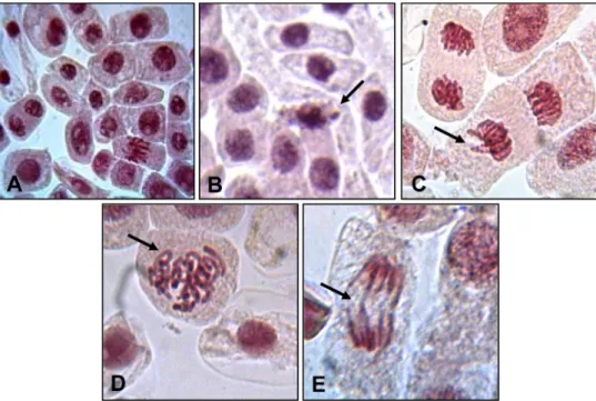
Marinobufagin caused a significant increase of chromosomal al-terations at concentrations of 2.5μM (15.8 ± 1.1), 25μM (36.5 ± 3.6) and 62.5μM (47.0 ± 3.4 changes) in relation to the negative control (7.3 ± 0.8 changes). Chromosomal changes included micronuclei (12.5 ± 0.6 and 23.3 ± 3.1), delays (9.0 ± 2.1 and 10.8 ± 1.1) and C-metaphases (10.5 ± 1.4 and 10.5 ± 0.5) at 25 and 62.5μM, respectively, but only micronuclei increasing was concentra-tion-dependent (Table 3,p <0.05). Copper sulphate also increases all forms of chromosomal changes [micronuclei (11.5 ± 0.7), delays (13.0 ± 0.7), C-metaphases (16.3 ± 0.9), bridges (6.3 ± 0.6) and breaks (8.0 ± 10)] in comparison with negative control (2.3 ± 0.3, 2.0 ± 0.4, 1.3 ± 0.3, 1.3 ± 0.3 and 0.5 ± 0.3, respectively, p <0.05). Representative images of the most commonly seen chro-mosomal changes are shown inFig. 6: micronucleus (Fig. 6B), delay (Fig. 6C), C-metaphase (Fig. 6D) and bridge (Fig. 6E).
4. Discussion
The World Health Organization (WHO) believes that until 2030 will raise about 21.4 million new cancer cases and 13.2 million cancer deaths. Oceania (Australia and New Zealand) has about 313 cases per 100,000 inhabitants, the highest incidence of cases among all con-tinents. South America and Brazil have an incidence of 172 cases per 100 thousand inhabitants (WHO, 2014). The main treatments against Fig. 2.Effects of marinobufagin on HL-60 leukemia cells after 24 h of incubation. A–
cancers involve surgery, chemotherapy, and/or radiotherapy. Nowa-days, most of the chemotherapeutic possibilities are natural, derived or synthesized molecules based on natural compounds (Srivastava et al., 2005;Greenlee, 2012;Newman and Cragg, 2012;Ferreira et al., 2016). More recently, studies have showed antiproliferative (Cunha-Filho et al., 2010;Gao et al., 2011;Moreno Y Banuls et al., 2013;Ferreira et al., 2013;Sciani et al., 2013;Wang and Bi, 2014) and antiangiogenic (Cunha-Filho et al., 2010) actions of toad dry poisons. Herein, wefirstly showed the potent capacity of the compound marinobufagin extracted fromR. marinaon several tumor lines, whose IC50values are compar-able to those observed with doxorubicin, a drug routinely used in the treatment of several cancers including breast, lung, gastric, ovarian, thyroid, non-Hodgkin’s and Hodgkin’s lymphoma, multiple myeloma, sarcoma, and pediatric cancers (Thorn et al., 2011).
Marinobufagin belongs to the class of bufadienolides, polyhydroxy steroids with 24 carbons related to cholesterol with one unsaturated lactone ring and a 2-pyrone group at the C-17 position of the perhy-drophenanthrene nucleus (Sousa et al., 2017). Previously,in vitro pre-vious antitumor studies performed with Rhinella, Bufo and Rhaebo species, including skin secretions and isolated bufadienolides, alkaloids, metabolic derivatives and bufadienolides from the traditional Chinese
drug (Chan Su), showed activity against several human tumor cell lines such as glioblastoma (U-373), osteosarcoma (MG-63), colon (26-L5), leukemia (K-562, U-937 ML-1, Jukart T, HL-60), melanoma (MDA/MB-435, SKMEL-28), bladder (BIU-87, J-82), breast (MCF-7, MDA/MB-231, MCF10A), oligodendrogliome (Hs-683), gastric adenocarcinoma (AGS), prostate (DU-145, PC-3, LNCaP), stomach (hepatocellular carcinoma (HepG2), lung carcinoma (A-549, SK-MES-1) and primary liver carci-noma (PLC/PRF/5) (Nogawa et al., 2001; Ogasawara et al., 2001; Kamano et al., 2002;Yeh et al., 2003;Su et al., 2009;Cunha-Filho et al., 2010;Dong et al., 2011;Qi et al., 2011;Banuls et al., 2013a;Ferreira et al., 2013;Sciani et al., 2013;Lee et al., 2014;Schmeda-Hirschmann et al., 2014; Wang and Bi, 2014;Schmeda-Hirschmann et al., 2016, 2017).
We also realized that marinobufagin was cytotoxic for human but not for mice cells. Similarly, a variety of chemical as bufotalin, hel-lebrin, and 5α-bufalin, were inactive on murine lines (Banuls et al., 2013b), but Banuls et al. (2013a) demonstrated that gamabufotalin rhamnoside was thefirst compound with cytotoxic action on murine neoplastic lines [(CT26.WT (colon), B-16/F-10 (melanoma)].
associated with direct membrane damages, corroborating our previous outcomes that showed venom extracts from R. marina as non-lytic samples (Ferreira et al., 2013). To understand its mechanism, HL-60 line was used as a biomedical cellular tool (Ferreira et al., 2014; Monção et al., 2015; Sousa et al., 2017). This line originated from a female patient with acute myeloid leukaemia after culture of her per-ipheral blood leukocytes and display predominant myeloblastic/pro-myelocytic morphology; the remaining cells display morphologies re-sembling those of more mature myeloid cells (mainly myelocytes, with some neutrophils and monocytes) (Collins, 1987).
Marinobufagin-treated HL-60 cells presented cell viability reduction in trypan exclusion tests, confirming results found in MTT analysis. AO/ BE fluorescence microscopy examinations also displayed viability re-duction of leukemia cells in association with expanding of apoptotic cells. Equivalent concentrations that reduced cell viability also caused morphological alterations in HL-60 cells, including binucleation, cel-lular shrinking, nuclear/chromatin condensation, nuclear fragmenta-tion, karyolysis, cellular shrinking and rarefacfragmenta-tion, occurrence of
cytoplasmic vacuoles and membrane disintegration (this latter in the highest concentration tested). Previously, some studies have shown that skin secretions obtained fromRhinellacrucifer,R. marina, R. major, R. schneideri, R. margaritifer, Phyllomedusa hypocondrialis, Rhaebo guttatus, R. margaritifer, R. major and P. hypocondrialisare a fascination source of telocinobufagin, hellebrin, marinobufagin and bufagin, substances with capacity of reducing cell viability, DNA synthesis and causing mor-phological changes (chromatin condensation, nuclear fragmentation, cytoplasm shrinkage, cytoplasmic vacuoles, stickiness reduction, blis-tering membrane and apoptotic bodies) (Yeh et al., 2003; Qi et al., 2011;Ferreira et al., 2013;Sciani et al., 2013).
Subsequently, our investigations were performed by flow cyto-metry, a very useful apparatus to evaluate cellular functions including mitototic capacity, metabolic activity, and integrity and potential of membranes (Paparella et al., 2008). Using such technology tool, we discovered that marinobufagin decreased membrane integrity and caused DNA fragmentation, cell cycle disruption and PS externalization, especially, in higher concentrations. These results supported the Fig. 4.Flow cytometry analysis of leukemia HL-60 cells after 24 h of incubation with marinobufagin isolated fromRhinella marinatoad venom. A–Cell membrane integrity evaluated by the exclusion of propidium iodide; B–DNA fragmentation determined by nuclearfluorescence using propidium iodide; C–Phases of the cell cycle detected by nuclearfluorescence using propidium iodide, triton X-100 and citrate; D–Phosphatidylserine externalization stained Annexin-V and 7-amino-actinomycin-D. Negative control (C) was treated with the vehicle used for diluting the tested substance. Doxorubicin (0.6μM) was used as positive control (Dox). Results are expressed as mean ± standard error of measurement (S.E.M.) from two in-dependent experiments. *p< 0.05 compared to control by ANOVA followed by Student Newman-Keuls test.
morphological changes and suggest that marinobufagin may cause cell death by apoptosis, since sub-diploid G0/G1, fragmented DNA and PS on cell surfaces are indicative of apoptosis (Krysko et al., 2008).
In the traditional Chinese medicine,Chan Su, an ethanolic extract from skin and parotid venom glands ofBufo bufo gargarizansCantor, is extensively used for cancer therapy and presented compounds as bu-falin and cinobufagin, whose cellular studies revealed cytotoxic action on MCF-7 (breast), A-549 (lung) and Jurkat T (leukemia) tumor cells after 48 h of treatment. They caused decreasing in cellular viability, augment of apoptotic cells, DNA single- and double-strand breaks, mi-cronuclei induction and slight effects on PBMC viability (Xu-Tao et al., 2009;Lee et al., 2014). Here, dividing leukocytes were also not a de-sired target for marinobufagin, since its cytotoxic activity was up to 72.5-fold more selective against proliferating leukemia cells. This high selectivity has already shown with extracts from R. marina venoms (Ferreira et al., 2013) and previous reports also described that com-pounds isolated from frogs decrease [ATP]i in cancer lines, while weaker effects were seen in normal cells (Lefranc et al., 2008;Mijatovic et al., 2012;Banuls et al., 2013b), though murine normal cells is not commonly attacked by bufadienolides.
Since cardenolides (ouabain and digoxin) and bufadienolides (are-nobufagin, bufalin, telocinobufagin and hellebrin) bind specifically to the subunits of the sodium/potassium pump (Na+/ K+-ATPase) and such subunits are distinctive between humans and mice (Bick et al., 2002;Gao et al., 2011;Touza et al., 2011;Banuls et al., 2013a;Laursen et al., 2015;Córdova et al., 2016;Sousa et al., 2017), it is likely that apoptosis activating by marinobufagin should be associated with a species-specific antiproliferative action.
In addition to thein vitromodels with animal cells, plant cytotoxi-city bioassays using A. cepa have been widely used to detect in situ genotoxic and mutagenic compounds of synthetic and natural products and have been validated by the International Chemical Safety Program (IPCS, WHO) and the United Nations Environment Program (UNEP) to assess chromosome changes (Bagatini et al., 2007; Pesnya and Romanovsky, 2013;Misik et al., 2014). However, to date, there are no reports regarding cytotoxicity studies with animal secretions, such as amphibian poisons and/or their constituents, with this vegetal system.
Then, our investigations revealed, for thefirst time, that marinobufagin reduced the MI and caused chromosomal changes (micronuclei, C-me-taphases, delays and bridges) in dividing meristematic cells ofA. cepa roots.
Mitotic Index (IM) is an important indicator of cell proliferation and the level of cytotoxicity of an agent can be determined by the increase or decrease of this parameter (Leme and Marin-Morales, 2009;Neves et al., 2014;Prajitha and Thoppil, 2016). Mitotic index reduction may be related to the inhibition of DNA synthesis, blockade or arrest of cell cycle at any stage or cell output induction from the cycle to the G0 phase, avoiding the cell returning to mitosis. Indeed, many compounds with antiproliferative action usually cause cell cycle arrest at some stage of mitosis as part of the cytotoxic mechanism due to the induction of aneugenic or clastogenic chromosome damages. Such arrest occurs to give to the cell machinery the opportunity to repair genoma injuries and to escape from cell death by apoptosis (Ferreira et al., 2014;Pandey et al., 2014; Santana et al., 2016). We noted marinobufagin-induced genotoxic and clastogenic properties on meristematic cells ofA. cepa roots and it is likely that clastogenicfindings (micronuclei, delays, C-metaphases and bridges) caused by marinobufagin led to cell arrest at interphase (G0) as a chance for“cellular escape”from death.
Micronucleus is a common injury caused by environmental sub-stances (xenobiotics) and represent loss of chromatin as a consequence of structural chromosomal damages or mitotic apparatus failure, pro-ducing acentric fragments or entire chromosomes that are not included in the main nucleus during the telophase (Fernandes et al., 2007; Fenech et al., 2011). Nevertheless, genotoxic agents do not necessarily cause mutagenic consequences, since some DNA lesions can be re-paired. An example is the inspection of the acute toxic and genotoxic properties of cytostatic drugs widely used in clinical practice, such as 5-fluorouracil, etoposide, cisplatin, carboplatin, vincristine sulfate and cyclophosphamide monohydrate, whose effects (inhibition of root growth and cell division) onA. cepameristematic cells were observed in lower concentrations when compared to those that lead to DNA da-mage, suggesting acute toxic effects (antiproliferative activity, for ex-ample) are not obligatory associated with genetic material injuries (Misik et al., 2014).
Table 2
Cytotoxic activity of marinobufagin on meristematic cells ofAllium ceparoots after 72 h exposure.
Treatment Concentration (μM) Interphase Mitosis Mitotic Index** (%)
Prophase Metaphase Anaphase Telophase
Negative control – 433.3 ± 10.1 398.8 ± 3.9 77.3 ± 7.8 58.0 ± 2.9 32.8 ± 3.5 56.7 ± 1.0 Copper sulphate 3 861.0 ± 5.6* 104.5 ± 3.0* 12.0 ± 1.9* 11.0 ± 1.5* 11.5 ± 3.2* 13.9 ± 0.6* Marinobufagin 1.25 558.0 ± 18.1* 390.8 ± 16.0 14.5 ± 2.1* 13.3 ± 2.2* 20.3 ± 4.3* 43.8 ± 1.6* 2.5 602.5 ± 14.1* 358.0 ± 15.5 11.5 ± 1.6* 12.8 ± 1.6* 15.3 ± 1.8* 39.7 ± 1.4* 25 689.8 ± 15.8* 259.0 ± 16.5* 17.8 ± 3.5* 15.8 ± 2.0* 17.8 ± 2.4* 31.0 ± 1.6* 62.5 779.3 ± 5.1* 182.3 ± 6.5* 11.8 ± 0.9* 10.8 ± 1.1* 16.0 ± 0.8* 22.1 ± 0.5*
Results are expressed as mean ± standard error of measurement (S.E.M.) from two independent experiments. *p <0.05 compared to control by ANOVA followed by Student Newman-Keuls test. Copper sulphate 3μM was used as positive control. ** Mitotic index was calculated as follow: Prophase + Metaphase + Anaphase + Telophase/Total number of cells x 100.
Table 3
Chromosomal changes induced by marinobufagin on meristematic cells ofAllium ceparoots after 72 h exposure.
Treatment Concentration (μM) Chromosomal alterations Total of chromosomal alterations
Micronuclei Delays C-metaphases Bridges Breaks
Negative control – 2.3 ± 0.3 2.0 ± 0.4 1.3 ± 0.3 1.3 ± 0,3 0.5 ± 0.3 7.3 ± 0.8 Copper sulphate 3 11.5 ± 0.7* 13.0 ± 0.7* 16.3 ± 0.9* 6.3 ± 0,6* 8.0 ± 1.0* 55.0 ± 2.0* Marinobufagin 1.25 1.0 ± 0.4 5.5 ± 1.0 4.3 ± 0.6* 0.5 ± 0,3 0.5 ± 0.5 11.8 ± 0.9
2.5 3.5 ± 0.6 5.0 ± 1.2 4.8 ± 0.8* 2.0 ± 0.0 0.5 ± 0.3 15.8 ± 1.1* 25 12.5 ± 0.6* 9.0 ± 2.1* 10.5 ± 1.4* 4.5 ± 0.5* 0.0 ± 0.0 36.5 ± 3.6* 62.5 23.3 ± 3.1* 10.8 ± 1.1* 10.5 ± 0.5* 2.0 ± 0.8 0.5 ± 0.5 47.0 ± 3.4*
Indeed, some injuries on chromosomes are time- and concentration-dependent and we showed this here withA. cepabioassays, since an-tiproliferative activity of marinobufagin was early detected in lower concentrations (1.25 and 2.5μM) and clastogenic action was noted only in higher ones (25 and 62.5μM). Moreover, DNA strand breaks were not observed by alkaline Cometa assays in HL-60 and polymorphic blood cells, suggesting, once again, that the antiproliferative action of marinobufagin on human cells was not necessarily related to a geno-toxic capacity. However, it is worth mentioning that many antitumor drugs does not display disassociated mechanisms and most of them are antiproliferative, genotoxic and mutagenic agents in similar con-centrations. In this situation, reducing mutagenicity would mean re-ducing the clinical efficacy of the drug (Misik et al., 2014;Prajitha and Thoppil, 2016).
Even with large differences about acute toxic potencies of drugs on vegetals’and mammals’cellular models, such as absorption capacity, species-specific differences in DNA repair and detoxification rate (Majer et al., 2005), we noted that marinobufagin has antiproliferative po-tentiality both on human andA. cepacells, suggesting it probably acts on common cellular processes of eukaryotic cells. Indeed,A. cepatest is a sensitive technique, and it indicates excellent correlation to other test systems (Fiskesjö, 1985;Bagatini et al., 2007). Herein, however, in a very interesting way, marinobufagin showed a comparable action to hellebrigenin, and these both molecules were highly cytotoxic to HL-60 cells and induce cell death by apoptosis but alkaline Cometa bioassays did not detect DNA damage (Soares, 2013). Maybe, this genotoxic ab-sence on human cells can be explained by technical conditions fre-quently established for studies with bufadienolides. These are very active compounds and this discourages scientists test them in higher concentrations. As described above, only the highest concentrations (25 and 62.5μM) caused specific clastogenic effects on vegetal cells, which indicates extreme exposure conditions are necessary to cause acti-vating-chromosomal damages of cytotoxicity.
In summary, marinobufagin displayed a remarkable anti-proliferative action on human tumor cells triggered by apoptotic signals in leukemia cells; it was up to 72.5-fold more selective against pro-liferating leukemia cells, has no genotoxic effects on human normal leukocytes or leukemia cells and it revealed antimitotic action, cell cycle arrest at interphase and concentration-dependent chromosomal alterations on meristematic cells of A. cepa roots. Moreover, mar-inobufagin was not cytotoxic upon murine lines. Tofind out partially
targeted natural molecules on human leukemia cells, as marinobufagin, is an amazing and stimulating way to continue the battle against cancer. In vivo assessments are in progress to confirm such in vitro findings.
Conflict of interest
The authors declare that there are no conflicts of interest.
Acknowledgments
We are grateful to the Brazilian agencies“Conselho Nacional de Desenvolvimento Científico e Tecnológico”[CNPq (#305086/2016-2)] and“Fundação de Amparo à Pesquisa do Estado do Piauí” [FAPEPI (#011/2016)] forfinancial support in the form of grants and fellow-ship.
References
Bagatini, M.D., Silva, A.C.F., Tedesco, S.B., 2007. The use ofAllium cepatest as a bioin-dicator of genotoxicity of medicinal plants infusions. Braz. J. Pharmacogn. 17, 444–447.
Banuls, L.M., Urban, E., Gelbcke, M., Dufrasne, F., Kopp, B., Beijo, R., Zehl, M.E., 2013a. Structure-activity relationship analysis of bufadienolide-inducedin vitrogrowth in-hibitory effects on mouse and human cancer cells. J. Nat. Prod. 76, 1078–1084. Banuls, L.M., Katz, A., Miklos, W., Cimmino, A., Tal, D.M., Ainbinder, E., Zehl, M., Urban,
E., Evidente, A., Kopp, B., Berger, W., Feron, O., Karlish, S., Kiss, R., 2013b. Hellebrin and its aglycone form hellebrigenin display similarin vitrogrowth inhibitory effects in cancer cells and binding profiles to the alpha subunits of the Na+/K+-ATPase. Mol. Cancer 12, 1–14.
Bick, R.J., Poindexter, B.J., Sweney, R.R., Dasgupta, A., 2002. Effects ofChan Su, a tra-ditional Chinese medicine, on the calcium transients of isolated cardiomyocytes: cardiotoxicity due to more than Na, K-ATPase blocking. Life Sci. 72, 699–709. Córdova, W.H.P., Leitão, S.Z., Cunha-Filho, G., Bosch, R.A., Alonso, I.P., Pereda-Miranda,
R., Gervou, R., Touza, N.A., Quintas, L.E.M., Noel, F., 2016. Bufadienolides from parotoid gland secretions of Cuban toadPeltophryne fustiger(Bufonidae): inhibition of human kidney Na+/K+-ATPase activity. Toxicon 110, 27–34.
Clarke, B.T., 1997. The natural history of amphibian skin secretions: their normal func-tioning and potential medical applications. Biol. Rev. Camb. Philos. Soc. 72, 365–379.
Collins, S.J., 1987. The HL-60 promyelocytic leukemia cell line: proliferation, diff er-entiation, and cellular oncogene expression. Blood 70, 1233–1244.
Costa-Neto, E.M., 2005. Animal-based medicines: biological prospection and the sus-tainable use of zootherapeutic resources. An. Acad. Bras. Cienc. 77, 33–43. Cunha-Filho, G.A., Resck, I.S., Cavalcanti, B.C., Pessoa, C.O., Moraes, M.O., Ferreira, J.R.,
Rodrigues, F.A., Santos, M.L., 2010. Cytotoxic profile of natural and some modified bufadienolides from toadRhinella schneideriparotoid gland secretion. Toxicon 56,
339–348.
Cunha-Flho, G.A., Schwartz, C.A., Resck, I.S., Murta, M.M., Lemos, S.S., Castro, M.S., Kyaw, C., Pires Jr., O.P., Leite, J.R.S., Bloch Jr., C., Schwartz, E.F., 2005. Antimicrobial activity of the bufadienolides marinobufagin and telocinobufagin iso-lated as major components from skin secretion of theBufo rubescens. Toxicon 45, 777–782.
Daly, J.W., 1995. The chemistry of poisons in amphibian skin. Proc. Natl. Acad. Sci. U. S. A. 92, 9–13.
Darzynkiewicz, Z., Bruno, S., Del Bino, G., Gorczyca, W., Hotz, M.A., Lassota, P., Traganos, F., 1992. Features of apoptotic cells measured byflow cytometry. Cytometry 13, 795–808.
Dmitrieva, R.I., Bagrov, A.Y., Lalli, E., Sassone-Corsi, P., Stocco, D.M., Doris, P.A., 2000. Mammalian bufadienolides is synthesized from cholesterol in the adrenal cortex by a pathway that is independent of cholesterol side-chain cleavage. Hypertension 36, 442–448.
Dong, Y., Yin, S., Li, J., Jiang, C., Ye, M., Hu, H., 2011. Bufadienolide compounds sensitize human breast cancer cells to TRAIL-induced apoptosis via inhibition of STAT3/Mcl-1 pathway. Apoptosis 16, 394–403.
Duellman, W.E., Trueb, L., 1996. The Biology of Amphibians 1. MacGraw-Hill, New York, pp. 670.
Fenech, M., Kirsch-Volders, H., Natarajan, A.T., Surralles, J., Crott, J.W., Parry, J., Norppa, H., Eastmond, D.A., Tucker, J.D., Thomas, P., 2011. Molecular mechanisms of micronucleus, nucleoplasmic bridge and nuclear bud formation in mammalian and human cells. Mutagenesis 26, 125–132.
Fernandes, T.C.C., Mazzeo, M.E.C., Marin-Morales, M.A., 2007. Mechanism of micro-nuclei formation in polyploidizated cells ofAllium cepaexposed to trifluralin herbi-cide. Pestic. Biochem. Phys. 88, 252–259.
Ferreira, P.M.P., Lima, D.J.B., Debiase, B.W., Soares, B.M., Machado, K.C., Noromha, J.C., Rodrigues, D.J., Shinhorin, A.P., Pessoa, C., Vieira-Junior, G.M., 2013.
Antiproliferative activity ofRhinella marinaandRhaebo gutatusvenom extracts fom Southerm Amazon. Toxicon 72, 43–51.
Ferreira, P.M.P., Militão, G.C.G., Lima, D.J.B., Costa, N.D.J., Machado, K.C., Santos, A.G., Cavalheiro, A.J., Bolzani, V.S., Silva, D.H.S., Pessoa, C., 2014. Morphological and biochemical alterations activated by antitumor clerodane diterpenes. Chem-Biol. Interact. 222, 112–125.
Ferreira, P.M.P., Bezerra, D.P., Silva, J.N., Costa, M.P., Ferreira, J.R.O., Alencar, N.M.N., Figueiredo, I.S.T., Cavalheiro, A.J., Machado, C.M.L., Chammas, R., Alves, A.P.N.N., Moraes, M.O., Pessoa, C., 2016. Preclinical anticancer effectiveness of a fraction from Casearia sylvestrisand its component Casearin X:in vivoandex vivomethods and microscopy examinations. J. Ethnopharmacol. 186, 270–279.
Fiskesjö, G., 1985. The Allium test as a standard in environmental monitoring. Hereditas 102, 99–112.
Gao, H., Popescu, R., Kopp, B., Wang, Z., 2011. Bufadienolides and their antitumor ac-tivity. Nat. Prod. Rep. 28, 953–969.
Geng, C.X., Zeng, Z.C., Wang, J.Y., 2003. Docetaxel inhibits SMMC-7721 human hepa-tocellular carcinoma cells growth and induces apoptosis. World J. Gastroenterol. 9, 696–700.
Greenlee, H., 2012. Natural products for cancer prevention. Semin. Oncol. Nurs. 28, 29–44.
Hartmann, A., Agurell, E., Beevers, C., Brendler-Schwaab, S., Burlinson, B., Clay, P., Collins, A., Smith, A., Speit, G., Thybaud, V., Tice, R.R., 2003. Recommendations for conducting thein vivoalkaline Comet assay. 4th International Comet Assay Workshop 45–51 (Mutagenesis 18).
Hickman-Júnior, C.P., Roberts, L.S., Larson, A., 2009. Integrated Principles of Zoology, 11 ed. Guanabara Koogan, Rio de Janeiro.
Imai, S., Murase, H., Katori, M., Okada, M., Shigei, T., 1965. A study on the structure-activity relationship of the cardiotonic steroids. Jpn. J. Pharmacol. 15, 62–71. Kamano, Y., Yamashita, A., Nogawa, T., Morita, H., Takeya, K., Itokawa, H., Segawa, T.,
Yukita, A., Saito, K., Katsuyama, M., Pettit, G.R., 2002. QSAR evaluation of the Chan’Suand related bufadienolides against the colchicine-resistant primary liver carcinoma cell line PLC/PRF/5. J. Med. Chem. 45, 5440–5447.
Kerkhoff, J., Noronha, J.D., Bonfilio, R., Sinhorin, A.P., Rodrigues, D.J., Chaves, M.H., Vieira Júnior, G.M., 2016. Quantification of bufadienolides in the poisons ofRhinella marinaandRhaebo guttatusby HPLC-UV. Toxicon 119, 311–318.
Krenn, L., Kopp, B., 1998. Bufadienolides from animal and plant sources. Phytochemistry 48, 1–29.
Krysko, D.V., Vanden, B.T., D'herde, K., Vandenabeele, P., 2008. Apoptosis and necrosis: detection, discrimination and phagocytosis. Methods 44, 205–221.
Laursen, M., Gregersen, J.L., Yatime, L., Nissen, P., Fedosova, N.U., 2015. Structures and characterization of digoxin- and bufalin-bound Na+/K+-ATPase compared with the
ouabain-bound complex. Proc. Natl. Acad. Sci. U. S. A. 112, 1755–1760. Lee, S., Lee, Y., Choi, Y.J., Han, K.-S., Chung, H.W., 2014. Cyto-/genotoxic effects of the
ethanol extract ofChan Su, a traditional Chinese medicine, in human cancer cell lines. J. Ethnopharmacol. 152, 372–376.
Lefranc, F., Mijatovic, T., Kondo, Y., Sauvage, S., Roland, I., Debeir, O., Krstic, D., Vasic, V., Gailly, P., Kondo, S., Blanco, G., Kiss, R., 2008. Targeting the alpha-1 subunit of the sodium pump to combat glioblastoma cells. Neurosurgery 62, 211–221. Leme, D.M., Marin-Morales, M.A., 2009.Allium cepatest in environmental monitoring: a
review on its application. Mutat. Res. Rev. Mutat. Res. 682, 71–81. Li, B.J., Tian, H.Y., Zhang, D.M., Lei, Y.H., Wang, L., Jiang, R.W., Ye, W.C., 2015.
Bufadienolides with cytotoxic activity from the skins ofBufo bufo gargarizans. Fitoterapia 105, 7–15.
Lichtstein, D., Gati, I., Babila, T., Haver, E., Katz, U., 1991. Effect of salt acclimation on digitalis-like compounds in the toad. Biochim. Biophys. Acta 1073, 65–68. Majer, B.J., Grummt, T., Uhl, M., Knasmüller, S., 2005. Use of plant bioassays for the
detection of genotoxins in the aquatic environment. Acta Hydrochim. Hydrobiol. 33,
45–55.
McGahon, A.J., Martin, S.M., Bissonnette, R.P., Mahboubi, A., Shi, Y., Mogil, R.J., Nishioka, W.K., Green, D.R., 1995. The end of the (cell) line: methods for the study of apoptosisin vitro. Met. Cell Biol. 46, 153–185.
Mijatovic, T., Dufrasne, F., Kiss, R., 2012. Na+/K+-ATPase and cancer. Pharm. Pat. Analyst 1, 91–106.
Misik, M., Pichler, C., Rainer, B., Filipic, M., Nersesyan, A., Knasmueller, S., 2014. Acute toxic and genotoxic activities of widely used cytostatic drugs in higher plants: pos-sible impact on the environment. Environ. Res. 135, 196–203.
Monção, N.B.N., Araújo, B.Q., Silva, J.N., Lima, D.J.B., Ferreira, P.M.P., Airoldi, F.P.S., Pessoa, C., Citó, A.M.G.L., 2015. Assessing chemical constituents ofMimosa cae-salpiniifoliastem bark: possible bioactive components accountable for the cytotoxic effect of M. caesalpiniifolia on human tumour cell lines. Molecules 20, 4204–4224. Moreno Y Banuls, L., Urban, E., Gelbcke, M., Dufrasne, F., Kopp, B., Kiss, R., Zehl, M.,
2013. Structure-activity relationship analysis of bufadienolide-inducedin vitro growth inhibitory effects on mouse and human cancer cells. J. Nat. Prod. 76, 1078–1084.
Mosmann, T., 1983. Rapid colorimetric assay for cellular growth and survival: application to proliferation and cytotoxicity assays. J. Immunol. Methods 16, 55–63. Neves, E.S.B., Ferreira, P.M.P., Lima, L.H.G.M., Peron, A.P., 2014. Action of aqueous
extracts ofPhyllanthus niruriL. (Euphorbiaceae) leaves on meristematic root cells of Allium cepaL. An. Acad. Bras. Cienc. 86, 1131–1136.
Newman, D.J., Cragg, G.M., 2012. Natural products as sources of new drugs over the 30 years from 1981 to 2010. J. Nat. Prod. 75, 311–335.
Nogawa, T., Kamano, Y., Yamashita, A., Pettit, G.R., 2001. Isolation and structure offive new cancer cell growth inhibitory bufadienolides from the Chinese traditional drug Ch’an Su. J. Nat. Prod. 64, 1148–1152.
Ogasawara, M., Matsubara, T., Suzuki, H., 2001. Screening of natural compounds for inhibitory activity on colon cancer cell migration. Biol. Pharm. Bull. 24, 720–723. Pandey, H., Kumar, V., Roy, B.K., 2014. Assessment of genotoxicity of some common food
preservatives usingAllium cepaL as a test plant. Toxicol. Rep. 1, 300–308. Paparella, A., Taccogna, L., Aguzzi, I., Chaves-Lopez, C., Serio, A., Marsilio, F., Suzzi, G.,
2008. Flow cytometric assessment of the antimicrobial activity of essential oils againstListeria monocytogenes. Food Control 19, 1174–1182.
Pesnya, D.S., Romanovsky, A.V., 2013. Comparison of cytotoxic and genotoxic effects of plutonium-239 alpha particles and mobile phone GSM 900 radiation in theAllium cepatest. Mutation Res. 750, 27–33.
Prajitha, V., Thoppil, J.E., 2016. Genotoxic and antigenotoxic potential of the aqueous leaf extracts ofAmaranthus spinosusLinn using Allium cepa assay. South Afr. J. Bot. 102, 18–25.
Qi, F., Inagaki, Y., Gao, B., Cui, X., Xu, H., Kokudo, N., Li, A., Tang, W., 2011. Bufalin and cinobufagin induce apoptosis of human hepatocellular carcinoma cells via Fas- and mitochondria-mediated pathways. Cancer. Sci. 102, 951–958.
Renzi, D., Valtolina, M., Foster, R., 1993. The evaluation of the multi-endpoint cyto-toxicity assay system. ATLA 21, 89–96.
Riera, A.S., Daud, A., Gallo, A., Genta, S., Ybar, M.A., Sanchez, S., 2003. Antibacterial activity of lactose-binding lectins fromBufo arenarumskin. Biocell 27, 37–46. Santana, G.M., Deus, M.S.M., Sousa, J.M.C., Ferreira, P.M.P., Fernandes, H.B., Peron,
A.P., 2016. Antimitotic and antimutagenic action of theHymenaea stigonocarpabark on dividing cells. Braz. J. Biol. 76, 520–525.
Schmeda-Hirschmann, G., Quispe, C., Theoduloz, C., Sousa Junior, P.T., Parizotto, C., 2014. Antiproliferative activity and new argininyl bufadienolide esters from the Cururú toadRhinella(Bufo)schneideri. J. Ethnopharmacol. 155, 1076–1085. Schmeda-Hirschmann, G., Quispe, C., Arana, G.V., Theoduloz, C., Urra, F.A., Cárdenas,
C., 2016. Antiproliferative activity and chemical composition of the venom from the Amazonian toadRhinella marina(Anura: Bufonidae). Toxicon 121, 119–129. Schmeda-Hirschmann, G., Gomez, C.V., Arias, A.R., Burgos-Edwards, A., Alfonso, J.,
Rolon, M., Brusquetti, F., Netto, F., Urra, F.A., Cárdenas, C., 2017. The Paraguayan Rhinellatoad venom: implications in the traditional medicine and proliferation of breast cancer cells. J. Ethnopharmacol. 199, 106–118.
Sciani, J.M., de-Sá-Júnior, P.L., Ferreira, A.K., Pereira, A., Antoniazzi, M.M., Jared, C., Pimenta, D.C., 2013. Cytotoxic and antiproliferative effects of crude amphibian skin secretions on breast tumor cells. Biomed. Prev. Nutr. 1, 10–18.
Singh, N.P., McCoy, M.T., Tice, R.R., Schneider, E.L., 1988. A simple technique for quantitation of low levels of DNA damage in individual cells. Exp. Cell Res. 175, 184–191.
Soares, B.M., 2013. Hellebrigenina, um bufodienolídeo com potencial ação compatível de inibidor catalítico da topoisomerase II. Universidade Federal do Ceará, Departamento de Fisiologia e Farmacologia, Fortaleza, Brasil.
Sousa, L.Q., Machado, K.C., Oliveira, S.F.C., Araújo, L.D., Monção-Filho, E.D., Melo-Cavalcante, A.A.C., Vieira-Júnior, G.M., Ferreira, P.M.P., 2017. Bufadienolides from amphibians: a promising source of anticancer prototypes for radical innovation, apoptosis triggering and Na+/K+-ATPase inhibition. Toxicon 127, 63–76. Srivastava, V., Negi, A.S., Kumar, J.K., Gupta, M., Khanuja, S.P.S., 2005. Plant-based
anticancer molecules: a chemical and biological profile of some important leads. Bioorg. Med. Chem. 13, 5892–5908.
Steyn, P.S., Heerden, F.V., 1998. Bufadienolides of plant and animals origin. Nat. Prod. Rep. 15, 397–413.
Su, J., Xu, Z.J., Ye, M.S., 2009. An experimental study of bladder cancer cell apoptosis induced by cinobufacin. Chinese J. Cell. Mol. Immunol. 25, 351–353.
Supratman, U., Fugita, T., Akiyama, K., Hayashi, H., 2000. New insecticidal bufadieno-lide, bryophyllin C, from Kalanchoe pinnata. Biosci. Biotechnol. Biochem. 64, 1310–1312.
secretion. Toxicon 52, 13–21.
Thorn, C.F., Oshiro, C., Marsh, S., Hernandez-Boussard, T., McLeod, H., Klein, T.E., Altman, R.B., 2011. Doxorubicin pathways: pharmacodynamics and adverse effects. Pharmacogenet. Genom. 21, 440–446.
Tice, R.R., Agurell, E., Anderson, D., Burlinson, B., Hartmann, A., Kobayashi, H., Miyamae, Y., Rojas, E., Ryu, J.C., Sasaki, Y.F., 2000. Single cell gel/comet assay: guidelines forin vitroandin vivogenetic toxicology testing. Environ. Mol. Mutagen 35, 206–221.
Toledo, R.R., Jared, R., 1995. Cutaneous granular glands and amphibian venoms. Comp. Biochem. Physiol. Part A: Mol. Integr. Physiol. 111, 1–29.
Touza, N.A., Póças, E.S., Quintas, L.E., Cunha-Filho, G., Santos, M.L., Novel, F., 2011. Inhibitory effect of combinations of digoxin and endogenous cardiotonic steroids on Na./K.-ATPase activity in human kidney membrane preparation. Life Sci. 88, 39–42. World Health Organization (WHO), 2014. All Cancers (Excluding Non-Melanoma Skin
Cancer). Estimated Incidence, Mortality and Prevalence Worldwide in 2012. http://
globocan.iarc.fr/Pages/fact_sheets_cancer.aspx.
Wang, D., Bi, Z., 2014. Bufalin inhibited the growth of human osteosarcoma MG-63 cells via down-regulation of Bcl-2/Bax and triggering of the mitochondrial pathway. Tumor Biol. 35, 4885–4890.
Wang, D.L., Qi, F.H., Tang, W., Wang, F.S., 2011. Chemical constituents and bioactivities of the skin ofBufo bufo gargarizansCantor. Chem. Biodivers. 8, 559–567. Xu-Tao, C., Dong, W., Na, W., Zheng, C., 2009. Water-soluble constitutions from the skin
ofBufo bufo gargarizansCantor. Chinese J. Nat. Med. 7, 181–183.
Ye, M., Qu, G., Guo, H., Guo, D., 2004. Novel cytotoxic bufadienolides derived from bufalin by microbial hydroxylation and their structure-activity relationships. J. Steroid Biochem. Mol. Biol. 91, 87–98.