Presence of
Mycobacterium avium
subsp.
paratuberculosis
(MAP) in Brazilian patients with inlammatory bowel
diseases and in controls
Presença de
Mycobacterium avium
subsp.
paratuberculosis
(MAP) em pacientes
brasileiros com doença inlamatória intestinal e em controles
Isabel Azevedo Carvalho
I, David Germano Gonçalves Schwarz
II, Pricila Aparecida Grasse Pietralonga
III, Ana Carolina Silva Faria
IV,
Isis Freitas Espechit Braga
II, Gabriel Domingos Carvalho
V, Fabrício Luciani Valente
VI, João Paulo Machado
I, Lize Maciel Pinheiro
Guimarães
VII, Maria de Lourdes Abreu Ferrari
VIII, Abelardo Silva Júnior
IX, Maria Aparecida Scatamburlo Moreira
IXPreventive Veterinary Medicine and Public Health, Veterinary Department, Universidade Federal de Viçosa (UFV), Viçosa, and Instituto
Alfa de Gastroenterologia, Hospital das Clínicas, Universidade Federal de Minas Gerais (UFMG), Belo Horizonte, Minas Gerais, Brazil
ABSTRACT
CONTEXT AND OBJECTIVE:Mycobacterium avium subsp. paratuberculosis (MAP) has attracted the inter-est of researchers because of similarities between paratuberculosis and Crohn’s disease (CD). The aim of this study was to evaluate the frequency of MAP through cultures, histology and polymerase chain re-action (PCR) on intestinal biopsies from Brazilian CD patients. Quantitative real time PCR (qRT-PCR) was performed on positive samples.
DESIGN AND SETTING: Analytical cross-sectional study with control group at two federal universities.
METHODS: Fresh samples were collected from 25 patients; ive with CD, eight with ulcerative colitis (UC) and 12 controls with non-inlammatory bowel disease (nIBD). Formalin-ixed parain-embedded (FFPE) samples from 143 patients were also collected: 44 CD, 49 UC and 56 nIBD.
RESULTS: None of the fresh samples was positive for MAP. Five FFPE samples (one CD, two UC and two nIBD) and three fresh samples (one in each group) were positive through IS900-PCR. qRT-PCR was per-formed on these eight samples. Among the FFPE samples, there were 192.12 copies/μl in the CD group, 72.28 copies/μl in UC and 81.43 copies/μl in nIBD. Among the fresh samples, there were 432.99 copies/μl, 167.92 copies/μl and 249.73 copies/μl in the CD, UC and nIBD groups, respectively. The highest bacterial load was in the CD group.
CONCLUSION: This study does not provide evidence for a role of MAP in the etiology of CD, although MAP DNA was detected in all three patient groups. This is the irst report of MAP presence in human intestinal biopsies in Brazil.
RESUMO
CONTEXTO E OBJETIVO:Mycobaterium aviumsubsp. paratuberculosis(MAP) tem atraído o interesse de pesquisadores devido às semelhanças entre a paratuberculose e a doença de Crohn (CD). Este estudo objetivou avaliar a frequência de MAP por meio de cultura, histologia e reação da polimerase em cadeia (PCR), em biópsias intestinais de pacientes brasileiros com CD. PCR quantitativa em tempo real (qRT-PCR) foi realizada nas amostras positivas.
TIPO DE ESTUDO E LOCAL: Estudo transversal analítico com grupo controle realizado em duas universi-dades federais.
MÉTODOS: Amostras frescas foram coletadas de 25 pacientes; cinco com CD, oito com colite ulcerativa (UC) e 12 controles sem doença inlamatória intestinal (nIBD). Também foram coletadas 149 amostras ixadas em paraina (FFPE): 44 CD, 49 UC e 56 nIBD.
RESULTADOS: Nenhuma das amostras frescas foi positiva para MAP. Cinco amostras FFPE (uma CD, duas UC e duas nIBD) e três amostras frescas (uma de cada grupo) foram positivas por IS900-PCR. qRT-PCR foi realizada nessas oito amostras. Nas amostras FFPE, havia 192,12 cópias/μl no grupo CD, 72,28 cópias/μl no UC e 81,43 cópias/μl no nIBD. Nas amostras frescas, havia 432,99 cópias/μl, 167,92 cópias/μl e 249,73 có-pias/μl nos grupos CD, UC e nIBD, respectivamente. A maior carga bacteriana foi encontrada no grupo CD.
CONCLUSÃO: Este estudo não fornece evidências do papel de MAP na etiologia da CD, embora DNA de MAP tenha sido detectado em pacientes dos três grupos. Este é o primeiro relato da presença de MAP em biópsias intestinais humanas no Brasil.
IDVM, PhD. Collaborator, Veterinary Department, Universidade Federal de Viçosa (UFV), Minas Gerais, Brazil.
IIDVM. Doctoral Student, Veterinary Department, Universidade Federal de Viçosa (UFV), Viçosa, Minas Gerais, Brazil.
IIIDVM, MSc. Collaborator, Veterinary Department, Universidade Federal de Viçosa (UFV), Viçosa, Minas Gerais, Brazil.
IVDVM. Master’s Student, Veterinary Department, Universidade Federal de Viçosa (UFV), Viçosa, Minas Gerais, Brazil.
VDVM, PhD. Professor, Veterinary Department, Instituto Federal do Norte de Minas (IFNMG), Salinas, Minas Gerais, Brazil.
VIDVM, PhD. Postdoctoral Student, Veterinary Department, Universidade Federal de Viçosa (UFV), Viçosa, Minas Gerais, Brazil.
VIIMD. Resident Physician, Medical School, Universidade Federal de Minas Gerais (UFMG), Belo Horizonte, Minas Gerais, Brazil. VIIIMD, PhD. Professor, Medical School, Universidade Federal de Minas Gerais (UFMG), Belo Horizonte, Minas Gerais, Brazil. IXDVM, PhD. Professor, Veterinary Department, Universidade Federal de Viçosa (UFV), Viçosa, Minas Gerais, Brazil.
KEY WORDS:
Mycobacterium aviumsubsp.paratuberculosis. Crohn disease.
Inlammatory bowel diseases. Colitis, ulcerative.
Brazil.
PALAVRAS-CHAVE:
Mycobacterium avium subsp. paratuberculosis. Doença de Crohn.
Doenças inlamatórias intestinais. Colite ulcerativa.
INTRODUCTION
Crohn’s disease (CD) is a chronic inlammatory disease of the human gastrointestinal tract. It has increasing incidence
world-wide, and unknown etiology.1 Paratuberculosis is a form of
chronic granulomatous enteritis, caused by Mycobacterium
avium subsp. paratuberculosis (MAP), which afects all species of
ruminants worldwide.2 Because of the similarities between
para-tuberculosis and CD, the possibility of an infectious etiology for this disease has been widely discussed and MAP has attracted the
interest of many researchers.3,4
MAP has been detected by means of cultures and the
poly-merase chain reaction (PCR) in patients with CD.3,5-8 Numerous
theories about a possible cause for CD have been postulated over
the years.9 Scientiic evidence supports the theory of an
inter-action between a persistent environmental stimulus (such as a microbial antigen) and genetic factors that regulate the immune
response and/or function of intestinal mucosa.10,11 However, it
would be simplistic to conclude that one agent is solely
responsi-ble for the etiology of CD: a multifactorial cause is more likely.4,12
Several studies investigating these causes have been con-ducted, and they suggest that, although further research is required, an association between MAP and CD cannot be ruled out.3,4,13 Some researchers believe that most CD cases are not
caused by “infection with MAP” although they do not neglect the extremely strong suspicion that MAP plays a role in the
patho-genesis of CD.14
OBJECTIVE
he aim of this study was to evaluate the frequency of MAP in fresh and formalin-ixed parain-embedded intestinal biopsies, through culture and molecular techniques. he samples were col-lected from CD patients attending a referral center for treatment of intestinal diseases in the state of Minas Gerais, Brazil.
METHODS
Patients and samples
Fresh tissues
Fresh samples were collected prospectively and randomly, using sterile biopsy forceps, from patients undergoing routine ileocolo-noscopy as part of their normal clinical treatment in the Instituto Alfa de Gastroenterologia (IAG), Hospital das Clínicas (HC), Universidade Federal de Minas Gerais (UFMG), in 2011-2012. Prior to sample collection, informed consent was obtained from each individual. he conirmation of CD in each patient was based on clinical, radiological, endoscopic and histopathological indings. Patients with other inlammatory bowel diseases were included in the ulcerative colitis (UC) group. Patients diagnosed with non-inlammatory bowel disease (nIBD) were those who
underwent ileocolonoscopy without a clinicopathological diag-nosis of any inlammatory disease. he inclusion criteria for this study were identiication of the patient’s condition and informed consent received from the patient.
Samples were collected from 25 patients, comprising ive patients with CD, eight with UC and 12 with nIBD. From each patient, six biopsy specimens were collected: three samples from the terminal ileum and three samples from the ascending colon. Four samples from each patient (two ileum and two colon sam-ples) were placed in 1.5 ml microcentrifuge tubes containing Middlebrook OADC broth supplemented with 20% autoclaved glycerol, and were then stored in liquid nitrogen for subsequent microbiological culturing and DNA extraction. he remaining two samples (one ileum and one colon sample) were placed in 1.5 mL microtubes containing bufered formalin for subsequent histological analysis. For patients with CD and UC, samples were
collected from inlamed and uninlamed parts of the mucosa.6
Formalin-ixed parain-embedded tissues
We obtained 149 parain blocks from biopsies on 143 patients who were attended at HC-UFMG between 2009 and 2011. his material was produced in the Pathology Laboratory of IAG-HC-UFMG. In the same way as with the fresh samples, the paraf-in blocks were also categorized paraf-into three groups: 56 samples from patients with nIBD, 44 from patients with CD and 49 from patients with UC. From each block, two slides for histopathologi-cal analysis were prepared and three slices of 10-20 μm thickness were used for DNA extraction.
Samples were selected by convenience and their power to detect diferences was estimated to be between 50% and 80%,
according to Pocock’s table15 (p1 = 0.04; p2 = 0.10; f(α,β) = 4.17).
Tissue processing and MAP cultures
Samples for cultivation were taken to the Bacterial Diseases Laboratory, Universidade Federal de Viçosa (LDBAC-UFV), where they were macerated and decontaminated as described by
Bull et al.6 and Sechi et al.8 Briely, for decontamination, 0.5 ml
of 2% NaOH was added to the samples, and they were let to rest for 20 minutes at room temperature. Subsequently, the samples
were centrifuged at 3000 x g for 30 minutes; the supernatant was
discarded, and the pellet was washed with 10 ml of phosphate-bufered saline (PBS). Ater washing, the pellet was resuspended in 0.5 ml of TEN bufer (50 μM of Tris-HCl, 100 mM of EDTA and 150 mM of NaCl; pH 8). Aliquots of 100 μl of the suspension were inoculated into four tubes of Herrold Egg Yolk Medium
(HEYM): two with mycobactin J and two without
mycobac-tin J. he remaining 100 μl were inoculated into a tube
contain-ing Middlebrook 7H9 broth, supplemented with Middlebrook
OADC and mycobactin J, and all the tubes were incubated at
DNA extraction and PCR
For DNA extraction, the Wizard genomic DNA puriication kit was used in accordance with the manufacturer’s recommenda-tions and the extracted DNA was stored at 8 °C for later use. PCR reactions were performed on all samples. Go Taq Green Master Mix was used in accordance with the manufacturer’s instruc-tions and the primers BN1 (5’-GTT ATT AAC GAC GCC CAG
C-3’) and BN2 (5’-ACG ATG CTG TGT TGG GCG TTA G-3’),16
based on the insertion sequence IS900, which ampliies a fragment
of 626 bp, were used. Each reaction had a total volume of 25 μl, comprising: 12.5 μl of mix, 1 μl of each primer at the initial con-centration of 10 pmol/μl, 6.5 μl of ultrapure water and 4 μl of DNA extracted at a concentration of approximately 200 μg/μl. PCR was
carried out as recommended by Sivakumar et al.,16 i.e. initial
dena-turation at 94 °C for four minutes, 30 cycles of 94 °C for one min-ute, 60 °C for one minmin-ute, 72 °C for one minute and a inal exten-sion step at 72 °C for four minutes.
To conirm the DNA extraction process, PCR reactions were
performed to target a region of the human APC gene, using the
primers F (5’-CCC CTC CAA ATG AGT TAG CTG C-3’) and R
(5’-CTCTGC TTT ATT GTC ATC CAA TTC A-3’).17 Ampliied
fragments were viewed by means of electrophoresis, on 1% aga-rose gel in tris-borate-EDTA (TBE) stained with GelRed nucleic
acid gel stain, using an ultraviolet transilluminator. A100 bp
lad-der was used as a molecular size marker and ultrapure water was used as a negative control.
Sequencing and genetic analysis
Ampliied fragments were extracted and puriied from agarose gel using the Wizard SV gel and PCR clean-up system, in accordance with the manufacturer’s instructions. Subsequently, both strands were sequenced in triplicate. he sequences were edited using the DNAMAN sotware, and then compared with the sequence of the MAP K-10 strain, which has been deposited in GenBank, using the Basic Local Alignment Search Tool (BLAST) sotware, which is available from the National Center for Biotechnology Information (NCBI; http://www.ncbi.nlm.nih.gov).
Histopathological analysis
he samples stored in bufered formalin were processed in accordance with the routine procedures of the Histopathology Laboratory, Veterinary Department, Universidade Federal de Viçosa (DVT/UFV). Parain-embedded material was used in preparing slides, which were then stained with Ziehl-Neelsen (ZN) to ascertain whether acid-fast bacilli were present.
Quantitative real time PCR (qRT-PCR)
qRT-PCR was performed on the samples that were found to be positive through PCR. Reactions with absolute quantitation were performed in duplicate on plates with 48 wells using the TaqMan
Universal Master Mix II (Applied Biosystems, Foster City, CA, USA). Genomic quantitation of each sample was generated by means of the detection sotware through the Eco real-time PCR system, and this was compared with the standard curve of the
bacterial genome (106 to 101 copies) using the values of the
quan-tiication cycle (Cq) for each reaction. he reaction used the primers MPF (5’-CCG CTA ATT GAG AGA TGC GAT T-3’) and MPR (5’-CCA GAC AGG TTG TGC CAC AA-3’), which
were based on the IS900 insertion sequence and the speciic
probe (5’-FAM-ACC TCC GTA ACC GTC ATT GTC CAG ATC
A-TAMRA-3’).18 he initial concentration of the sample for
con-structing the standard curve was determined by using the follow-ing formula, in accordance with the QuantiFast SYBR green PCR handbook (Qiagen, Valencia, CA, USA):
molecules/ml = concentration of DNA (g/ml) size of DNA (bp) x 660 x 6.022 x 10-23
For each reaction (total of 20 μl), we used 10 μl of Mix II, 1 μl of each primer at the initial concentration of 10 pmol/μl, 5.5 μl of nuclease-free water, 2 μl of DNA and 0.5 μl of speciic probe at the initial concentration of 10 pmol/μl. Ampliications
were performed as described by Herthnek et al.,18 and are briely
described here: incubation for two minutes at 50 °C followed by activation of polymerase for 10 minutes at 95 °C; ater this pre-treatment, the samples were subjected to 45 cycles of 95 °C for 15 seconds and 60 °C for one minute.
Statistical analysis
All the statistical analyses were performed using the Statistica 7.0 sotware (StatSot Inc, 2007). he data were subjected to analy-sis of variance (ANOVA) and means were compared using the F test. In cases of signiicant diferences Tukey’s test was used at 5% probability (P < 0.05).
Ethical considerations
his study was approved by the Research Ethics Committee of Universidade Federal de Minas Gerais (UFMG) (ETIC no. 0471.0.203.000-10). All participants provided documented informed consent prior to taking part in this study.
RESULTS
Patients
Fresh samples were collected from 14 male and 11 female patients,
whose mean age was 46.5 years, (range 23-74 years; Table 1). One
two diferent times. he samples were divided into three groups: 44 from patients with CD, 49 from patients with UC and 56 from
patients with nIBD (Table 2).
MAP cultures
Fresh samples from CD, UC or nIBD patients did not provide any positive result for viable MAP with any culture medium used in this study, even ater 30 weeks.
PCR
DNA was extracted successfully from all samples. In ive formalin-ixed parain-embedded samples, fragments of a size similar to what was expected were ampliied by means of PCR: 1/44 (2.3%) from patients with CD, 2/49 (4%) from patients with UC and 2/56 (3.5%) from patients with nIBD. However, these diferences between the
groups were not statistically signiicant (Table 3). In three fresh
sam-ples, fragments of a size similar to what was expected were ampliied: 1/5 (20%) from patients with CD, 1/8 (12.5%) from patients with UC and 1/12 (8.3%) from patients with nIBD. hese diferences between
the groups were not statistically signiicant (Table 3).
Sequencing and genetic analysis
All the amplicons of size 626 bp were sequenced; genetic analysis revealed that these amplicons were 97-99% identical to the sequence of the MAP K-10 strain, which is available in the NCBI database.
Histopathological analysis
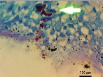
Among the formalin-ixed parain-embedded samples, acid-fast
bacilli were identiied (Figure 1) on 15/149 (10%) of the slides
stained with ZN. hese comprised 9/44 (20.4%) from patients with CD, 1/49 (2%) from patients with UC and 5/56 (8.9%) from patients with nIBD. he mean detection rate for acid-fast bacilli in the CD group was 10 times higher than in the UC group (P < 0.01); there were no statistically signiicant diferences between the CD and nIBD groups, or between the UC and nIBD
groups (Table 4). Among the fresh samples, acid-fast bacilli were
detected on 1/25 (4%) of the slides, from one patient with UC. Acid-fast bacilli were not identiied on slides from patients with CD or nIBD. Among the fresh samples, the diferences between
the groups were not statistically signiicant (Table 4).
Figure 1. Acid-fast bacilli in the intestinal mucosa of a patient with Crohn’s disease, with Ziehl-Neelsen staining.
Group Parain-embedded samples Fresh samples Total n acid-fast bacilli + n acid-fast bacilli +
CD 44 9 (20.4%)a 5 0 (0%)c 49 UC 49 1 (2%)b 8 1 (12.5%)c 57 nIBD 56 5 (8.9%)a,b 12 0 (0%)c 68
Total 149 15 25 1 174
Table 4. Relationship between presence of acid-fast bacilli and clinical groups of patients included in the study
Results followed by the same letters did not difer statistically according to Tukey’s test at 5% probability. Parain-embedded samples: F = 4.5879, P = 0.0117; fresh samples: F = 1.0686, P = 0.3607. CD = Crohn’s disease; UC = ulcerative colitis; nIBD = non-inlammatory bowel disease.
Group Parain-embedded samples Fresh samples Total n IS900PCR + n IS900PCR +
CD 44 1 (2.3%)a 5 1 (20%)b 49 UC 49 2 (4%)a 8 1 (12.5%)b 57 nIBD 56 2 (3.5%)a 12 1 (8.3%)b 68 Total 149 5 (3.3%) 25 3 (12%) 174
Results followed by the same letters did not difer statistically according to Tukey’s test at 5% probability. Parain-embedded samples: F = 0.1211, P = 0.8860; fresh samples: F = 0.2051, P = 0.8161. CD = Crohn’s disease; UC = ulcerative colitis; nIBD = non-inlammatory bowel disease; PCR = polymerase chain reaction.
Table 3. Relationship between IS900 PCR results and clinical groups of patients
CD UC nIBD Total
n 44 49 56 149
Age at time of biopsy,
mean (range) 40.5 (11-77) 37 (2-77) 44.5 (13-83) Female sex 24 (54.5%) 32 (65.3%) 34 (60.7%) 87
Male sex 20 (45.5%) 17 (34.7%) 22 (39.3%) 56 Table 2. Characteristics of patients with Crohn’s disease (CD), ulcerative colitis (UC) and non-inlammatory bowel disease (nIBD) (controls): formalin-ixed parain-embedded samples included in the study
CD UC nIBD Total
n 5 8 12 25
Age at time of biopsy,
mean (range) 26 (23-29) 49.2 (31-74) 49.5 (32-70) Female sex 3 (60%) 4 (50%) 4 (33%) 11
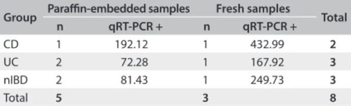
qRT-PCR
qRT-PCR was performed on eight samples that were positive according to PCR (ive formalin-ixed parain-embedded and three fresh samples). Among the formalin-ixed parain-embed-ded samples, we observed values of 192.12 copies/μl in the CD group, 72.28 copies/μl in the UC group and 81.43 copies/μl in the nIBD group. Among the fresh samples, we observed values of 432.99 copies/μl, 167.92 copies/μl and 249.73 copies/μl in the CD,
UC and nIBD groups, respectively (Table 5).
DISCUSSION
Studies that included children have been more likely to report
a positive result for MAP than those with an adult population.3
Dell’Isola et al.19 suggested that if the initial MAP infection
occurred during childhood, detection of this infection would be more likely in studies among children. However, in the present study, inclusion of children (formalin-ixed parain-embedded samples) did not inluence our results.
MAP culturing from human intestinal biopsy material is quite diicult, even under optimal conditions. However, several research groups have been able to grow MAP from tissues from patients with CD, using classical culturing methods, with success
rates ranging from 0-40%.13
MAP isolates from humans not only present the usual sam-ple decontamination requirements and have very slow growth, but also occur in spheroplasts (a cell wall-deicient form), which are extremely hard to isolate, recover and maintain in suicient
numbers for studies.20
Additionally, MAP isolation may also have been negatively afected by the freezing of the samples. It was not possible to work with fresh samples because of the distance between HC/UFMG and LDBAC. Freezing of samples before processing was there-fore necessitated and even though a cryoprotectant was used, the ideal would have been for the tissues to have been processed
immediately.6
Although MAP was not isolated in this study, it is important to highlight that MAP has only been recovered from human tis-sues with CD. his microorganism has never been isolated from
patients with UC or nIBD.13
he PCR results regarding MAP detection have been incon-clusive and conlicting. Reports have ranged from 0-100% detection in each group (CD, UC and nIBD) using a variety
of diferent methodologies and target sequences.3,4 Contrary to
other studies in this ield,5-7 MAP was not detected more
fre-quently among our patients with CD than among those with UC or nIBD. However, other research groups have shown that MAP detection in patients with CD occurs more frequently
than in patients with UC or nIBD.21,22
In this study, we observed the presence of MAP DNA in intestinal biopsy specimens from eight patients among the 174 samples tested. Previous studies have demonstrated that MAP is diicult to detect reliably and reproducibly by means of PCR
on DNA extracted from human tissues.6 hus, the use of
non-optimal procedures in processing the samples may result in
false-negative results. Bull et al.6 indicated several important steps
that should be followed in the DNA extraction method, such as processing of fresh tissues (i.e. which have never been frozen); mechanical disturbance to ensure access to MAP DNA; resus-pension of the DNA overnight at 4 °C; and nested PCR. In this study, we followed these recommendations wherever possible. However, as previously mentioned, it was necessary for us to freeze the samples, and this could be one reason for the low MAP detection rate in our results.
Few studies have shown higher frequencies of detection
of MAP or acid-fast bacilli in patients with CD.23-26 his may
be due to the diiculty in detecting MAP in tissue, given that MAP occurs in spheroplast form, and only small quantities of the microorganism are present in the tissues. For as long as these technical limitations remain unresolved, it will continue to be a challenge to demonstrate the presence of MAP in tissues of
patients with CD.13
Considering the small number of PCR-positive samples that were tested for qRT-PCR, we could not make any statistical infer-ences about the quantities of DNA found in the three groups (CD, UC and nIBD), although we observed that CD patients had higher bacterial loads in both formalin-ixed parain-embedded and fresh samples.
Some studies have supported the theory that MAP is present in most individuals. However, it is found in greater quantities in people with CD, thus suggesting that MAP is an organism that is ubiquitous in the environment and that it is an opportunistic
pathogen and not a primary cause of CD.13
In this study, the frequency of MAP detection by means of PCR did not difer between CD, UC and nIBD patients, and although the bacterial load was higher in patients with CD, it is not known whether higher bacterial loads cause higher inlam-mation scores or whether higher inlaminlam-mation scores cause higher bacterial loads. One possible explanation may be that the
Group Parain-embedded samples Fresh samples Total
n qRT-PCR + n qRT-PCR +
CD 1 192.12 1 432.99 2
UC 2 72.28 1 167.92 3
nIBD 2 81.43 1 249.73 3
Total 5 3 8
Table 5. Relationship between qRT-PCR results and clinical groups of patients
microorganism inds better conditions for replication in patients with CD than in patients with UC or nIBD. Immunological fac-tors relating to MAP, in susceptible patients, may allow MAP to replicate in larger quantities, thereby increasing the bacterial
load in patients with CD. his is corroborated by Lee et al.,27 who
showed that MAP gave rise to general colonization of the mucosa and suggested that there was simply an increase in mucosal sur-face colonization (dysbiosis) in CD cases that was unassociated with causality. Dysbiosis and reduced bacterial diversity of the intestinal microbiome in CD are likely to promote MAP growth
and detection.13
Further investigations into the etiological role of MAP in CD are needed. Analysis on the human intestinal microbiome in healthy and CD patients would establish whether MAP belongs to the nor-mal human microbiota or not. CD remains a debilitating disease that severely afects the quality of life of its suferers. Further research is required in order to deinitively answer the questions regarding the etiological nature of the disease.
CONCLUSION
MAP was present in all the groups of patients analyzed, although the greatest bacterial loads were observed in the CD group. his study supports the view that MAP is a ubiquitous organism that colo-nizes the mucosal surfaces of the gut, thereby resulting in increased detection in CD patients. his study does not provide evidence for any role played by MAP in Crohn’s disease; its role remains contro-versial and inconclusive. his is the irst report on the presence of MAP in biopsy specimens from the human gut in Brazil.
REFERENCES
1. Shanahan F. Crohn’s disease. Lancet.2002;359(9300):62-9.
2. Chiodini RJ, Van Kruiningen HJ, Merkal RS. Ruminant paratuberculosis (Johne’s disease): the current status and future prospects. Cornell Vet. 1984;74(3):218-62.
3. Abubakar I, Myhill D, Aliyu SH, Hunter PR. Detection of Mycobacterium avium subspecies paratuberculosis from patients with Crohn’s disease using nucleic acid-based techniques: a systematic review and meta-analysis. Inlamm Bowel Dis.2008;14(3):401-10.
4. Feller M, Huwiler K, Stephan R, et al. Mycobacterium aviumsubspecies paratuberculosis and Crohn’s disease: a systematic review and meta-analysis. Lancet Infect Dis. 2007;7(9):607-13.
5. Autschbach F, Eisold S, Hinz U, et al. High prevalence of Mycobacterium aviumsubspecies paratuberculosis IS900 DNA in gut tissues from individuals with Crohn’s disease. Gut.2005;54(7):944-9. 6. Bull TJ, McMinn EJ, Sidi-Boumedine K, et al. Detection and veriication
of Mycobacterium aviumsubsp. paratuberculosis in fresh ileocolonic mucosal biopsy specimens from individuals with and without Crohn’s disease. J Clin Microbiol.2003;41(7):2915-23.
7. Naser SA, Ghobrial G, Romero C, Valentine JF. Culture of Mycobacterium aviumsubspecies paratuberculosis from the blood of patients with Crohn’s disease. Lancet.2004;364(9439):1039-44. 8. Sechi LA, Scanu AM, Molicotti P, et al. Detection and Isolation of
Mycobacterium avium subspecies paratuberculosis from intestinal mucosal biopsies of patients with and without Crohn’s disease in Sardinia. Am J Gastroenterol.2005;100(7):1529-36.
9. Prantera C. Mycobacteria and Crohn’s disease: the endless story. Dig Liver Dis.2007;39(5):452-4.
10. Chamberlin W, Graham DY, Hulten K, et al. Review article: Mycobacterium avium subsp. paratuberculosis as one cause of Crohn’s disease. Aliment Pharmacol Ther.2001;15(3):337-46.
11. Chiodini RJ, Hermon-Taylor J. The thermal resistance of Mycobacterium paratuberculosis in raw milk under conditions simulating pasteurization. J Vet Diagn Invest.1993;5(4):629-31. 12. Sibartie S, Scully P, Keohane J, et al. Mycobacterium aviumsubsp.
Paratuberculosis (MAP) as a modifying factor in Crohn’s disease. Inlamm Bowel Dis.2010;16(2):296-304.
13. Chiodini RJ, Chamberlin WM, Sarosiek J, McCallum RW. Crohn’s disease and the mycobacterioses: a quarter century later. Causation or simple association? Crit Rev Microbiol.2012;38(1):52-93.
14. Momotani E, Romona NM, Yoshihara K, et al. Molecular pathogenesis of bovine paratuberculosis and human inlammatory bowel diseases. Vet Immunol Immunopathol.2012;148(1-2):55-68.
15. Pocock SJ. Clinical trials. A practical approach. New York: Wiley; 1988. 16. Sivakumar P, Tripathi BN, Singh N. Detection of Mycobacterium
aviumsubsp. paratuberculosis in intestinal and lymph node tissues of water bufaloes (Bubalus bubalis) by PCR and bacterial culture. Vet Microbiol.2005;108(3-4):263-70.
17. Rivero ER, Neves AC, Silva-Valenzuela MG, Sousa SO, Nunes FD. Simple salting-out method for DNA extraction from formalin-ixed, parain-embedded tissues. Pathol Res Pract. 2006;202(7):523-9. 18. Herthnek D, Englund S, Willemsen PT, Bölske G. Sensitive detection of
Mycobacterium aviumsubsp. paratuberculosis in bovine semen by real-time PCR. J Appl Microbiol.2006;100(5):1095-102.
19. Dell’Isola B, Poyart C, Goulet O, et al. Detection of Mycobacterium paratuberculosis by polymerase chain reaction in children with Crohn’s disease. J Infect Dis.1994;169(2):449-51.
20. Hines ME 2nd, Styer EL. Preliminary characterization of chemically generated Mycobacterium aviumsubsp. paratuberculosis cell wall deicient forms (spheroplasts). Vet Microbiol.2003;95(4):247-58. 21. Rath T, Roderfeld M, Blöcher S, et al. Presence of intestinal
Mycobacterium aviumsubspecies paratuberculosis(MAP) DNA is not associated with altered MMP expression in ulcerative colitis. BMC Gastroenterol.2011;11:34.
23. Ellingson JL, Cheville JC, Brees D, Miller JM, Cheville NF. Absence of Mycobacterium avium subspecies paratuberculosis components from Crohn’s disease intestinal biopsy tissues. Clin Med Res. 2003;1(3):217-26.
24. Jeyanathan M, Boutros-Tadros O, Radhi J, et al. Visualization of Mycobacterium aviumin Crohn’s tissue by oil-immersion microscopy. Microbes Infect. 2007;9(14-15):1567-73.
25. Romero C, Hamdi A, Valentine JF, Naser SA. Evaluation of surgical tissue from patients with Crohn’s disease for the presence of Mycobacterium aviumsubspecies paratuberculosis DNA by in situ hybridization and nested polymerase chain reaction. Inlamm Bowel Dis.2005;11(2):116-25.
26. Sechi LA, Mura M, Tanda F, et al. Identiication of Mycobacterium aviumsubsp. paratuberculosis in biopsy specimens from patients with Crohn’s disease identiied by in situ hybridization. J Clin Microbiol.2001;39(12):4514-7.
27. Lee A, Griiths TA, Parab RS, et al. Association of Mycobacterium aviumsubspecies paratuberculosis with Crohn Disease in pediatric patients. J Pediatr Gastroenterol Nutr.2011;52(2):170-4.
Acknowledgements: We would like to thank the staf of Instituto Alfa de Gastroenterologia (IAG), Hospital das Clínicas (HC), Universidade Federal de Minas Gerais (UFMG) for providing the bowel biopsies
Sources of funding: Isabel Azevedo Carvalho and Isis Freitas Espechit Braga are supported by Coordenação de Aperfeiçoamento de Pessoal de Nível Superior (CAPES) and Conselho Nacional de Desenvolvimento Cientíico e Tecnológico (CNPq); David Germano Gonçalves Schwarz, Pricila Aparecida Grasse Pietralonga, Fabrício Luciani Valente and João Paulo Machado are supported by CAPES; Abelardo Silva Júnior and Maria Aparecida Scatamburlo Moreira are supported by CNPq. This work was supported by FAPEMIG, Brazil (Grant no. APQ-00236-11)
Conlict of interest: None
Date of irst submission: January 14, 2014
Last received: August 21, 2014
Accepted: September 18, 2014
Address for correspondence: Maria Aparecida Scatamburlo Moreira Av. Peter Henry Rolfs, s/no
Campus Universitário — Viçosa (MG) — Brasil CEP 36570-900