Action of
Action of
Action of
Action of
Action of
tacrolimus
tacrolimus
tacrolimus in arginine induced experimental acute
tacrolimus
tacrolimus
in arginine induced experimental acute
in arginine induced experimental acute
in arginine induced experimental acute
in arginine induced experimental acute
pancreatitis
pancreatitis
pancreatitis
pancreatitis
pancreatitis
Ação do
Ação do
Ação do
Ação do
Ação do tacrolimus
tacrolimus
tacrolimus
tacrolimus
tacrolimus na pancreatite aguda experimental induzida pela arginina
na pancreatite aguda experimental induzida pela arginina
na pancreatite aguda experimental induzida pela arginina
na pancreatite aguda experimental induzida pela arginina
na pancreatite aguda experimental induzida pela arginina
MARLUS MOREIRA1; JORGE EDUARDO FOUTO MATIAS ACBC-PR2; CARLOS JOSÉ FRANCODE SOUZA, ACBC-PR3; JOÃO EDUARDO LEAL NICOLUZZI
TCBC-PR3; PEDRO ERNESTO CARON TCBC-PR3; JOÃO CARLOS DOMINGUES REPKA4
A B S T R A C T A B S T R A C T A B S T R A C T A B S T R A C T A B S T R A C T
Objective Objective Objective Objective
Objective: To determine whether tacrolimus administered to rats, in the presence of pancreatitis induced by L-Arginine, interferes with the serum levels of amylase and glucose and the histological pattern of the pancreatic parenchyma. MethodsMethodsMethodsMethods:Methods Forty Wistar rats were divided into four groups with 10 rats each: control group (C), tacrolimus group (T), pancreatitis group (P) and pancreatitis-tacrolimus group (PT). We evaluated serum levels of amylase, glucose, and tacrolimus and made histological assessments of the pancreas. Induction of pancreatitis was made by inoculation of L-Arginine at a dose of 500mg/100g body weight intraperitoneally, and tacrolimus treatment at a dose of 1ìg/kg subcutaneously for four days. ResultsResultsResultsResultsResults: Serum amylase was higher (p = 0.0000) in groups PT, P and T than in the control group. The PT group mean was higher (p = 0.0009) than in the T group, but did not differ (p = 0.6802) from the average of the P group. There was no difference between groups P and T (p = 0.2568). Neither in mean blood glucose between the groups (p = 0.4920); serum levels of tacrolimus were similar in PT and T groups (p = 0.7112). There were no histological changes in groups T and C and no hemorrhage in the pancreas of rats in groups P and PT. In group P, there was no edema in 30%, mild edema in 20% and in 50%, moderate; as for inflammatory infiltration, it was moderate in 80% and absent in 20%, and atrophy of the parenchyma was moderate in 60% and severe in 40%. In the PT group, there was edema, inflammatory infiltration or atrophy in the pancreas in all rats. ConclusionConclusionConclusionConclusionConclusion: treatment with Tacrolimus induced an increase in serum amylase in normal mice, but did not affect blood glucose or the histological pattern of the pancreatic parenchyma. In the presence of pancreatitis induced by L-Arginine tacrolimus induced edema, inflammatory infiltration and more severe atrophy in the pancreatic parenchyma.
Key words Key words Key words Key words
Key words: Immunosuppressants. Tacrolimus. Pancreatitis. Arginine. Amylase. Glucose. Disease / pathology. Animal experimentation. Rats.
Paper presented to the post-graduation program in Clinical Surgery, Federal University of Parana (UFPR), as a partial requirement for obtaining the Master’s degree.
1. Master’s Graduate, Post-Graduation Program in Clinical Surgery, Universidade Federal do Paraná - UFPR (Curitiba - PR-BR); 2. Assistant Professor, Department of Surgery, Universidade Federal do Paraná - UFPR (Curitiba - PR-BR); 3. Surgeon, General Surgery and Transplant Service, Hospital Angelina Caron (Campina Grande do Sul - PR - BR); 4. Coordinator, Education and Research, Hospital Angelina Caron (Campina Grande do Sul - PR - BR).
INTRODUCTION
INTRODUCTION
INTRODUCTION
INTRODUCTION
INTRODUCTION
T
he improved outcomes in solid organ transplantation, demonstrated by the increase in graft and patient survival, is due primarily to improvement in surgical techniques, progress in immunological techniques for selection of donors, effectiveness of solutions for organ preservation and a new immunosuppressive drugs1.Among these stands out tacrolimus, which has a macrolide structure originally isolated from culture of Streptomyces tsukubaensis2. Its immunosuppressive effect
is mediated by its binding to a cytosolic protein (FKBP12) responsible for its intracellular accumulation. The tacrolimus-FKBP12 complex binds specifically and competitively to calcineurin, with consequent calcium-dependent inhibition of transduction of cytotoxic T lymphocytes and activation
of B lymphocytes proliferation. It also inhibits the transcription of different genes of lymphokines, such as interferon-gamma, and expression of interleukin-2 receptors, thus leading to immunosuppression3.
The continuous use of immunosuppressive drugs require regular blood monitoring to perform individual adjustments and maintain blood levels stable enough to prevent rejection and, at the same, below the toxic threshold to avoid adverse effects4. The main adverse effects
observed with tacrolimus are tremors, headache, hypertension, nausea, diarrhea and renal dysfunction, which can be controlled by dose reducing5.
Little known, and still of hypothetical character, is the occurrence of acute pancreatitis (AP) associated with the chronic use of tacrolimus6-8. The AP is a disease caused
phenomenon, intraparenchymal activation of pancreatic enzymes, which induce a process of autodigestion manifested by edema, hemorrhage, and pancreatic or peripancreatic necrosis, accompanied by systemic repercussions, with involvement of multiple organs and systems and, in some cases, death9. However, its
pathophysiological mechanisms are not yet completely understood. It is known that excessive doses of basic amino acids, such as arginine, cause injury to the pancreas of rats10,11.
Due to the high protein metabolism in pancreatic acinar cells, it is likely that these cells are the first target of excess arginine, resulting in degeneration, atrophy or necrosis with, severe mitochondrial damage and reduction in cellular energy supply12. Experimental models of
pancreatitis have contributed to the understanding of cell biology and pathophysiology of this disease, but for the best interpretation of experimental results it should always be considered that the pathogenesis of the disease in humans may differ from the one of the animal model13.
Regarding the occurrence of acute pancreatitis with the use of tacrolimus there is no consensus7,8,14,15. Its
possible pancreatic toxicity is not considered a side effect of immunosuppressive treatment, but the incidence of these problems is high among patients who underwent transplantation of the organ. There is no background for this drug to be appointed as a trigger point of the process, since the target organ undergoes intense manipulation and ischemia during transplantation.
This study aims to determine whether tacrolimus administered to rats in the presence of pancreatitis induced by L-Arginine interferes in serum amylase and glucose and the histological pattern of the pancreatic parenchyma.
METHODS
METHODS
METHODS
METHODS
METHODS
Sample characterization Sample characterization Sample characterization Sample characterization Sample characterization
Were used 40 Wistar rats (Rattus norvegicus, Rodentia, Mammalia), not inbred, weighing 282.4 ± 12.6g, obtained from the vivarium of the Universidade Federal do
Paraná. The rats were kept in groups of five in polypropylene boxes suitable for the species, in an environment with controlled temperature and humidity, under cycles of light automatically set every 12 hours. They were fed diets specific to the species (Nuvilab, Nuvital®) and water ad libitum. Ethical Principles in Animal Experimentation of the Colégio Brasileiro de Experimentação Animal (COBEA) were followed16, and the Walker Nomina Anatomica17. The project
was approved by the Ethics Committee for Animal Research of the Hospital Angelina Caron, according to the protocol 023/08.
Experimental design Experimental design Experimental design Experimental design Experimental design
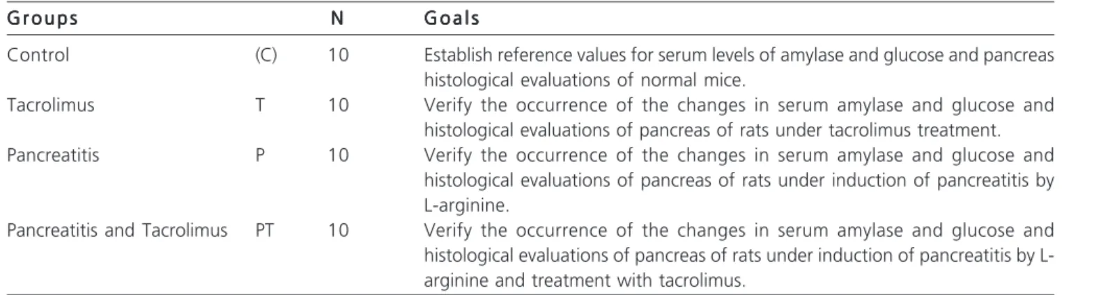
The sample was separated into four groups (Table 1).
Induction of pancreatitis by arginine Induction of pancreatitis by arginine Induction of pancreatitis by arginine Induction of pancreatitis by arginine Induction of pancreatitis by arginine Pancreatitis was induced by intraperitoneal inoculation of L-arginine solution (Merck 1.01542 Articleâ) with rats under inhalation sedation with halothane (Tanohalo®, Cristália). The 20% (w/v) solution was prepared in phosphate buffer pH 6.8 and inoculated at a dose of intarperitoneal 500mg/100g of weight for the groups P and PT. C and T groups received phosphate buffer pH 6.8, at a dose proportional to weight, the same route of inoculation of the other groups18,19,20.
Treatment by tacrolimus Treatment by tacrolimus Treatment by tacrolimus Treatment by tacrolimus Treatment by tacrolimus
We used the product Prograf® (Janssen-Cilag/ Fugisawa), in the form of 5mg/ml injectable tacrolimus. The dose used was the 1ìg/kg weight and the solution was prepared from dilutions in phosphate buffer pH 6.8. The rats in groups T and PT subcutaneously received this treatment for four days from the day of induction of pancreatitis and the rats of groups C and P were treated with phosphate buffer pH 6.8, at a dose proportional to weight, also subcutaneously.
Sample collection and laboratory analysis Sample collection and laboratory analysis Sample collection and laboratory analysis Sample collection and laboratory analysis Sample collection and laboratory analysis On the fourth day of evolution, the rats were anesthetized by inhalation with halothane in closed circuit
Table 1 Table 1 Table 1 Table 1
Table 1 - Groups of rts in the study. G r o u p s
G r o u p s G r o u p s G r o u p s
G r o u p s NNNNN G o a l sG o a l sG o a l sG o a l sG o a l s
Control (C) 10 Establish reference values for serum levels of amylase and glucose and pancreas histological evaluations of normal mice.
Tacrolimus T 10 Verify the occurrence of the changes in serum amylase and glucose and histological evaluations of pancreas of rats under tacrolimus treatment. Pancreatitis P 10 Verify the occurrence of the changes in serum amylase and glucose and
histological evaluations of pancreas of rats under induction of pancreatitis by L-arginine.
and subjected to transthoracic cardiac puncture. We collected volumes of blood sufficient to induce cardiac arrest. After separation of the serum samples were allocated for the measurement of tacrolimus, amylase and glucose. Once death of the mice was evidenced, we performed laparotomies and resections of the pancreas for histopathological evaluations.
Measurement of amylase and glucose Measurement of amylase and glucose Measurement of amylase and glucose Measurement of amylase and glucose Measurement of amylase and glucose We performed the measurements in an automated equipment - COBAS System - MIRA “S”, with specific reagents, following the manufacturer’s guidelines and included positive and negative standards21.
Dosing of tacrolimus Dosing of tacrolimus Dosing of tacrolimus Dosing of tacrolimus Dosing of tacrolimus
We used the IMx Tacrolimus II assay (Abbott csc 0800-11-90-99), which is based on microparticle immunoenzymatic methodology (MEIA). The results are expressed in ng/ml22.
Histopathological evaluations Histopathological evaluations Histopathological evaluations Histopathological evaluations Histopathological evaluations
The removed pancreas was fixed in formaldehyde 10% for at least 48 hours, and sections were made through the long axis of the gland, stained with hematoxylin-eosin and examined by light microscopy. All sections were examined without knowledge of the group and period. We considered the following histological patterns: edema, inflammatory infiltration, hemorrhage and parenchymal necrosis. The histologic pattern (Table 2) was defined according to the presence and predominance of microscopic changes19.,23.
Statistical analysis Statistical analysis Statistical analysis Statistical analysis Statistical analysis
We applied statistical methods of ANOVA and Student-T and adopted the significance level of p £ 0.05. During induction of pancreatitis in the arginine group there were two deaths, so the sample of this group had n = 8. In order to assess whether there were differences in serum amylase, glucose, and tacrolimus between groups C, T, P and PT, we tested the null hypothesis that the average level of amylase in the groups would be equal to the average
Group C versus the alternative hypothesis that these levels were different.
RESULTS
RESULTS
RESULTS
RESULTS
RESULTS
Measurement of amylase Measurement of amylase Measurement of amylase Measurement of amylase Measurement of amylase
As shown in figure 1, the amylase of the rats was significantly higher (p = 0.0000) in the PT group (2788.1 ± 531.1 U/L), T (2009.7 ± 310.8 U/L) and P (2577.5 ± 1501.5 U/L) compared to the control group (1000.0 ± 87.14 U/L). The group PT mean was significantly higher (p = 0.0009) than the average of the group T, but did not differ (p = 0.6802) from the average of the group P. Among the groups P and T there was no significant difference (p = 0.2568).
Levels of glucose Levels of glucose Levels of glucose Levels of glucose Levels of glucose
Blood glucose was similar in all groups (p = 0.4920) and, when compared to the control group, the groups P, PT and T also displayed no significant difference, as shown in figure 2.
Levels of tacrolimus Levels of tacrolimus Levels of tacrolimus Levels of tacrolimus Levels of tacrolimus
Serum levels of tacrolimus in rats was similar in PT and T groups (p = 0.7112), as shown in figure 3.
Figure 1 -Figure 1 -Figure 1
-Figure 1 -Figure 1 - Means and standard deviations of the amylase measurements (units/liter).
Table 2 Table 2Table 2 Table 2
Table 2 - Histological Criteria for the evaluation of pancreatitis.
Histological Findings Histological FindingsHistological Findings Histological Findings
Histological Findings C l a s s i f i c a t i o nC l a s s i f i c a t i o nC l a s s i f i c a t i o nC l a s s i f i c a t i o nC l a s s i f i c a t i o n
Atrophy Absent Mild Moderate Severe
Edema Absent Mild Moderate Severe
Inflammatory infiltration Absent Mild Moderate Severe
Inflammatory Population Absent Mild Moderate Severe
Neutrophils Macrophages Lymphocytes Mista
Haemorrhage Absent Mild Moderate Severe
Histological examinations Histological examinations Histological examinations Histological examinations Histological examinations
No histological changes were observed in the tacrolimus and control groups and no cases of hemorrhage in the pancreatic parenchyma of rats in groups P and PT. As shown in figure 4, in the pancreatitis group there was no edema in 30%, and edema occurred in 20% as mild and 50% moderately; inflammatory infiltration occurred moderately in 80% and did not occur in 20% of the animals; atrophy of the parenchyma was moderate in 60% and severe in 40%. In the PT group, there was edema, inflammatory infiltration and atrophy of pancreatic parenchyma in all rats, with moderate edema in 60% and severe in 40%; inflammatory infiltrate occurred in 90% moderately and in 10%, severely; and atrophy of pancreatic parenchyma occurred moderately in 30% and severely in 70%.
DISCUSSION
DISCUSSION
DISCUSSION
DISCUSSION
DISCUSSION
The experimental model of this study was based on previous experiments that have described a new form of acute necrotizing pancreatitis induced in rats by a single intraperitoneal injection of arginine at a concentration of 500mg/100g body weight18,19. During the pilot study of this
work it was observed that the inoculation of the total dose given by the authors caused immediate death of all animals and at necropsy a widespread occurrence of mesenteric thrombosis was observed. For this reason, the proposed dose was split in two 250mg/100g interval of thirty minutes. This non-invasive model proved to be easily reproduced and affordable when compared to invasive experimental models of acute pancreatitis20. The
experi-mental model used herein displays, as its basic features, the elevation of serum amylase in the first 24 hours, with return to normal until the seventh day; it does not induce changes in glucose levels and the histological changes of the pancreatic parenchyma (edema and necrosis) are more severe in the period between 72 and 96 hours of onset23.
As seen in figure 2, injection of L-arginine did not alter the endocrine function of the pancreas because the serum glucose levels were maintained in groups P, PT and T levels similar to those of group C. For this reason, in this study we chose the collecting samples within 96 hours of onset (fourth day) to coincide with the timing of more severe pancreatic histopathological changes, allowing time for the action of tacrolimus, which was administered daily during the four days of evaluation.
As shown in figure 3, groups T and PT – treated with tacrolimus –had similar serum levels, which endorses the procedure and demonstrates the bioavailability of the drug. It is known that the increase of serum amylase does not correlate with the severity of acute pancreatitis and also with the histological changes observed in the pancreas23,24. In this study, as shown in figure 1, serum
amylase levels remained normal in group C (1000 ± 87.1
U/L) but rose significantly in groups P (2577.5 ± 1501.5 U/ L) and PT (2788.7 ± 531.1 U/L), injected with L-arginine (p = 0.0000). Noteworthy is the amylase average observed in rats of group T, receiving treatment with tracrolimus (2009.7 ± 310.8 U/L), which was also significantly higher than group C (p = 0.0000). Although the efficacy of tacrolimus in the suppression of chronic pancreatitis in Wistar rats, variant Bonn/Kobori, was documented25, these results cannot be
compared to the present study because they were obtained
Figure 2 Figure 2 Figure 2 Figure 2
-Figure 2 - Means and standard deviations of blood glucose (mg/ dl).
Figure 3 Figure 3 Figure 3 Figure 3
Figure 3 - Means and standard deviations of the doses of tacrolimus (ng/ml).
Figure 4 Figure 4 Figure 4 Figure 4
in a distinct animal model, whose autoimmune mechanism is of chronic evolution and inducer of fibrosis of the pancreatic parenchyma. In this pathophysiological context, an immunosuppressive drug such as tacrolimus has effects on the autoimmune mechanism, which explains those results. Still on the results of measurements of amylase in the present study, it appears that tracrolimus treatment for four days at a dose of 1 mg/kg effectively induced an increase in serum enzyme, showing, however, a protective effect against the action of arginine because the results of the T groups (2009.7 ± 310.8 U/L) are significantly smaller than the PT group (2577.5 ± 1501.5 U/L). The justification for these results can be based on the fact that tacrolimus at therapeutic doses may increase the secretion of pancreatic enzymes, with deteriorating effect on the organ, culminating in acute pancreatitis when the pancreas is stimulated24,25.
It is known that the pancreas is considered the tissue that has the highest level of protein synthesis. There are reasons to believe that damaged, but still viable, acinar cells may cause greater and longer lasting elevation of serum amylase than necrotic cells, unable to maintain production of enzymes26,27.
From the results obtained in the histological analysis of this study, it can be argued that tracrolimus, when inoculated into normal mice, did not alter the pancreatic histological architecture (group T), these results being consistent with another study28.
There was no bleeding from the pancreatic parenchyma of rats in groups Pancreatitis and Pancreatitis-Tacrolimus, however, as the other criteria of histological assessment, there were differences between these groups, as in the PT group, edema, inflammatory infiltration and atrophy of pancreatic parenchyma was more severe than in the P group, as shown in figure 4. Few reports observed pancreatic histological changes caused by tacrolimus and described the occurrence of nuclear pyknosis and cytoplasmic vacuolation as a cause of necrotic degeneration of the acinar cells28.
Treatment by tacrolimus induced a significant increase in serum amylase in normal mice and did not alter blood glucose levels and histological pattern of the pancreatic parenchyma. In the presence of pancreatitis induced by L-Arginine, tacrolimus induced edema, inflammatory infiltration and atrophy more severe in the pancreatic parenchyma.
R E S U M O R E S U M O R E S U M O R E S U M O R E S U M O
Objetivo: Objetivo: Objetivo: Objetivo:
Objetivo: verificar se o tacrolimus administrado em ratos, em vigência de pancreatite induzida pela L-Arginina, interfere nos níveis séricos da amilase e glicose e no padrão histológico do parênquima pancreático. Métodos:Métodos:Métodos:Métodos:Métodos: quarenta ratos Wistar foram distribuídos em quatro grupos com 10 ratos cada. Grupo controle (C), grupo tacrolimus (T), grupo pancreatite (P) e grupo pancreatite-tacrolimus (PT). Foram avaliados os níveis séricos de amilase, glicose e tacrolimus e feitas avaliações histológicas do pâncreas, A indução de pancreatite foi feita pela inoculação de L-Arginina na dose de 500mg/100g de peso corporal por via intraperitoneal e o tratamento com tacrolimus na dose de 1ìg/kg por via subcutânea durante quatro dias. Resultados:Resultados:Resultados:Resultados:Resultados: a amilasemia estava mais elevada (p=0,0000) nos grupos PT, T e P do que no grupo controle. A média do grupo PT foi maior (p=0,0009) que a do grupo T, mas não diferiu (p=0,6802) da média do grupo P. Entre os grupos P e T não houve diferença (p=0,2568). Não houve diferença nas médias de glicemia entre os grupos (p=0,4920) e os níveis séricos de tacrolimus foram similares nos grupos PT e T (p=0,7112). Não ocorreram alterações histológicas nos grupos T e C e não ocorreu hemorragia no pâncreas dos ratos dos grupos P e PT. No grupo P, em 30% não se observou edema, em 20% observou-se a forma leve e em 50%, a moderada; quanto à infiltração inflamatória, em 80% moderada e em 20% não ocorreu, e a atrofia do parênquima foi de 60% moderada e 40% acentuada. No grupo PT, houve ocorrência de edema, infiltração inflamatória e atrofia do pâncreas em todos os ratos. Conclusão:Conclusão:Conclusão:Conclusão:Conclusão: o tratamento pelo tacrolimus induziu aumento nos níveis séricos de amilase em ratos normais, não alterou a glicemia nem o padrão histológico do parênquima pancreático. Na vigência de pancreatite induzida pela L-Arginina o tacrolimus induziu edema, infiltração inflamatória e atrofia com maior gravidade no parênquima pancreático.
Descritores Descritores Descritores Descritores
Descritores: Imunossupressores. Tacrolimo. Pancreatite. Arginina. Amilase. Glicemia. Doença/patologia. Experimentação animal. Ratos.
REFERENCES
REFERENCES
REFERENCES
REFERENCES
REFERENCES
1. Nicoluzzi J, Silveira F, Porto F, Macri M. One hundred pancreas transplants performed in a Brazilian institution. Transplant Proc 2009; 41(10):4270-3.
2. Kino T, Hatanaka H, Miyata S, Inamura N, Nishiyama M, Yajima T, et al. FK-506, a novel immunosuppressant isolated from a Streptomyces. II. Immunosuppressive effect of FK-506 in vitro. J Antibiot 1987; 40(9):1256-65.
3. Jordan ML, Shapiro R, Fung J, Tzakis A, Todo S, Kusne S, et al. Inicial studies of FK506 in renal transplantation. Cleve Clin J Med 1991; 58(5):444-6.
4. Macleod AM, Thomson AW. FK 506: an imunosupressant for the 1990s ? Lancet 1991; 337(8732):25-7.
5. Verleden GM, Besse T, Maes B. Successful conversion from cyclosporine to tacrolimus for gastric motor dysfunction in a lung transplant recipient. Transplantation 2002; 73(12):1974-6. 6. Nieto Y, Russ P, Everson G, Bearman SI, Cagnoni PJ, Jones RB, et
al. Acute pancreatitis during immunosuppression with tacrolimus following an allogeneic umbilical cord blood transplantation. Bone Marrow Transplant 2000; 26(1):109-11.
8. Ogunseinde BA, Wimmers E, Washington B, Iyob M, Cropper T, Callender CO. A case of tacrolimus (FK506)-induced pancreatitis and fatality 2 years postcadaveric renal transplant. Transplantation 2003; 76(2):448.
9. Ohara K, Billington R, James RW, Dean GA, Nishiyama M, Noguchi H. Toxicologic evaluation of FK 506. Transplant Proc 1990; 22(1):83-6.
10. Hegyi P, Rakonczay Z Jr, Sári R, Góg C, Lonovics J, Takács T, et al. L-arginine-induced experimental pancreatitis. World J Gastroenterol 2004; 10(14):2003-9.
11. Kishino Y, Kawamura S. Pancreatic damage induced by injecting a large dose of arginine. Virchows Arch B Cell Pathol Incl Mol Pathol 1984; 47(2):147-55.
12. Banks PA. Practice guidelines in acute pancreatitis. Am J Gastroenterol 1997; 92(3):377-86.
13. Husain SZ, Grant WM, Gorelick FS, Nathanson MH, Shah AU. Caerulein-induced intracellular pancreatic zymogen activation is dependent on calcineurin. Am J Physiol Gastrointest Liver Physiol 2007; 292(6):G1594-9.
14. Peters DH, Fitton A, Plosker GL, Faulds D. Tacrolimus. A review of its pharmacology, and therapeutic potential in hepatic and renal transplantation. Drugs 1993; 46(4):746-94.
15. Loss M, Winkler M, Schneider A, Brinkmann C, Manns M, Ringe B, Pichlmayr R. Influence of long-term cyclosporine or FK 506 therapy on glucose and lipid metabolism in stable liver graft recipients. Transplant Proc 1995; 27(1):1136-9.
16. Sociedade Brasileira de Ciência de Animais de Laboratório (SBCAL) – C OBEA. Bem-estar em Animais de Laboratório [online]. Acessado em 08 de fevereiro de 2011. Disponível em: h t t p : / / w w w . c o b e a . o r g . b r / index.php?option=com_content&view=article&id=64%3Abem-estar-em-animais-de-laboratorio&catid=44&Itemid=69. 17. Walker WF, Homberger DG. Digestive and Respiratory Systems.
In: Walker WF, Homberger DG. Anatomy and Dissection of the Rat. 3a ed. New York: WH Freeman; 1997; p.37-48.
18. Mizunuma T, Kawamura S, Kishino Y. Effect of injecting excess arginine on rat pancreas. J Nutr 1984; 114(3):467-71.
19. Tani S, Itoh H, Okabayashi Y, Nakamura T, Fujii M, Fujisawa T, et al. New model of acute necrotizing pancreatitis induced by excessive doses of arginine in rats. Dig Dis Sci 1990; 35(3):367-74. 20. Banerjee AK, Galloway SW, Kingsnorth AN. Experimental models
of acute pancreatitis. Br J Surg 1994; 81(8):1096-103.
21. Markin RS. Automação do laboratório clínico. In: Henry JB. Diag-nósticos clínicos e tratamentos por métodos laboratoriais. 20a ed.
São Paulo: Manole. 2008. p. 91-123.
22. Winkler M, Wonigeit K, Undre N, Ringe B, Oldhafer K, Christians U, et al. Comparison of plasma vs whole blood as matrix for FK 506 drug level monitoring. Transplant Proc 1995; 27(1):822-5. 23. Ramos Jr O, Leitão OR, Repka JCD, Barros SGS. Pancreatite
agu-da experimental induziagu-da pela L-arginina: avaliação histológica e bioquímica. Arq Gastroenterol 2005; 42(1):55-9.
24. Dubick MA, Mar G, Mayer AD, Majumdar AP, McMahon MJ, Geokas MC. Digestive enzymes and protease inhibitors in plasma from patients with acute pancreatitis. Pancreas 1987; 2(2):187-94.
25. Yamada T, Hashimoto T, Sogawa M, Kobayashi S, Kaneda K, Nakamura S, et al. Role of T cells in development of chronic pancreatitis in male Wistar Bonn/Kobori rats: effects of tacrolimus. Am J Physiol Gastrointest Liver Physiol 2001; 281(6):G1397-404. 26. Ito T, Kimura T, Furukawa M, Yamaguchi H, Goto M, Nakano I, et
al. Protective effects of gabexate mesilate on acute pancreatitis induced by tacrolimus (FK-506) in rats in wich the pancreas was stimulated by caerulein. J Gastroenterol 1994; 29(3):305-13. 27. Gullick HD. Relation of the magnitude of blood enzyme elevation
to severity of exocrine pancreatic disease. Am J Dig Dis1973; 18(5):375-83.
28. Echigo Y, Inoue K, Kogire M, Doi R, Higashide S, Sumi S, et al. Effects of cyclosporine and tacrolimus (FK 506) on acute pancreatitis in mice. Arch Surg 1995; 130(1):64-8.
Received: 18/07/2010
Accepted for publication: 21/09/2010 Conflict of interest: none
Source of funding: none
How to cite this article: How to cite this article: How to cite this article: How to cite this article: How to cite this article:
Moreira M, Matias JEF, Souza CJF, Nicoluzzi JEL, Caron PE, Repka JCD. Action of tacrolimus in arginine induced experimental acute pancreatitis . Rev Col Bras Cir. [periódico na Internet] 2011; 38(4). Disponível em URL: http://www.scielo.br/rcbc