633
Communication/Comunicação
1. Department of Morphology, Stomatology and Physiology, Ribeirão Preto Dental School, University of São Paulo, Ribeirão Preto, SP. 2. Division of Dermatology, Department of Internal Medicine, Ribeirão Preto Medical School, University of São Ribeirão Preto, SP.
Address to: Dra. Ana Carolina Fragoso Mota. Depto Morfologia, Estomatologia e Fisiologia/FORP/USP. Av. do Café s/n, 14040-904 Ribeirão Preto, SP, Brasil. Phone/Fax: 55 16 3633-0236
e-mail: [email protected]
Received in 12/08/2010
Accepted in 26/10/2010
Revista da Sociedade Brasileira de Medicina Tropical 44(5):633-635, set-out, 2011
Could leprosy reaction episodes be exacerbated by oral infections?
Episódios reacionais da hanseníase podem ser exacerbados por infecções orais?
Ana Carolina Fragoso Mota
1, Renata Bazan Furini
2, João Carlos Lopes Simão
2, Mariana Bellini Vieira
2,
Maria Aparecida Nunes Ferreira
2, Marilena Chinali Komesu
1and Norma Tiraboschi Foss
2ABSTACT
Introduction: his study evaluated whether leprosy reactions could be
associated with oral infection. Methods: Leprosy patients (n = 38) with (group I) and without (group II) oral infections were selected. Reactions were identiied from the clinical and histopathological features associated with serum C-reactive protein (CRP) and10kDa interferon-gamma-induced protein (IP-10) levels, determined before and ater elimination of the foci of infection. Results: group I presented more reactions than group II did, and improvement of the reactions ater dental treatment. Serum CRP and IP-10 did not difer before and ater the dental treatment, but difered between the groups. Conclusions: Oral infection could be an exacerbating factor in leprosy reactions.
Keywords: Leprosy reaction episodes. Oral infection. Inflammatory
markers.
RESUMO
Introdução: Este estudo avaliou se as reações hansênicas podem estar
associadas a infecções orais. Métodos: Pacientes com hanseníase (n=38) com (grupo I) e sem (grupo II) infecções orais foram selecionados. As reações foram identiicadas pelas características clínicas, histopatológicas, associadas a proteína-C-reativa (PCR) e proteína indutora de interferon-gamma de 10kDa (IP-10) séricos determinados antes e após a eliminação dos focos de infecção. Resultados: grupo I apresentou mais reações que o grupo II, e melhora das reações após o tratamento odontológico. PCR e IP-10 séricos não diferiram antes e após o tratamento odontológico, entretanto diferiram entre os grupos. Conclusões: As infecções orais podem ser exacerbadores das reações hansênicas.
Palavras-chaves: Episódios reacionais da hanseníase. Infecções orais.
Marcadores inlamatórios.
Reactional episodes are a serious problem during the course of leprosy since they may be responsible for much of the permanent nerve damage, thus leading to disability and deformities1. hese episodes represent an exacerbation of the inlammatory process that can occur before, during or ater leprosy treatment [multidrug therapy/World Health Organization (MDT - WHO)]2,3. here are two well recognized main types of reaction: reversal reaction (RR) and erythema nodosum leprosum (ENL). Reversal reactions may be due to an increase in the cell-mediated response to Mycobacterium
leprae characterized by a h1 response. Erythema nodosum leprosum
is a systemic inlammatory process characterized by intralesional neutrophilic iniltration and by a h2 response3,4. Since both types of reaction are accompanied by increased release of the inlammatory markers5-7, it is reasonable to consider the possibility that these episodes may be associated with an infectious process, such as dental abscess or periodontal diseases. hese may induce overstimulation of the host immune system through the release of numerous inlammatory markers, including cytokines, acute-phase proteins and chemokines8-10. he present study aimed to determine whether the presence of leprosy reaction episodes might be associated with dental and periodontal infection, and to determine the serum C-reactive protein (CRP)and IP-10 levels before and ater the elimination of oral infections in leprosy patients.
We selected 38 leprosy patients (29 men and 9 women; mean age ± SD: 43.92 ± 2.12 years; range 18-81 years) with and without oral infection (OI) before, during or ater speciic leprosy treatment. hese patients were divided into two groups: group I consisted of leprosy patients presenting some oral infections, and group II consisted of leprosy patients without oral infections. he diagnosis of leprosy was made based on clinical and histopathological indings, and on the Ridley & Jopling classiication11, bacilloscopy, biopsy and determination of the presence of antibodies to phenolic glycolipid-1 (anti-PgL-1). he oral infections considered were: periodontal diseases (PD), irreversible pulpitis (IP), pulpal necrosis (PN) and inlammatory periapical lesions (IPL). Subjects were excluded if they presented a coexisting local or systemic infection or diabetes mellitus, or if they had received antimicrobial treatment for any infections over the previous six months. he trial was approved by the local Ethics Commitee and all subjects gave writen informed consent to participate.
634
Mota ACF et al - Leprosy reaction episodes and oral infections
by each patient between the two data collection times. Mean CRP and serum IP-10 were calculated for each patient, as well as for each group. he results from the two groups were compared using the Mann-Whitney test, and baseline values (before) were compared with those obtained seven days later, by means of the Wilcoxon rank-sum test with the aid of the graphPad Prism sotware (San Diego, CA, USA). Signiicance was set at p < 0.05.
group I consisted of 19 leprosy patients (13 men and 6 women; mean age 45.05 ± 6.3 years; range 18–72 years) presenting some oral infections: seven patients presented dental diseases (IP, PN and IPL), three presented PD and nine presented a combination of dental diseases and PD. In this group, nine patients presented lepromatous leprosy (LL), ive were borderline lepromatous (BL), three were borderline borderline (BB) and two were borderline tuberculoid (BT). group II consisted of 19 patients (16 men and 3 women; mean age 42.8 ± 13.4 years; range 21–81 years) without oral infections. In this group, four patients presented LL, one BL, one BB, nine BT and four, the tuberculoid (T) form. Fiteen (78.9%) of the 19 patients in group I presented ENL at the time of diagnosis, three (15.8%) had reversal reactions (two BT and one BL patient) and only one (5.3%) patient did not present reactional episodes. Ater the dental and/or periodontal disease had been treated, 68.4% (13/19) of group I patients presented a clinical improvement in reactional episodes, as determined by decreases in skin lesions and symptoms, whereas no clinical change was detected in group II.
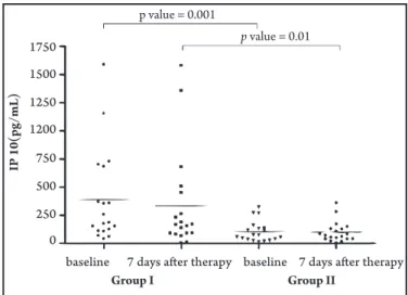
C-reactive proteinand IP-10 levels did not difer signiicantly between before and ater dental treatment in group I, even though they tended to become reduced after dental and periodontal treatment. No diferences were observed between the two data collection times regarding clinical findings or any monitored mediators in group II. Serum CRP levels (at baseline) and IP-10 levels (at baseline and seven days ater OI control) were signiicantly higher in group I than in group II (Figures 1 and 2).
This study attempted to clarify whether chronic OI could represent a maintenance factor in leprosy reaction episodes. A relationship between focal infection and dermatoses such as psoriasis12 and atopic dermatitis13 has been reported, but none concerning reactional episodes. It was observed that ENL was more
p value = 0.006
baseline 7 days after therapy baseline 7 days after therapy
2.0
1.5
1.0
0.5
0.0
C
R
P (
m
g/dL
)
Group I Group II
FIGURE 1 -Serum C-reactive protein levels of leprosy patients presenting chronic oral infections (Group I) and of leprosy patients without oral infection (Group II) before and ater chronic oral infection therapy.
1750
1500
1250
1200
750
500
250
0
baseline 7 days after therapy baseline 7 days after therapy Group I Group II
p value = 0.001
p value = 0.01
IP 10(
pg/mL
)
FIGURE 2 -Serum IP-10 levels of leprosy patients presenting chronic oral infections (Group I) and of leprosy patients without oral infection (Group II) before and ater oral infection therapy.
frequent in patients with OI (78.9%) than in patients with absence of OI (15.2%). his may have been due to the fact that the elevation in inlammatory marker expression by cells in the presence of OI can cause a spillover of these markers into the circulation, where they can act as a maintenance factor in leprosy reactions, as described for other diseases7,8,12,13.
After OI treatment, there was an improvement of clinical symptoms in 68.4% (13/19) of the patients in group I. In addition, there was an improvement in all of the periodontal clinical parameters monitored. However, no improvement was detected in peripheral serum levels of CRP or IP-10. he period of seven days ater OI treatment was probably not suicient to determine whether there were any decreases in serum CRP and IP-10 levels, although there was a tendency towards a reduction (Figures 1 and 2).
We found a signiicantly higher CRP level in patients with OI than in patients without these infections at baseline (p= 0.006). No statistically signiicant diference in serum CRP levels was observed between the two groups ater OI therapy. We believe that CRP may be a diagnostic biomarker of interest, although the signiicance of this inding should be considered with caution since CRP is a general inlammatory marker.
Similarly to CRP, we found higher levels of IP-10 in patients with OI than in patients without these infections (p = 0.001). his may be related to the activity of IP-10, which atracts h1 cells as a delayed hypersensitivity response14. Furthermore, IP-10 expression induced by IFNγ has been demonstrated in the skin of a leprosy patient 24 hours ater administration of the classic puriied protein derivative of tuberculin10. hus, during the inlammatory reaction of the ENL, the increases in serum CRP and IP-10 may be associated with higher cell activity and with some bacterial destruction, as determined by peripheral inlammatory manifestations.
635
he authors declare that there are no conlict of interest.CONFLICT OF INTEREST
REFERENCES FINANCIAL SUPPORT ACKNOWLEDGMENTS
Revista da Sociedade Brasileira de Medicina Tropical 44(5):633-635, set-out, 2011
We thankMr Mario Ignácio Neto for assistance with sample analysis.
National Council for Scientiic and Technological Development (CNPq) (grant 154806/2006-4), São Paulo State Foundation against Leprosy (grant 110)and the Teaching, Research and Assistance Support Foundation of HCFMRP-USP (FAEPA).
1. Jopling WH. Classiication of reaction in leprosy. Leprosy Rev 1970; 41:62-63. 2. Seghal VN, Sharma V. Reactions in leprosy - a prospective study of clinical,
bacteriological, immunological and histopathological parameters in thirty-ive Indians. J Dermatol 1998; 15:412-419.
3. Rea TH, Modlin RL. Immunopathology of leprosy skin lesions. Semin Dermatol 1991; 10: 188-193.
4. Cuevas J, Rodríguez-Peralto JL, Carrillo R, Contreras F. Erythema nodosum leprosum: reactional leprosy. Sem Cutan Med Surg 2007; 26:126-130. 5. Foss NT, Oliveira EB, Silva CL. Correlation between TNF production, increase
of plasma C-reactive protein level and suppression of T lymphocyte response to concanavalin A during erythema nodosum leprosum. Int J Lepr Other Mycobact Dis 1993; 61:218-226.
6. Silva EA, Iyer A, Ura S, Lauris JR, Naafs B, Das PK, et al. Utility of measuring serum levels of anti-PgL-1 antibody, neopterin and C-reactive protein in monitoring leprosy patients during multi-drug treatment and reactions. Trop Med Int Health 2007; 12:1450-1458.
7. Jefcoat MK, geurs NC, Reddy MS, Cliver SP, goldenberg RL, Hauth JC. Periodontal infection and preterm birth: results of a prospective study. J Am Dent Assoc 2001; 132:875-880.
8. Rodrigues DC, Taba MJ, Novaes AB, Souza SL, grisi MF. Efect of non-surgical periodontal therapy on glycemic control in patients with type 2 diabetes mellitus. J Periodontol 2003; 74:1361-1367.
9. Mota AC, Furini RB, Simão JC, Ferreira MA, Komesu MC, Foss NT. he recurrence of leprosy reactional episodes could be associated with oral chronic infections and expression of serum IL-1, TNF-α, IL-6, IFN-γ and IL-10. Braz Dent J 2010; 21:158-164.
10. Kaplan g, Luster AD, Hancock g, Cohn ZA. he expression of a γ interferon-induced protein (IP-10) in delayed immune responses in human skin. J Exp Med 1987; 166:1098-1108.
11. Ridley DS, Jopling WH. Classiication of leprosy according to immunity: a ive-group system. Int J Lepr Other Mycobact Dis 1966; 34:255-273.
12. Mizutani H, Ohmoto Y, Mizutani T, Murata M, Schimizu M. Role of increased production of monocytes TNF-alpha, IL-1 beta and IL-6 in psoriasis: relation to focal infection, disease activity and responses to treatments. J Dermatol Sci 1997; 14:145-153.
13. Igawa K, Nishioka K, Yokozeki H. Odontogenic focal infection could be partly involved in the pathogenesis of atopic dermatitis as exacerbating factor. Int J Dermatol 2007; 46:376-379.