Thoracic damage control surgery
Cirurgia de controle de danos torácico
RobeRto Gonçalves, tCbC-sP1; RobeRto saad JR, tCbC-sP1.
INTRODUCTION
T
he damage control strategy emerged in the Allied navy campaign during World War II. It was observed that in ships severely hit during the fighting, there were minor casualties and shipwrecks where efforts focused on extinguishing fires and keeping the boat running, retreating while covered by other friendly fleet ships, rather than continuing to fight. Then, in friendly territory, on construction sites, far from hostile areas, definitive repairs were held.The concept in the field of trauma surgery emerged in the nineties, for patients in near exhaustion of their physiological reserves, with manifestation of hy-pothermia, metabolic acidosis and coagulopathy (“dead-ly triad”). The adopted strategy consisted in making the minimum necessary (bleeding and contamination con-trol) in the shortest time, the patients being referred to the intensive care unit to be heated and stabilized, and only then, have their injuries definitely repaired in the operating environment. There were thus three defined stages through which the survivors would pass before recovery, accompanied by a multidisciplinary team, al-ways led by surgeon1,2.
Shortly after the first Damage Control publica-tions, arose the concept of the so-called phase 0, where-in even before startwhere-ing the damage control operation (Phase 1), the surgeon should have in mind which pa-tients would be candidates for this type of therapy, even
in the pre hospital setting and emergency room. That is, aim to “control the damage” before the lethal triad en-sues and when the chances of survival are very low. The so shortened surgery, without definitive correction of in-juries, implies an increase in morbidity in exchange for decreased mortality3.
In thoracic trauma, most patients are treated by simple measures, such as supplementation with oxygen therapy and pleural drainage as effective and definitive measures. However, in a fraction of patients, this strategy can be employed. Some patients evolve with continuous bleeding from the drain with maintained shock despite resuscitation, with air leak impairing ventilation or with vascular or esophageal injury, which are classical indications for intervention. These situations may be associated with injuries to other body compartments, adding in the consumption of the traumatized physiological reserves. In these individuals, or when the surgical time and bleeding extend, the surgeon must know thoracic damage control surgery.
SPECIFIC THORACIC INJURIES
(temporary and final control)
HEART
Most patients who have cardiac injuries die at the trauma scene. Of those who live to arrive at the emergency room, penetrating trauma is the most common and often the patient is unstable, a thoracotomy being
1 - Faculty of Medical Sciences of the São Paulo Holy Home, Department of Surgery - São Paulo - SP - Brazil
The damage control surgery came up with the philosophy of applying essential maneuvers to control bleeding and abdominal contamina-tion in trauma patients who are within the limits of their physiological reserves. This concept was extended to thoracic injuries, where rela-tively simple maneuvers can shorten operative time of in extremis patients. This article aims to revise the various damage control techniques in thoracic organs that must be known to the surgeon engaged in emergency care.
Keywords: Multiple Trauma. Thoracic Injuries. Emergency Medicine.
required already in the emergency room. Despite the high mortality rate associated with this procedure, due to the extreme patient’s conditions, this type of approach has indications and set goals. The emergency thoracotomy may represent the biggest paradigm of damage control in surgery, as it involves aggressive, fast and often temporary control of lesions that are consuming the patient’s physiological reserves, in an inhospitable environment (emergency room). Then the patient is sent to the operating room, and finally, to the most appropriate environment, the ICU, for compensation of acidosis and coagulopathy, and where he/she will be monitored, heated and reanimated.
The objectives of an emergency thoracotomy in-clude: cardiac tamponade relief that is causing diastolic restriction, control of heart wound or exsanguinantion due to vascular injury, descending aortic clamping to increase coronary and brain blood flow, internal cardiac compressions and occlusion of pulmonary hilum to re-duce possibility of air embolism and decreased bleeding in severe lung injury4.
In ventricular wounds, the temporary initial con-trol should be performed by digital occlusion or introduc-tion of a Foley catheter through the wound and inflaintroduc-tion of its cuff5. The ventricular suture must be performed
with nonabsorbable suture and vascular needle (4-0 poly-propylene) in “U” or “Halsted”-shape stitches. In injuries close to the coronary arteries, the stitches must be passed beneath these structures. In the event of coronary lesions,
distal lesions should be treated with ligation. Proximal le-sions display high mortality, since most often myocardial infarction is already installed. These require a cardiovas-cular surgeon for correction and extracorporeal circula-tion may be necessary, which in most cases is impractical for this type of patient. The option then is ligation, with high mortality rates6.
Atrial wounds have higher friability, demanding a more careful suture. Their initial control can be accom-plished with lateral application of a vascular clamp, and then holding the repair with continuous nonabsorbable suture.
Some maneuvers can be employed in an attempt to stabilize the cardiac movements during cardiorraphy. Among these, there are the application of stitches in the heart apex, which are then tractioned, or the application of a Satinsky vascular clamp in the right ventricular angle with gentle traction to stabilize the movements and get a more accurate suture7.
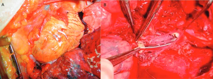
In recent years there have descriptions of the use of skin staplers for primary control of ventricular le-sions in the emergency room with good results8 (Figure
1). Extensive cardiac lacerations may require interrup-tion of blood flow, but this tactic should be the last re-sort, as it will reduce and even stop the heartbeats, and will often be difficult to restart them. There are at least two maneuvers for this purpose: the first is to clamp the superior vena cava and the inferior vena cava and thereby reduce the preload, with cardiac emptying and
arrest; the second technique is done by a right atrium manual compression against the ventricular mass9. The
principle is the same as the first technique, as it will lead to decreased blood flow and eventually cardiac arrest. After these maneuvers, suturing will be possible for a few minutes. Following suture, the heart must be heated with saline while performing direct compres-sions. At the moment that there is fibrillation, defibril-lation can be applied directly to the heart with energy up to 30-50 Joules.
LUNG
The surgeon will hardly come across a lung inju-ry where there is difficult bleeding control. This is main-ly due to two factors: the pulmonary vasculature houses a low-pressure system and the lung parenchyma is rich in thromboplastin. However, there are situations where bleeding may be profuse, both due to the anatomy where the lesion is located, and to the patient’s metabolic exhaus-tion. Among these types of injury, we can mention the tunnelling or transfixing lesions that do not reach the hi-lum, the hilar lesions, and the diffuse parenchymal lesions. Tunnelling or transfixing injuries: patients who have this type of injury possibly have bleeding from the
inlet and outlet of the lung. Simple closure of these injuries would imply a large lung dissecting hematoma or infection in the postoperative period, with lung abscess formation. The technical solution for this type of injury occurs through pulmonary tractotomy. It consists in applying two long vascular clamps or parts of a linear cutting stapler through the “path” of the lesion and sectioning the parenchymal bridge between the clamps, which exposes the interior of injury to the outside, and then holding selective hemostasis and aerostasis inside the wound. When using clamps, aerostasis and hemostasis are terminated with a running absorbable suture over the clamps, which is unnecessary when using the stapler10,11 (Figure 2). Peripheral bleeding and
air-leaking lesions may be quickly resected using the mechanical suture12.
Hilar injuries: in this type of injury, often lethal, the victim is extremely ill on admission to the emergency room. It classically presents with profuse bleeding into the pleural cavity or pericardial tamponade, if the injury is within the pericardial sac, which in both cases will require an emergency thoracotomy. The primary control of hilar bleeding should be manual, through the fastening between the thumb and index finger, followed
by pulmonary mobilization by section of the pulmonary ligament and only then the placement of a large vascular clamp around the hilum13. This type of patient generally
tolerates badly this maneuver, developing severe right ventricular dysfunction, which requires rapid diagnosis of the extent to which the hilum was affected. Partial arterial or venous lesions should be treated with lateral sutures. Venous transection imply corresponding lobectomy, while the arterial lesion will usually require pneumonectomy, with a high degree of mortality. When this is unavoidable, it can be quickly made using a stapler with a vascular load13,14. The technique consists in applying the stapler as
distal as possible so that one can perform a reinforcement suture, and after firing, holding the stump with two Allis clamps. Only then one opens the stapler and completes hemostasis with nonabsorbable suture.
Some injuries are central, but spare the pulmonary hilum. In those situations where bleeding is profuse and air embolization may occur through some larger caliber pulmonary vein, one can use Lung Twist Maneuver for the rapid control of the wound. This consists in quickly releasing the pulmonary ligament and rotating the lung 180 degrees around the hilum. With this done the hilar vessels undergo a “sprain”, immediately stopping the bleeding15.
Diffuse lung injuries: sometimes the lungs may be diffusely bruised or lacerated and the patient already has
coagulopathy. In such dramatic situations, the options are pneumonectomy, which can be devastating for the pa-tient. Alternatively, one can set up a selective ventilation of the non-traumatized lung, when possible, and packaging of the traumatized one16, a technique that has emerged
for control of extensive lesions of the liver parenchyma, an organ that has also an extensive, low-pressure vascular network. One should take care that packs do not cause car-diac diastolic restriction or mediastinal shift, which would lead to a low cardiac output. Packs are to be removed after the correction of coagulopathy (Figure 3).
Figure 3. Chest radiography in the postoperative period of thoracic packaging for coagulopathy. Note the presence of radiopa-que stripes of seven packs in the left hemithorax.
THORACIC VESSELS
Within the concept of damage control, in thoracic vascular trauma there are the following situations: active bleeding externalizing through an open wound; active bleeding into some thoracic compartment (mediastinal or pleural); or bleeding contained as a extrapleural or mediastinal hematoma. The first situation can be exemplified by a penetrating wound in the cervicothoracic region with active bleeding. The temporary control of the bleeding can be done by introducing a Foley catheter in the wound, balloon inflation and traction of the catheter. If the wound is extensive in the skin, one can apply stitches around the catheter. During the surgical exploration one can again use a Foley or Fogarty catheter for compressing bleeding coming from a deep spot of the thoracic cavity. One should keep in mind which vessels could be ligated17.
Theoretically, one can perform ligation of all thoracic venous vessels, with the exception of the venae cavae; amongst the arterial branches, the ligation of the innominate artery can result in extensive stroke. The subclavian arteries, however, ultimately may be ligated because they have a rich collateral circulation network in the shoulder girdle. An alternative to arterial ligation is construction of an intravascular shunt (Figure 1) through a silicone conduit tied to the arterial ends, particularly in the subclavian ones. In the event of a stable mediastinal or extrapleural hematoma due to a greater vascular injury, the conduct may be initially conservative
in a seriously injured patient in whom a thoracic operation would involve major trauma and bleeding, consuming the last physiologic reserves. The classic case to illustrate this is a patient with multi-systemic trauma and a contained injury in the descending aorta. This patient may benefit from a conservative approach, with resolution of the other injuries (brain, orthopedic and abdominal), and only then be submitted to aortic endovascular treatment18.
ESOPHAGUS
Early diagnosed lesions have been treated by primary surgical synthesis associated with nasogastric catheterization and pleural drainage. In iatrogenic perforation during endoscopic procedures, there have descriptions of endoscopic treatments with clipping, positioning of esophageal stents, endoscopic suturing and even conservative treatment for minor injuries in stable patients19,20.
However, injuries of the thoracic esophagus from external causes (i.e. stabbing of gunshot wounds) are often silent lesions that go unnoticed, with late diagnosis performed by pleural effusion and by sepsis installed due to associated mediastinitis. In these critically ill patients with low physiological reserve for arepair attempt (with high rates of fistula), or to perform esophagectomy (high mortality), the choices left are the bypass and exclusion,through cervical esophagostomy, associated with gastrostomy
or jejunostomy, or a T-tube drainage (either a large Kehr drain or a Montgomery tube), introduced inside the esophageal lumen, coming out through the organ lesion and exteriorized through the skin, thus creating a controlled fistula21. A nasogastric catheter may be passed into the
tube for subsequent feeding. An abundant pleural drainage should be associated with this procedure (Figure 4).
CHEST WALL
Situations involving damage control in the chest wall can be divided into control of parietal bleeding and temporary closure of the chest wall. One of the most common causes of review and re-operating of the thoracic cavity is in a parietal bleeding vessel. Intercostal and internal thoracicarteries havetheir flow greatly diminished in a patient with hypovolemic shock. However, when the individual returns to the hemodynamically normal state, these vessels
can reach a flow rate of 300ml per minute. Hence, the cautious ligature of these vessels becomes an important part in the first approach. Sometimes an intercostal vessel may present bleeding at the time of surgical exploration and, after sectioned, retract and become difficult to ligate. The options in this case are: “U” stitches parallel to the ribs; stitches circulating the entire rib and intercostal region into two segments and piercing of the wall through the bleeding point, and positioning of a catheter balloon (Foley or Fogarty) and fixing it to the skin, leaving it for a few days until the thrombosis of the affected vessel. In multiple traumato the ribs and thoracic wall, in which there is diffuse bleeding, one can apply thoracic packaging22.
The temporary closure (abbreviated thoracoto-my) is indicated in two situations. The first one, in which there is temporary control of the injuries, with coagulop-athy and requiring rapprochement to remove packs or to
repair the injuries permanently. The other indication is in the event of an anterolateral thoracotomy or sternotomy where, after opening the pericardium, the heart is very dilated and the closing of the wall will restrict the heart-beats23,24 (Figure 5).
The temporary closure technique may be per-formed only by approximation of the skin or, in extreme cases where there is still cardio-respiratory restriction, by suturing a Smarchtape to the skin. One can also use an open urine collection bag (similar to the “Bogota bag”
principle), or placing packs, covered by adhesive surgi-cal drapes in the skin, like steri-drape, with application of vacuum (VAC – vacuum-assisted closure– technique), in particular in infected wounds25,26 (Figure 6). The
fi-nal synthesis of the chest wall may take several days to complete.
In conclusion, we believe that the general sur-geon who serves in the emergency should know the tho-racic damage control techniques, so as they can serve as therapeutic management tools in critically ill patients.
REFERENCES
1. Rotondo MF, Schwab CW, McGonigal MD, Phillips GR 3rd, Fruchterman TM, Kauder DR, et al. ‘Damage control’: an approach for improved survival in exsan-guinating penetrating abdominal injury. J Trauma. 1993;35(3):375-82; discussion 382-3.
2. Hirshberg A, Walden R. Damage control for abdomi-nal trauma. Surg Clin North Am. 1997;77(4):813-20. 3. Schwab CW. Introduction: damage control at the start
of 21st century. Injury. 2004;35(7): 639-41.
4. Gonçalves R, Saad Júnior R. Vias de acesso aos grandes vasos medistinais no trauma torácico. Rev Col Bras Cir. 2012;39(1):64-73.
5. Rotondo MF, Bard MR. Damage control surgery for thoracic injuries. Injury. 2004;35(7):649-54.
6. Wall MJ Jr, Soltero E. Damage control for thoracic in-juries. Surg Clin North Am. 1997;77(4):863-78. 7. Grabowski MW, Buckman RF Jr, Goldberg A,
Badelli-no MM. Clamp control of the right ventricular angle to facilitate exposure and repair of cardiac wounds. Am J Surg. 1995;170 (4):399-400.
8. Macho JR, Markinson RE, Schecter WP. Cardiac sta-pling in the management of penetrating injuries of the heart: rapid control of hemorrhage and decreased risk
of personal contamination. J Trauma. 1993;34(5):711-5; discussion 715-6.
9. Ellertson DG, Johnson SB. Total inflow occlusion to repair a penetrating cardiac injury: case repor. J Trau-ma. 2008;64(6):1628-9.
10. Wall MJ Jr, Hirshberg A, Mattox KL. Pulmonary trac-totomy with selective vascular ligation for penetrating injuries to the lung. Am J Surg. 1994;168)6):665-9. 11. Asensio JA, Demetriades D, Berne JD, Velmahos G,
Cornwell EE 3rd, Murray J, et al. Stapled pulmonary tractotomy: a rapid way to control hemorrhage in penetrating pulmonary injuries. J Am Coll Surg. 1997;185(5):486-7.
12. Velmahos GC, Baker C, Demetriades D, Goodman J, Murray JA, Asensio JA. Lung-sparing surgery af-ter penetrating trauma using tractotomy, partial lobectomy, and pneumonorrhaphy. Arch Surg. 1999;134(2):186-9.
13. Van Natta TL, Smith BR, Bricker SD, Putnam BA. Hi-lar control in penetrating chest trauma: a simplified approach to an underutilized maneuver. J Trauma. 2009;66(6):1564-9.
14. Huh J, Wall MJ Jr, Estrera AL, Soltero ER, Mattox KL. Surgical management of traumatic pulmonary inju-ry. Am J Surg. 2003;186(6):620-4.
A cirurgia de controle de danos surgiu com a filosofia de se aplicar manobras essenciais para controle de sangramento e contaminação abdominal, em doentes traumatizados, nos limites de suas reservas fisiológicas. Este conceito se estendeu para as lesões torácicas, onde manobras relativamente simples, podem abreviar o tempo operatório de doentes in extremis. Este artigo tem como objetivo, revisar as diversas técnicas de controle de dano em órgãos torácicos, que devem ser de conhecimento do cirurgião que atua na emergência.
Descritores: Traumatismo Múltiplo. Traumatismos Torácicos. Medicina de Emergência.
15. Wilson A, Wall MJ Jr, Maxson R, Mattox K. The pul-monary hilium twist as a thoracic damage control procedure. Am J Surg. 2003;186(1):49-52.
16. Caceres M, Buecher KJ, Tillou A, Shih JA, Liu D, Steeb G. Thoracic packing for uncontrolled bleed-ing in penetratbleed-ing thoracic injuries. South Med J. 2004;97(7):637-41.
17. Mattox KL, Walkes JC. Advances in the man-agement of thoracic vascular injury. Scan J Surg. 2002;91(1):46-51.
18. Demetriades D, Velmahos GC, Scalea TM, Jurkovich GJ, Karmy-Jones R, Teixeira PG, et al. Diagnosis and treatment of blunt thoracic aortic injuries: changing perspectives. J Trauma. 2008;64(6):1415-18; discus-sion 1418-9.
19. Plott E, Jones D, McDermott D, Levoyer T. A state-of-the-art review of esophageal trauma: where do we stand? Dis Esophagus. 2007;20(4):279-89. 20. Dickinson KJ, Blackmon SH. Endoscopic
tech-niques for the management of esophageal per-furation. Operat Tech Thorac Cardiovasc Surg. 2016;20(3):251-78.
21. Wu JT, Mattox KL, Wall MJ Jr. Esophageal perfora-tions: new perspectives and treatment paradigms. J Trauma. 2007;63(5):1173-84.
22. Vargo DJ, Battistella FD. Abbreviated thoracoto-my and temporary chest closure: an application of
damage control after thoracic trauma. Arch Surg. 2001;136(1):21-4.
23. Lang JL, Gonzalez RP, Aldy KN, Carroll EA, et al. Does temporary chest wall closure with or without chest packing improve survival for trauma patients in shock after emergent thoracotomy? J Trauma. 2011; 70(3):705-9.
24. Phelan HA, Patterson SG, Hassan MO, Gonzalez RP, et al. Thoracic damage-control operation: principles, techniques, and definitive repair. J Am Coll Surg. 2006;203(6):933-41.
25. O’Connor JV, Chi A, Joshi M, DuBose J, Scalea TM. Post-traumatic empyema: aetiology, surgery and outcome in 125 consecutive patients. Injury. 2013;44(9):1153-8.
26. O’Connor JV, DuBose JJ, Scalea TM. Damage-con-trol thoracic surgery: management and outcomes. J Trauma Acute Care Surg. 2014;77(5):660-5.
Received in: 18/08/2016
Accepted for publication: 29/09/2016 Conflict of interest: none.
Source of funding: none.
Mailing address:
Roberto Gonçalves