Recognition Particle to Regulate Protein Homeostasis in
E. coli
Bentley Lim1, Ryoji Miyazaki2., Saskia Neher3¤., Deborah A. Siegele4
, Koreaki Ito5, Peter Walter3, Yoshinori Akiyama2*, Takashi Yura5*, Carol A. Gross1,6*
1Department of Microbiology and Immunology, University of California at San Francisco, San Francisco, California, United States of America,2Institute for Virus Research, Kyoto University, Kyoto, Japan,3Department of Biochemistry and Biophysics and Howard Hughes Medical Institute, University of California at San Francisco, San Francisco, California United States of America,4Department of Biology, Texas A&M University, College Station, Texas, United States of America,5Faculty of Life Sciences, Kyoto Sangyo University, Kyoto, Japan,6Department of Cell and Tissue Biology, University of California at San Francisco, San Francisco, California, United States
Abstract
All cells must adapt to rapidly changing conditions. The heat shock response (HSR) is an intracellular signaling pathway that maintains proteostasis (protein folding homeostasis), a process critical for survival in all organisms exposed to heat stress or other conditions that alter the folding of the proteome. Yet despite decades of study, the circuitry described for responding to altered protein status in the best-studied bacterium,E. coli, does not faithfully recapitulate the range of cellular responses in response to this stress. Here, we report the discovery of the missing link. Surprisingly, we found thats32, the central transcription factor driving the HSR, must be localized to the membrane rather than dispersed in the cytoplasm as previously assumed. Genetic analyses indicate thats32localization results from a protein targeting reaction facilitated by the signal recognition particle (SRP) and its receptor (SR), which together comprise a conserved protein targeting machine and mediate the cotranslational targeting of inner membrane proteins to the membrane. SRP interacts withs32directly and transports it to the inner membrane. Our results show thats32must be membrane-associated to be properly regulated in response to the protein folding status in the cell, explaining how the HSR integrates information from both the cytoplasm and bacterial cell membrane.
Citation:Lim B, Miyazaki R, Neher S, Siegele DA, Ito K, et al. (2013) Heat Shock Transcription Factors32Co-opts the Signal Recognition Particle to Regulate
Protein Homeostasis inE. coli. PLoS Biol 11(12): e1001735. doi:10.1371/journal.pbio.1001735 Academic Editor:Arthur L. Horwich, Yale School of Medicine, United States of America
ReceivedJune 5, 2013;AcceptedOctober 23, 2013;PublishedDecember 17, 2013
Copyright:ß2013 Lim et al. This is an open-access article distributed under the terms of the Creative Commons Attribution License, which permits unrestricted use, distribution, and reproduction in any medium, provided the original author and source are credited.
Funding:This work was supported by National Institutes of Health Grant GM-36278 (to C.A.G.), a National Science Foundation Graduate Research Fellowship (to B.L.), and the Jane Coffin Child Memorial Fund (to S.N). The funders had no role in study design, data collection and analysis, decision to publish, or preparation of the manuscript.
Competing Interests:The authors have declared that no competing interests exist.
Abbreviations:HSPs, heat shock proteins; HSR, heat shock response; IM, inner membrane; pBPA,p-benzoylphenylalanine; PhoA, alkaline phosphatase; S.A., specific activity; SR, signal receptor; SRP, signal recognition particle.
* E-mail: [email protected] (Y.A.); [email protected] (T.Y.); [email protected] (C.A.G.)
¤ Current address: Department of Biochemistry and Biophysics, University of North Carolina at Chapel Hill, Chapel Hill, North Carolina, United States of America.
.These authors contributed equally to this work.
Introduction
The heat shock response (HSR) maintains protein homeostasis (proteostasis) in all organisms. The HSR responds to protein unfolding, aggregation, and damage by the rapid and transient production of heat shock proteins (HSPs) and by triggering other cellular protective pathways that help mitigate the stress. Although the specific HSR is tailored to each organism, chaperones that mediate protein folding and proteases that degrade misfolded proteins are almost always included in the core repertoire of induced protein and are among the most conserved proteins in the cell. These HSPs maintain optimal states of protein folding and turnover during normal growth, while decreasing cellular damage from stress-induced protein misfolding and aggregation. Malfunc-tion of the HSR pathway reduces lifespan and is implicated in the onset of neurodegenerative diseases in higher organisms [1–3].
InE. coliand other proteobacteria,s32 mediates the HSR by directing RNA polymerase to promoters of HSR target genes [4– 9]. Given the importance of this response and the necessity for a rapid but transient increase in expression of HSPs, it is not surprising that regulation of the HSR across organisms is complex. s32
is positively regulated by a feed-forward mechanism in which exposure to heat melts an inhibitory mRNA structure enabling high translation ofs32
mRNA [10,11] and is negatively regulated by two feedback loops [12] mediated through members of thes32
regulon (Figure 1A).s32
activity is coupled to the cellular protein folding state via a negative feedback loop executed by the two major chaperone systems, DnaK/J/GrpE and GroEL/S. There is extensive support for the model that free chaperones directly inactivates32
chaperone substrates leads to an increase in s32
activity, and conversely, overexpression of either chaperone system decreases s32 activity [13,14]. Inhibition is likely direct, as DnaK/J and GroEL/S binds32
in vitroand inhibit its activity in a purifiedin vitrotranscription system [13,15–17].s32
stability is controlled by the inner membrane (IM) protease FtsH: deletion of the protease stabilizess32[18–20], and FtsH degradess32in vitro, albeit slowly [18,20]. DnaK/J and GroEL/S also regulate stability, as their depletion leads tos32
stabilizationin vivo[13,14,21], although this finding has not yet been recapitulatedin vitro[22].
Despite the regulatory complexity of the current model, it inadequately addresses two issues that are central to our understanding of the circuitry controlling the HSR, motivating us to search for additional players in the response: (1) Exhaustive genetic screens for mutations ins32 that result in misregulation have identified a small cluster of four closely spaced amino acid residues (Leu47, Ala50, Lys51, and Ile54), of which three are surface exposed, as well as a somewhat distant fifth residue that abuts this patch in the foldeds32structure. When these residues are mutated, cells have both increased level and activity ofs32, indicating that this region is involved in a central process required for operation of the negative feedback loops that control both the activity and degradation ofs32
(Figure 1A) [23–25]. However, the phenotypes of these mutants are not recapitulatedin vitro, where both FtsH degradation and chaperone-mediated inactivation of mutant and WTs32
are experimentally indistinguishable [25,26]. Thus, we do not understand how this ‘‘homeostatic control region’’ ofs32
functions. (2)s32
is thought to monitor the folding status of IM proteins as well as cytoplasmic proteins, but the mechanism for this additional surveillance is unknown. Their close connection is indicated because (1) the IM protease, FtsH, not only degrades s32
, but also maintains quality control in the IM by
degrading unassembled IM proteins; (2) induction of the HSR is a very early response to perturbations in the co-translational membrane-trafficking system that brings ribosomes translating IM proteins to the membrane [27–29]; and (3) IM proteins are significantly overrepresented both in thes32
regulon [30] and in an unbiased overexpression screen for HSR inducers [30].
In this report, we identify the co-translational protein targeting machinery, comprised of the Signal Recognition Particle (SRP; Ffh protein in complex with 4.5S RNA; Figure 2A) and the SRP Receptor (SR; FtsY), as a regulator ofs32
. We show that SRP preferentially binds to WTs32
compared to a mutants32
with a defective homeostatic control region. We further show that a fraction ofs32is associated with the cell membrane and that both the SRP-dependent machinery and the homeostatic control region of s32
are important for this localization. Lastly, the regulatory defects in HSR circuitry caused by mutation of either the s32 homeostatic control region or the co-translational targeting machinery are circumvented by artificially tetherings32
to the IM. We propose that SRP-dependent membrane localization is a critical step in the control circuitry that governs the activity and stability ofs32
. Membrane localization is widely used to controls factors, but this is the first case where the IM-localized state is used for dynamic regulation rather than as a repository for an inactive protein.
Results
A Transposon Insertion Mutant at theftsY Promoter Region Is Defective in Feedback Control
To identify additional players involved in activity control ofs32
, we carried out a genetic screen for transposon mutants with increaseds32
activity under conditions that inactivates32
in wild-type cells (see Methods). To impose a condition that mimics the negative feedback control of s32
, the DnaK/J chaperones were overexpressed from an inducible promoter at their chromosomal locus. Under these conditions, as32-regulatedlacZchromosomal reporter (PhtpG-lacZ) is expressed so poorly that cells do not make
sufficientb-galactosidase to turn colonies blue on X-gal indicator plates. We screened for blue colonies, indicative of a defect ins32
inactivation. A conceptually similar screen previously identified mutations in the DnaK/J chaperones—key negative regulators of the s32
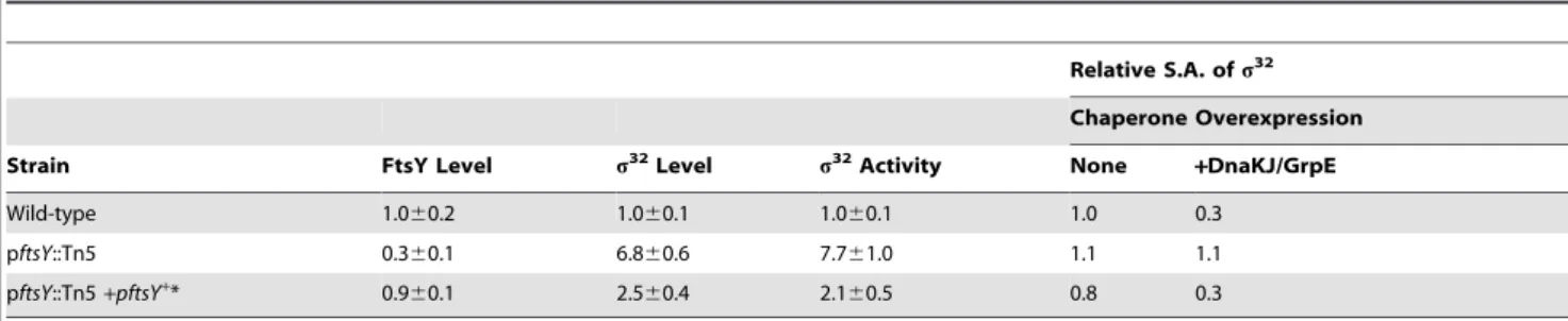
response [31]. In addition to re-identifying these components, we found an insertion in the promoter region of ftsY(pftsY::Tn5), located 39 bp upstream of theftsYopen reading frame. The pftsY::Tn5strain had a 3- to 4-fold reduction in the level of FtsY, the SR, and a,7-fold increase in the activity and amount of s32 relative to WT (Table 1). Defects were complemented by a plasmid carrying ftsY. Unlike WT, in the pftsY::Tn5 strain s32
activity did not respond to increased chaperone expression. Upon chaperone overexpression in WT cells, the specific activity (S.A.) ofs32
fell to 0.3, relative to that in cells growing without chaperone overexpression. In contrast, upon chaperone overexpression in pftsY::Tn5cells, the S.A. ofs32
did not change, suggesting a defect in chaperone-mediated activity control in that strain (Table 1). This finding raised the possibility that the high activity ofs32
in pftsY::Tn5resulted from disruption of activity control ofs32, rather than reflecting a cellular response to accumulation of unassembled membrane proteins.
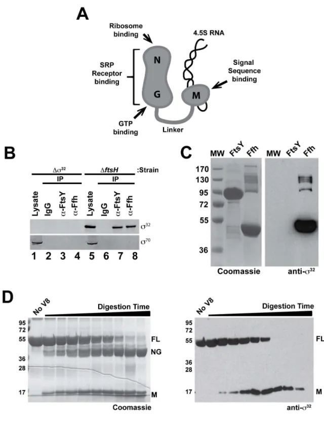
s32Directly Interacts with SRP
We tested whethers32
binds to either FtsY (SR) or to Ffh, the protein component of SRP. Ffh is a two-domain protein, comprised of an M-domain that binds the signal sequence and 4.5S RNA, and an NG-domain that binds to SR, the ribosome, Author Summary
All cells have to adjust to frequent changes in their environmental conditions. The heat shock response is a signaling pathway critical for survival of all organisms exposed to elevated temperatures. Under such conditions, the heat shock response maintains enzymes and other proteins in a properly folded state. The mechanisms for sensing temperature and the subsequent induction of the appropriate transcriptional response have been extensive-ly studied. Prior to this work, however, the circuitry described in the best studied bacteriumE. colicould not fully explain the range of cellular responses that are observed following heat shock. We report the discovery of this missing link. Surprisingly, we find that s32, a transcription factor that induces gene expression during heat shock, needs to be localized to the membrane, rather than being active as a soluble cytoplasmic protein as previously thought. We show that, equally surprisingly,s32
is targeted to the membrane by the signal recognition particle (SRP) and its receptor (SR). SRP and SR constitute a conserved protein targeting machine that normally only operates on membrane and periplasmic proteins that contain identifiable signal sequences. Intriguingly, s32
does not have any canonical signal sequence for export or membrane-integration. Our results indicate that mem-brane-associated s32, not soluble cytoplasmic s32, is the preferred target of regulatory control in response to heat shock. Our new model thus explains how protein folding status from both the cytoplasm and bacterial cell membrane can be integrated to control the heat shock response.
and GTP (Figure 2A). We first used co-immunoprecipitation analysis. Interacting proteins were immunoprecipitated with antibodies against either FtsY or Ffh and, following resolution on SDS-PAGE, antibodies againsts32
ors70
were used to probe for the presence of these proteins. s32
was detected in the immuno-precipitations (Figure 2B, lanes 7 and 8), and this signal was dependent on the presence ofs32
in the strain (Figure 2B, lanes 1– 4). By contrast,s70, although much more abundant thans32in the cell, did not interact with either SRP or SR (Figure 2B, Lanes 3,4 and 7,8), indicating that interaction with SRP is not a general property ofss. It was not surprising thats32
was co-immunopre-cipitated with both SRP and SR, as the latter two components interact in vivo. To determine the direct binding partner ofs32
, purified Ffh and FtsY were resolved on SDS-PAGE, transferred to nitrocellulose, and incubated with purifieds32
. Antibodies against s32
detecteds32
present at the molecular weight corresponding to Ffh but not SR (Figure 2C). In a reciprocal experiment, purifieds32
was resolved on SDS-PAGE, transferred to nitrocellulose, and incubated with purified Ffh or SR. Ffh, but not SR, bound s32 (unpublished data). Similar studies did not reveal an interaction between s70
and either Ffh or SR (unpublished data). We determined which Ffh domain bindss32
by partially-proteolyzing Ffh to produce an 18 kDa M-domain and a 38 kDa NG-domain, resolving the mixture by SDS-PAGE, transferring to nitrocellulose,
and probing withs32.s32was detected at the position of full-length Ffh and the M-domain, but not at the position of the NG-domain (Figure 2D), indicating that the M-domain contains the determi-nants mediating thes32
-interaction.
We usedin vivocrosslinking to validate the direct interaction of SRP (Ffh+4.5S RNA) ands32
. We created as32
derivative with an N-terminal 66HIS-tag and a photoreactive amino acid analog (pBPA) at amino acid position 52 (66HIS-s32
T52pBPA; see Methods), which is active as WTs32
in expression of the s32
reporter PhtpG-lacZ(activity is 150% that of WT; within the range
of the variability of the assay; unpublished data). Following UV irradiation of whole cells, anti-Ffh immunoblotting of the whole cell lysate detected one predominant crosslinked product, which was dependent on UV-irradiation (Figure 3A, lanes 1 and 2) and pBPA at position 52 (Figure 3A, lanes 2 and 4). This UV- and pBPA-dependent product was also detected with anti-s32
immu-noblotting (Figure 3A, lane 6). To determine whether the crosslinked product represented 66HIS-s32
T52pBPA-Ffh, we determined whether this product was identified both by co-immunoprecipitation with anti-Ffh antisera (Figure 3B) and by affinity purification of 66HIS-s32T52pBPA on a TALON resin (Figure 3C). Upon immunoprecipitation with anti-Ffh antisera, we detected a single higher molecular mass band, which reacted with both anti-Ffh (Figure 3B, lane 2) and -s32
(Figure 3B, lane 6).
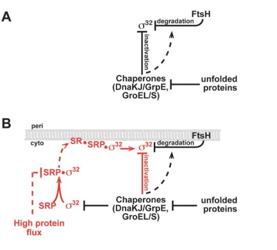
Figure 1. Homeostatic control circuits ofs32.(A) Current and (B) revised model for activity and degradation control ofs32
. The revised model incorporates SRP-mediated trafficking ofs32to the membrane. Interactions validatedin vitroare shown as solid lines; those inferred fromin vivodata are shown as dashed lines. Newly identified interactions are shown in red.
Figure 2.s32binds to Ffh.(A) Schematic representation ofE. coliSRP (Ffh+4.5S RNA), indicating experimentally confirmed functions associated
with each domain. (B)s32co-immunoprecipitates with Ffh and FtsYin vivo, buts70does not. Immunoprecipitations of Ffh or FtsY were carried out on lysates ofDs32andDftsHcells grown to exponential phase. Immunocomplexes were isolated, analyzed by SDS-PAGE, and immunoblotted with anti-s32and anti-s70antibodies. Proteins from approximately 15-fold more cells were loaded onto the gel for the immunoprecipitated samples againsts32ands70as compared with the lysate samples. (C) Protein–protein interaction analysis indicates thats32binds to Ffh, but not FtsY. Purified FtsY and Ffh were run on a 10% SDS-PAGE gel, transferred to nitrocellulose, re-natured, and incubated with purified WTs32. The Coomassie-stained gel (left) and the nitrocellulose blot probed with polyclonal anti-s32antibodies (right) are shown. (D)s32binds to the M-domain of Ffh. Ffh,
Upon affinity purification on a TALON resin, anti-Ffh identified the same predominant UV- andpBPA-dependent Ffh-containing crosslinked product (compare Figure 3B and 3C, lane 2). Importantly, no free Ffh was recovered following TALON purification, indicating that the recovery of the Ffh conjugate was mediated by the covalently linked 66HIS-s32, rather than interaction with either the TALON resin or another protein. These results strongly suggest thats32directly interacts with Ffhin vivo. Although only a faint band was seen at the same position using anti-s32 immunoblotting, this was likely a result of high background in this area of the gel, possibly because of extensive interaction between chaperones ands32(Figure 3C, lanes 5–8).
I54Ns32Is Defective in Interacting with SRP
The function of the homeostatic control region ofs32
is not known [25]. I54Ns32
is a mutation located in this region is severely compromised in both activity and degradation control, but the mechanism responsible for this phenotype had not yet been determined [25]. We therefore compared the binding of WTs32
and I54Ns32
to SRP using gel filtration. We incubated WTs32
or I54Ns32
either alone or in combination with SRP and subjected the mixture to gel filtration. Analysis of the elution profiles demonstrated that most WTs32
was shifted towards the higher molecular weight region in the presence of SRP, and additionally, a fraction ofs32
eluted at a higher molecular weight than that of SRP alone, indicative of an SRP–s32
complex [compare A280profiles ofs32, SRP, and SRP-s32(Figure 4A) with
immunoblotting fors32
(Figure 4B; rows 1,2)].s32
present at a molecular weight betweens32
and SRP likely represents transient forms of thes32
–SRP complex. In sharp contrast, an interaction between I54Ns32
with SRP was almost undetectable [compare A280profiles of I54Ns32and SRP (Figure 4A) with
immunoblot-ting for I54Ns32
(Figure 4B; rows 3,4)], indicating that I54Ns32
bound more weakly to SRP than WTs32
. Neither WTs32
nor I54Ns32
interacted detectably with Ffh, indicating that differential binding is dependent on the formation of SRP (Ffh+4.5S RNA), the biologically relevant cellular species of Ffh.
s32 Is Partially Membrane Associated in an SRP-Dependent Process
The biological function of SRP is co-translational protein targeting, leading us to test whethers32may be targeted to the IM
through an SRP-dependent mechanism. Rapid degradation by FtsH normally keepss32
levels very close to the detection limit (,20–50 molecules/cell; [8]), making reproducible detection following fractionation very difficult. Therefore, we performed fractionation experiments (Figure 5), either in cells expressing an enzymatically inactive mutant of the FtsH protease (FtsH E415A) or in cells lacking FtsH altogether (DftsH). Approximately 44% of s32
fractionated to the membrane in a DftsH strain, and this fraction was increased to,58% in the FtsH E415A strain, raising the possibility that FtsH itself may participate in retention ofs32at the IM. As the b9 subunit of RNA polymerase, a known interaction partner ofs32
, also fractionated with the membrane, we next tested whethers32association with the IM was dependent
on its association with RNA polymerase. To this end, we used s32D
21aa, which is defective in interacting with RNA polymerase [32]. We confirmed thats32D
21aa did not detectably interact with RNA polymerase (Figure S1A,B). Yet endogenous WTs32 and ectopically expresseds32D
21aa fractionated equivalently to the IM both inDftsHcells (,39%) and in FtsH E415A cells (,58%) (Figure S2), indicating that s32 transited to the membrane independent of RNA polymerase.
We next tested whether the pftsY::Tn5 mutation or the homeostatic control region mutation ofs32
disrupted membrane partitioning of s32. Both WTs32 and ectopically expressed s32D
21aa were defective in partitioning to the IM in pftsY::Tn5 cells (Figure 5). To look at the effect of disrupting the homeostatic control region on membrane fractionation, we expressed I54Ns32 as a s32D
21aa variant (I54Ns32D
21aa). The size difference allowed us to compare I54Ns32D
21aa and WTs32
in the same cells (Figure S2). Whereas WTs32exhibited normal fractionation, I54Ns32D
21aa showed a severe localization defect, comparable to that of pftsY::Tn5cells (Figure 5). We conclude thats32
targeting to the IM is dependent on both SRP/SR and thes32homeostatic
control region.
Both SecA and SecY Are Important for Membrane Association ofs32
SecA is an ATP-fueled motor protein that recognizes signal peptides, drives the translocation of secreted proteins through the Sec translocon [33–37], and collaborates with the SRP/SR for integration of a subset of IM proteins into the membrane [33,38]. We previously found thats32
activity is increased in a SecA(ts) strain [39]. This observation motivated us to explore the relationship of partially digested by endopeptidase V8, was resolved on a 10% SDS-PAGE gel, transferred to nitrocellulose, and incubated withs32. The Coomassie-stained gel (left) and the nitrocellulose membrane, containing transferred Ffh fragments, probed againsts32(right) are shown.
doi:10.1371/journal.pbio.1001735.g002
Table 1.The altereds32phenotypes of the Tn5 insertion mutant (pftsY::Tn5) are significantly complemented by anftsY+plasmid.
Relative S.A. ofs32
Chaperone Overexpression
Strain FtsY Level s32Level s32Activity None +DnaKJ/GrpE
Wild-type 1.060.2 1.060.1 1.060.1 1.0 0.3
pftsY::Tn5 0.360.1 6.860.6 7.761.0 1.1 1.1
pftsY::Tn5+pftsY+* 0.9
60.1 2.560.4 2.160.5 0.8 0.3
In this and all other experiments, protein levels were determined by SDS-PAGE followed by quantitative immunoblotting.s32activity was determined from a chromosomalb-galactosidase reporter (calculated as a differential rate of synthesis); values presented are from$3 experiments. Relative S.A. ofs32is defined as: [(s32
activity/s32level) normalized tos32S.A. of WT cells grown at 30uC].
*TheftsY+
SecA to IM trafficking ofs32. Indeed, using a SecA(ts) mutant with
general defects in protein export (SecAL43P) [40,41], we observed that cells displayed a significant defect in membrane localization of s32
(Figure 5), as well as increased s32
activity ([39] and unpublished data). In addition, purified SecA, resolved on SDS-PAGE and transferred to nitrocellulose, showed binding affinity for s32
, suggesting that these two proteins interact (Figure S3). We conclude that SecA participates in trafficking ofs32to the IM.
SecY forms the core of the SecYEG IM translocon. This multidomain protein has a large cytoplasmic domain (C5) that functionally interacts with SR [42], SecA, and the ribosome [43– 50] (Figure 6A). We tested whether 10 previously described secY mutations located in various domains of SecY (Figure 6A) [51] perturb chaperone-mediated control ofs32
activity and trafficking of s32
to the IM (Figure 6B). All mutants had enhanced s32
activity. This result was not surprising assecYmutants are expected to accumulate secretory protein precursors that titrate chaperones [52]. Importantly, four mutants (secY124,secY351,secY40,secY129) were also defective in chaperone-mediated control ofs32activity (Figure 6B), as indicated by a lack of down-regulation of s32
activity in response to overexpression of one or both of the
chaperone systems. We examined thesecY351mutant, which had both high s32
activity and a significant defect in chaperone-mediated inactivation, and found it to be defective in IM trafficking of s32 (Figure 5). secY40 and secY351 affect domain
C5 (Figure 6A), implicated in the interaction of SecY with SR, raising the possibility that this interaction is important for both homeostatic control and IM targeting ofs32.
An Independent Methodology Indicates Association of
s32with the IM
Alkaline phosphatase is active only in the periplasm, where it forms the disulfide bonds necessary for its activity. Therefore, translational fusions to alkaline phosphatase (PhoA) lacking its own export signal are commonly used as an indicator of membrane targeting by the appended N-terminal sequence [53]. If the appended N-terminal sequence has either an export or insertion sequence, the fusion protein will exhibit alkaline phosphatase activityin vivobecause it is partly transported to the periplasmic side of the membrane through the SecYEG translocon. Although s32
has neither a membrane insertion nor an export sequence, it
Figure 3.In vivocross-linking betweens32and Ffh.(A) Detection of a cross-linked product following UV irradiation in whole cells. Cells of CAG48238/pEVOL-pBpF/p6XH-rpoHT52amber(lanes 1, 2, 5, and 6) and CAG48238/pEVOL-pBpF/p6XH-rpoH(lanes 3, 4, 7, and 8) were grown at 30uC in L-medium supplemented with 0.02% arabinose, induced with 1 mM IPTG for 1 h, and UV-irradiated for 0 or 10 min as indicated. Total cellular proteins were acid-precipitated and analyzed by SDS-PAGE and immunoblotting with anti-Ffh and anti-s32antibodies. (B) Immunoprecipitation with anti-Ffh reveals a unique cross-linked product that interacts with anti-s32. Supernatants of sonically disrupted UV-irradiated cells were subjected to immunoprecipitation with anti-Ffh antibodies. Immunocomplexes were solubilized in SDS sample buffer, analyzed by SDS-PAGE, and immunoblotted with anti-Ffh and anti-s32antibodies. Proteins from approximately 4.4-fold more cells were loaded onto the gel for the immunoprecipitated samples as compared with the whole cell samples. (C) Purification of 66H-s32from UV-irradiated cells reveals a band that interacts with anti-Ffh. Supernatants of sonically disrupted UV-irradiated cells were subjected to TALON affinity chromatography, and bound proteins were eluted with 300 mM imidazole. Proteins in the eluate were acid-precipitated and analyzed by SDS-PAGE and immunoblotting with anti-Ffh antibodies. Proteins form approximately 20-fold more cells were loaded onto the gel for the TALON-affinity isolated samples as compared with the whole cell samples.
doi:10.1371/journal.pbio.1001735.g003
may contain a sequence that targets it to the cytosolic face of the IM. There is some evidence that the secretory apparatus can recognize the mature domains of exported proteins at low efficiency [54]. If so, proximity of PhoA to the translocon resulting from the IM targeting signal might enable transit of some fraction of PhoA to localize to the periplasmic side of the membrane, where it is active. By random insertion of the transposon probe TnphoAintorpoH, encodings32
(see Materials and Methods), we found that aphoAfusion to the first 52 amino acids ofs32
(N52-s32
-PhoA) showed,10-fold greater PhoA activity than signal-less PhoA itself, indicating that the N-terminus ofs32
facilitates PhoA export (Table 2). Moreover, PhoA activity enhancement is dependent both on the SRP/SR-dependent trafficking system and on SecY, as both pftsY::Tn5 and secY351 decreased the PhoA activity ,2-fold, whereas leaderless PhoA exhibited little response to these perturbations (Table 2). Thus, this assay is consistent with the idea that the N-terminus ofs32
carries an IM-trafficking sequence and that the targeting process is dependent on SRP and SecY.
Membrane-Tethering of Otherwise Deregulateds32
Restores Homeostatic Control
The I54Ns32
mutant and mutants in the IM-targeting machinery (pftsY::Tn5, secA(ts), secY351) were both defective in proper regulation ofs32
and ins32
association with the IM. This convergence motivated us to test whether artificially tetherings32
to the IM could restore homeostatic control. To this end, we exploited the bacteriophage Pf3 coat protein. With the addition of three leucine residues in its membrane-spanning region, 3L-Pf3 translocates spontaneously in an orientation-specific manner to the IM, where it inserts in an N-out/C-in orientation [55]. We modifiedrpoH(encodings32
) at its chromosomal locus to encode a s32
variant with the 3L-Pf3 membrane-insertion signal attached to its N-terminus (schematized in Figure S4A). Strains carrying
Figure 4. SRP (Ffh+4.5S RNA) preferentially interacts with WTs32.(A) A280elution profiles of WTs32, I54Ns32, Ffh, and SRP alone or in complex. WTs32or I54Ns32was incubated with a 10-fold molar excess of purified SRP on ice for 10 min, and complexes were analyzed by gel filtration on a Superdex 200 PC3.2/30 column. Protein elution was monitored by A280. Gel filtration of purified WTs32, I54Ns32, and SRP alone was carried out to determine the migration of each individual protein on the column. (B) Eluted fractions were separated on SDS-PAGE and probed with polyclonal antibodies against Ffh and s32; Western blots ofs32are shown. Experiments were performed at least four times, and a representative experiment is shown.
doi:10.1371/journal.pbio.1001735.g004
Figure 5.s32is partially membrane associated.The extent of association ofs32and theb9subunit of RNA polymerase with the membrane
fraction was determined by quantitative immunoblotting of the soluble and nonsoluble fractions. Membrane association ofs32andb9was assessed in several relevant strain backgrounds. In addition to endogenouss32, all strains contained a plasmid-encoded variant ofs32lacking its 21 C-terminal amino acids (s32D21aa). Ectopically expresseds32D21aa or I54Ns32D21aa were present at levels comparable to natives32and were distinguished from endogenouss32on a 12% SDS-PAGE gel. All fractionation experiments were performed$8 times, and % fractionation was calculated from experiments where probed cytoplasmic (RuvB) and membrane (RseA) proteins separated properly.
3L-Pf3-s32 (IM-WTs32) or 3L-Pf3-I54Ns32 (IM-I54Ns32) as their sole source of s32
were viable, even though 99% of IM-WTs32
was inserted in the membrane as judged by fractionation studies (Figure S4B). Thus,s32functions when it is tethered to the
IM.
We determined whether IM-WTs32was subject to homeostatic
control circuitry exhibited by WTs32.s32is maintained at a low level by FtsH degradation, and its activity is decreased by chaperone-mediated inactivation. Both phenotypes are evident by comparing the amount and activity ofs32in a WT versus a DftsHstrain. In aDftsHstrain, the level of WTs32
increases, 30-fold because the major protease degrading s32
is removed (Table 3; Figure S5 [compare lanes 1 and 3]; and [25]). However, the activity of s32 increases only 3-fold as a consequence of chaperone-mediated activity control, leading to a 10-fold reduc-tion in the S.A. ofs32
inDftsHcells relative to that in WT cells
(Table 3 and [56]). Both the level and S.A. of WTs32and IM-WTs32
were closely similar in aDftsHstrain, indicating that the chaperone-mediated activity control circuit is active in IM-WTs32
(Table 3 and Figure S5 [compare lanes 3 and 4]). Additionally, the level of IM-WTs32was significantly lower inftsH+than in aDftsH
strain, indicating that IM-WTs32
was efficiently degraded by FtsH (Table 3 and Figure S5 [compare lanes 2 and 4]). The presence of a contaminating band prevented absolute quantification of IM-WTs32levels via Western blot analysis (Figure S5). However, if the relative S.A. of IM-WTs32and WTs32are equivalent in the ftsH+
strain as we found in the DftsH strain, then the 2-fold decrease in activity of IM-WTs32
relative to WTs32
implies a slight increase in the rate of degradation of IM-WTs32relative to
WTs32. Note that the 3L-Pf3 membrane-insertion tag itself is not a signal for FtsH degradation, as the stability of the FliAsfactor, which is closely related tos32
, was unchanged when expressed as
Figure 6. The SecY translocon plays a role in chaperone-mediated activity control ofs32.(A) Schematic of SecY topology in the IM by highlighting in yellow the locations/allele names of the mutated residues used in this study [51]. The region that interacts with FtsY (Domain C5) is boxed in green. (B) Mutations insecYshow higher s32activity and affect chaperone-mediated activity control ofs32. The activity ofs32was measured in WT andsecYmutant cells growing at 30uC in LB medium (column 1) or in the same cells following induction of DnaK/J (column 2) or GroEL/S (column 3). Activity is calculated as the differential rate ofb-galactosidase synthesis from a chromosomal PtpG-lacZreporter in each cell type
relative to that of WT cells.
doi:10.1371/journal.pbio.1001735.g006
3L-Pf3-FliA (Figure S6). In summary, both the chaperone-mediated activity control circuit and the FtsH-chaperone-mediated degrada-tion control circuit are active on IM-tethereds32
.
Next, we asked whether the forced and stable tethering ofs32
to the IM bypassed the regulatory defects of I54Ns32
and the reduced-level SR mutant pftsY:::Tn5. I54Ns32
is degraded poorly by FtsH as its level was 11-fold higher than that of WTs32
(Table 3; Figure S5 [compare lanes 1 and 6] and [25]). I54Ns32
also had compromised chaperone-mediated activity control as the high chaperone levels in this strain did not reduce the S.A. of I54Ns32
(Table 3; and [25]). In stark contrast, both degradation and activity control were restored when I54Ns32was converted to
IM-I54Ns32. FtsH efficiently degraded the membrane-tethered variant: IM-I54Ns32was undetectable inftsH+cells but present at
a high level inDftsHcells (Table 3 and Figure S5 [compare lanes 5 and 7]). Additionally, IM-I54Ns32 and IM-WTs32 exhibited comparable reductions in relative S.A. of s32
in DftsH cells (Table 3). Stable tethering of s32
to the IM also bypassed the regulatory defects of pftsY::Tn5as IM-WTs32
in the reduced-level SR background was degraded and subject to chaperone-mediated activity control. Indeed, IM-WTs32
behaved identically in WT and pftsY::Tn5 strains, exhibiting comparable s32
activity at a protein level below detection (Table 3 and Figure S5 [compare lanes 8 and 9]). Finally, IM-tethering relieved the growth defects of both I54Ns32
(Figure S7A and C) and of pftsY::Tn5(Figure S7B, C, and D). In summary, stable tethering ofs32to the IM restored
both homeostatic control and normal growth to cells with a defective s32 homeostatic control region and to cells with a compromised SRP/SR co-translational targeting apparatus.
Discussion
Our work has led to a revised model of the HSR circuitry (Figure 1B).s32
first transits to the IM via an SRP/SR-dependent process and is then subjected to the chaperone-mediated activity control and FtsH-mediated degradation control that have been previously described. This revised model enables the homeostatic control circuit to integrate information on both cytosolic and IM status. Importantly, the efficiency of co-translational protein targeting depends on the cumulative effect of multiple SRP checkpoints including differences in cargo binding affinities, kinetics of SRP-SR complex assembly, and GTP hydrolysis [57]. Multiple checkpoints and the fact that SRP is sub-stoichiometric relative to translating ribosomes (,1:100; SRP molecules to translating ribosomes [58]) may allow SRP to modulate the extent
of IM-localization ofs32
during times of stress and/or increased protein flux. Thus,s32
down-regulation through its localization to the membrane could be alleviated when the IM is disturbed or SRP is overloaded in assisting membrane protein biogenesis. This feed-forward mechanism allows thes32
homeostatic control to sense the state of cytosolic and IM proteostasis before unfolded proteins accumulate to a significant extent. Interest-ingly, ffh (encoding the protein subunit of the SRP) is a s32
regulon member as its expression increases at least 3-fold following induction ofs32either by heat shock or by deletion of dnaK/J ([30] and unpublished data). This could provide an additional connection between s32
and protein flux to the IM. Finally, and more speculatively, given the demonstrated involvement of SecA in IM targeting of s32
and its direct interaction with s32
, the s32
homeostatic control circuit may also monitor protein flux through SecA to the periplasm and outer membrane.
The idea that the high activity of s32 in the I54Ns32
homeostatic control mutant and in SRP/SR mutants (eg. pftsY::Tn5) results from s32 mislocalization to the cytosol and consequent homeostatic dysregulation, rather than from chaper-one titration by a buildup of unfolded proteins, is supported by our data. First, forced IM-tethering overcomes the inviability of the I54Ns32
mutation in theDftsH strain background (Table 3), as well as the growth defects of I54Ns32and pftsY::Tn5(Figure S7), suggesting that high expression ofs32
is aberrant and deleterious to cells, rather than required to remodel misfolded proteins. This is reminiscent of previous findings that reduced-function s32
mutants suppress physiological defects of a DdnaK strain [59] and that overexpression of HSPs was deleterious to growth [13,60]. Second,secYmutants dysregulated in chaperone-mediated activity control were not distinguished by their extent of s32 induction. This is contrary to the prediction of the chaperone titration model, which posits thatsecYmutants with the highests32
induction would have the highest level of unfolded proteins. These mutants would then be refractory to activity control because the additional chaperones resulting from chaperone overexpression would actually be needed to remodel the misfolded protein burden. We conclude that homeostatic dysregulation ofs32
results froms32
mislocalization, rather than from the buildup of unfolded proteins.
Table 2.N-terminal segment ofs32directs activation of PhoA protein through the SRP-Sec pathway.
N52-s32 -PhoA
Signal-Less PhoA
Strain PhoA Activity Protein Level PhoA Activity Protein Level
Wild-type 1.0060.11* 1.0 0.1160.03 0.15
pftsY::Tn5 0.4360.05 0.8 0.1460.03 0.2
secY351 0.4660.07 1.5 0.3260.05 0.2
Wild-type strain (MG1655) and its derivatives carrying the mutation as indicated were transformed by the plasmid containing each PhoA construct. The resulting transformants were grown to log phase in LB medium at 30uC. PhoA activity and protein levels were determined by standard procedures (see Materials and Methods).
*The activity of N52-s32-PhoA was set to 1.00 in the wild-type strain.
doi:10.1371/journal.pbio.1001735.t002
Table 3.IM-insertion ofs32significantly restores homeostatic control to mutant cells.
s32Level s32Activity Relative S.A.ofs32
s32Variant WT DftsH WT DftsH WT DftsH
s32 1.0
60.1 32.165.0 1.060.1 3.160.1 1.0 0.1
IM-s32 a 26.764.5 0.760.1 2.060.1 N.D. 0.1 I54Ns32 11.4
62.1 b 6.7
60.5 b 0.6 N.D.
IM-I54Ns32 a 29.5
66.0 1.360.6 4.360.8 N.D. 0.1
pftsY::Tn5 6.860.6 7.761.0 1.1 pftsY::Tn5; IM-s32 a 0.7
60.1 N.D.
Protein levels were determined by SDS-PAGE followed by immunoblotting, and averages from 3–4 expts are presented. Relative S.A., relative Specific Activity, is calculated as described in Table 1. N.D., not determined, denotes that values could not be determined because ofaandb.
aLevels of thes32variants could not be measured accurately. bAn I54Ns32DftsHstrain is inviable.
The molecular nature of IM-localized s32
remains unclear. Prediction programs [61,62] do not detect either a signal peptide-like or transmembrane sequence in s32
. We favor the idea that following transit to the IM,s32is maintained at the membrane via interactions with other proteins and/or lipid head groups during its short half-life in the cell (30–600). Indeed, we have already demonstrated interactions between s32
and several membrane-associated or IM proteins, including SRP, SecA, and FtsH itself. Moreover, the chaperone systems regulatings32 (DnaK/J/GrpE and GroEL/S) show partial distribution to the membrane [63–68], whereas other potential membrane-associated protein partners have not yet been tested fors32
interaction (e.g., SecY and additional members of the Sec machinery). Each of these proteins could result in partial membrane localization ofs32, as was shown for FtsH where deletion of the protein decreased localization relative to cells with the protease-dead mutation FtsH E415A. Importantly, ifs32
is membrane associated via transient protein–protein and/or protein– lipid interactions, some s32 may dissociate from the membrane during cell lysis, as was demonstrated for FtsY, another peripheral membrane protein [69,70]. Therefore, although we report that ,50% of s32is membrane-associated, the fraction ofs32that is actually IM-localized may be significantly higher.
IM-associated s32
may provide regulatory flexibility not possible for IM-tethereds32
. For example, during times of high stress,s32may be able to dissociate from the membrane to escape homeostatic control. These excursions could be transient if SRP were able to transport s32
posttranslationally, a possibility suggested by the fact that full-length, fully folded s32
binds to SRP (Figures 2 and 3 and Figure S1). Additionally, IM-tethered s32is more rapidly degraded than IM-associateds32, suggesting that tethering makes s32
a better FtsH substrate. This could diminish the ability of the cell to regulate the rate at which FtsH degrades s32
, which is of physiological significance during temperature upshift [8]. The transient reduction ins32 degrada-tion following increased temperature contributes significantly to the rapid build-up ofs32
during heat shock [8].
Membrane localization is widely used to control s factors [71,72]. The inactive B. subtilis SigK pro-protein is membrane inserted; cleavage of its N-terminal pro-sequence releases SigK [73,74]. Cleavage is coordinated with passage of a checkpoint in spore development to provide just-in-time SigK activity [75]. Additionally, many sfactors are held in an inactive state at the membrane by cognate membrane-spanning anti-s factors and released as transcriptionally active proteins when stress signals lead to degradation of their anti-s [71,76]. IM-localization of s32 serves a conceptually distinct role ass32is equally active in the cytoplasm or at the IM. Instead, the localization process itself is the key regulatory step in two ways: localization is both regulated by protein folding status and is prerequisite for proper function of the homeostatic control circuit.
The SRP-SR co-translational targeting system has an important role in maintaining proteostasis. SRP-SR minimizes aggregation and misfolding of the approximately 20%–30% of proteins destined for the IM, by making their translation coincident with membrane insertion. Our finding, that SRP/SR-mediated transit ofs32
to the IM is also critical for proper control of the HSR, points to a significant new regulatory role for the co-translational targeting apparatus in protein-folding homeostasis. This finding also raises important mechanistic questions. Our in vitro interaction results suggest a direct, but weak, interaction between full-lengths32
and the M-domain of SRP. The prevailing paradigm suggests that the M-domain interacts only with nascent polypeptides with particu-larly hydrophobic signal sequences. It is possible thats32
is detected co-translationally, as the Region 2.1 N-terminala-helical structure,
which resembles a hydrophobic signal sequence, may be recognized by the SRP. Alternatively, we note that the SRP chloroplast homolog (cpSRP54) has a dedicated posttranslational targeting mechanism for several fully translated membrane proteins [77], and E. coli SRP, alone or in combination with additional accessory factors (e.g., others32
interactors, such as chaperones or SecA), may target mature s32
to the membrane in vivo. It remains to be determined whether an interaction between full-length s32
and SRP, or a novel co-translational targeting interaction by the SRP-SR system, mediates transit ofs32
to the membrane.
Materials and Methods
Strains, Plasmids, and Growth Conditions
All strains used were derivatives of the E. coli K-12 strain MG1655, CAG48238 [25,39]. For chaperone overexpression experiments, mutations were transduced with phage P1 into strains carrying chromosomal Para-groEL/S[78] or PA1/lacO-1-dnaK/J-lacIq
[14]. Mutant alleles insecY[51] andsecA[39] were transferred to various strain backgrounds through P1 transduction. The Se-cAL43P mutant used here is a SecA(ts) allele, with general defects in protein export [40,41]. For propagation and transfer of the R6Kpir plasmid, pKNG101, strains DH5s lpirand SM10lpirwere used, respectively. Plasmids pET21a and pTrc99A were used as expression plasmids. For construction of pRM5 (66HIS-rpoH), the
rpoH gene was PCR-amplified from the chromosomal DNA of W3110 and cloned into the EcoRI-SalI sites of pTTQ18 [79]. Then, the T52amber mutation was introduced into pRM5 by site-directed mutagenesis, yielding pRM17 (66HIS-s32T52amber). pEVOL-pBpF (Addgene) carried evolved Methanocaldococcus jan-naschiiaminoacyl-tRNA synthetase/suppressor tRNA for incorpo-ration of a photoreactive amino acid analog,p -benzoylphenylala-nine (pBPA), into the amber codon site. All strains were grown in LB medium. When required, antibiotics were added to the medium as follows: 100mg/ml ampicillin, 30mg/ml kanamycin, 20mg/ml chloramphenicol, and 25mg/mL streptomycin.
Isolation of pftsY::Tn5 Mutant
Strain CAG48275 [25], which is DlacX74, contains the prophage JW2 (PhtpG-lacZ), and a chromosomal dnaK/J locus
driven from PA1/lacO-1under control oflacI q
[14] was grown in LB, induced with 1 mM IPTG to overexpress DnaK/J chaperones, treated with Tn5, and plated at 30uC on X-gal indicator plates containing kanamycin to select for strains containing Tn5. Blue colonies were picked and tested for highers32 activity and for feedback resistance to excess DnaK/J [25]. Tn5 insertion sites were determined by DNA sequencing.
b-Galactosidase Assay
Overnight cultures (LB medium) were diluted 250-fold and grown to exponential phase (OD600= 0.05–0.5). Samples were
taken at intervals starting at OD600= 0.05, ands32activity was
monitored by measuring b-galactosidase activity expressed from thes32
-dependenthtpGpromoter, as done previously [25].
Protein Purification
The following proteins were purified essentially as described: 66H-tagged, Strep-66H-tagged, and untagged WTs32
or I54Ns32
[80], FtsY, Ffh, 4.5S RNA [81], and SecA [82]. Chaperones were removed from s32
with an additional wash containing 10 mM ATP, 10 mM MgCl2, and 25 uM of both
peptides, CALLLSAARR and MQERITLKDYAM, synthesized by Elim Biopharmaceuticals, Inc (Hayward, CA).
In VivoCo-Immunoprecipitations
Cells were grown to OD600,0.35 in LB medium at 30uC, harvested, washed two times with 16PBS, resuspended in Lysis Buffer (20 mM Hepes-KOH, 150 mM NaCl, 10 mM EDTA, 10% glycerol, pH 7.5), and lysed by passing 46through an Avestin EmulsiFlex-C5 cell homogenizer at 15,000 psi. Cellular debris was spun out and the supernatants were incubated with anti-Ffh or anti-FtsY antibodies at 4uC for 14 h by rotation. TrueBlot anti-Rabbit Ig IP Beads (eBioscience) were added and the supernatants rotated for an additional 2 h at 4uC. Immuno-complexes were isolated by centrifugation and washed 56in Lysis Buffer without EDTA, and eluted in TCA Resuspension Buffer (100 mM Tris (pH 11.0), 3% SDS) containing LDS Sample Buffer (Invitrogen). Proteins were separated by 10% SDS-PAGE, analyzed by immunoblotting using anti-s70and anti-s32
antibod-ies, and imaged using fluorescent secondary antibodies (as described below).
Identification of Direct Protein–Protein/Domain Interactions
Detection of a direct protein–protein/domain interaction was carried out exactly as previously described [83]. Proteins were separated on 10% SDS-PAGE. Partially proteolyzed Ffh was obtained by incubating 400mg of purified Ffh with 4mg of Glu-C endopeptidase (New England Biolabs) at 25uC in 10 mM Na-HEPES (pH 7.5), 150 mM NaCl, 1 mM DTT, 10 mM MgCl2,
and 10% glycerol. An aliquot of the reaction was taken out at various times (0, 5, 10, 15, 30, 45, 60, 120, 180, and 330 min) and stopped by addition of 56volume of 56SDS-sample loading buffer. The samples were then analyzed by blot overlay withs32
as the probe.
In VivoCrosslinking, 66HIS-tag Affinity Isolation and Co-Immunoprecipitation
In vivocrosslinking experiments were carried out essentially as described previously [84]. Strains of CAG48238 carrying pEVOL-pBpF were further transformed with pRM5 or pRM17. Cells were grown at 30uC in L medium containing 0.02% arabinose and 1 mMpBPA, induced with 1 mM IPTG for 1 h, and UV-irradiated for 0 or 10 min at 4uC. For analysis of whole cell samples, total cellular proteins were precipitated with 5% trichloroacetic acid, solublized in SDS sample buffer, and analyzed by 7.5% SDS-PAGE and immunoblotting.
Co-immunoprecipitations were carried out as follows: UV-irradiated cells were suspended in 10 mM Tris-HCl (pH 8.1) and disrupted by sonication at 0uC. After removal of total membranes by ultracentrifugation, proteins were precipitated with 5% trichloroacetic acid, washed with acetone, and solubilized in buffer containing 50 mM TrisHCl (pH 8.1), 1% SDS, 1 mM EDTA. The samples were then diluted 33-fold with NP40 buffer (50 mM TrisHCl (pH 8.1), 150 mM NaCl, 1% NP40). After clarification, supernatants were incubated with anti-Ffh antibodies and TrueBlot anti-Rabbit Ig IP Beads (eBioscience) at 4uC for 13 h with rotation. Immunocomplexes were isolated by centrifu-gation, washed 2 times with NP40 buffer and then once with 10 mM TrisHCl (pH 8.1), and dissolved in SDS sample buffer. Proteins were separated by 7.5% SDS-PAGE and analyzed by immunoblotting using anti-Ffh and anti-s32antibodies, TrueBlot anti-Rabbit IgG (eBioscience), and Can Get Signal immunoreac-tion enhancer soluimmunoreac-tion (TOYOBO Life Science, Japan).
For 66HIS-tag affinity isolation, UV-irradiated cells were suspended in 10 mM Tris-HCl (pH 8.1) containing 6 M urea and disrupted by sonication at 0uC. After clarification by
ultracentrifugation, the soluble fraction was loaded onto the TALON resin (TAKARA BIO, Inc., Japan). After washing the resin with wash buffer (50 mM TrisHCl (pH 7.0), 300 mM KCl, 6 M urea, 20 mM imidazole), bound proteins were eluted with wash buffer containing 300 mM imidazole. Proteins were precipitated with 5% trichloroacetic acid, solublized in SDS sample buffer, and analyzed by 7.5% SDS-PAGE and immuno-blotting.
Gel Filtration
Purified proteins were run on a Superdex 200 PC 3.2/30 column, pre-equilibrated with Buffer A (20 mM Tris-HCl pH 8.0, 150 mM NaCl, 10 mM MgCl2, 2 mM DTT). Purified proteins or
protein complexes were run with Buffer A at a flow rate of 40mL/ min, and collected fractions were analyzed by SDS-PAGE and immunoblotting fors32
. SRP was formed by incubating purified Ffh with 1.56molar excess of purified 4.5S RNA on ice for 10 min. To form SRP-s32
complexes, 3mM of purified WTs32
or I54Ns32
was mixed with 106molar excess of SRP; proteins were incubated on ice for 30 min before analysis by gel filtration.
Construction ofs32-PhoA Fusion
A 52-s32
-Tn5PhoA fusion was initially isolated by random screening for PhoA+
clones on PhoA indicator plates—using a strain carrying a TnphoA transposon probe [85] on a low-copy plasmid and Plac-rpoH(encodings32) on a multicopy plasmid. The
fusion used in this article (N52-s32
-PhoA lacking the transposon but containing the first 52 amino acids of WTs32) was
subsequently constructed by standard recombinant DNA tech-niques. Direct construction of fusions past amino acid 52 ofs32 was very unstable, precluding their analysis.
Cell Fractionation
Cells were grown to OD600= 0.3–0.4, harvested, and
resus-pended in ice-cold Buffer B (10 mM Tris-Acetate (pH 7.4), 10 mM Mg(OAc)2, 60 mM NH4Cl, 1 mM EDTA, supplemented
with 1 mM PMSF) to an OD600 of 15. Cells were immediately
lysed by passaging the extracts through an Avestin EmulsiFlex-C5 cell homogenizer at 15,000 psi, and subjected to low-speed centrifugation to remove cell debris and un-lysed cells. Membranes were collected by ultracentrifugation in an Optima benchtop centrifuge (Beckman–Spinco) with a TLA 100.3 rotor (60 min; 52,000 rpm; 4uC). The supernatant was saved as the soluble fraction, while the pellet was washed 36with Buffer B and then resuspended in Buffer C (50 mM HEPES-KOH pH 7.6, 50 mM KCl, 1 mM EDTA, 1 mM EGTA, 0.5%n-Dodecylb -D-malto-side, and 5% glycerol). Both the soluble and membrane fractions were precipitated in trichloroacetic acid (13% vol/vol), incubated on ice for 30 min, and then overnight at 4uC. Precipitated proteins were then washed with ice-cold acetone and analyzed by SDS-PAGE and immunoblotted fors32(Neoclone),b9(Neoclone),s70 (Neoclone), RseA [86], and RuvB (Abcam) with fluorescent secondary antibodies (LI-COR Biosciences) used for detection. The percentage ofs32
in each fraction was determined by direct scanning and analyzing bands with ImageJ software (National Institutes of Health).
RNA Polymerase Pull-Downs
Cells were grown to OD600= 0.35–0.45, harvested, and
resuspended in ice-cold Buffer D (50 Tris-HCl, pH 8.0, 0.1 mM EDTA, 150 mM NaCl, and 5% glycerol) to an OD600 of 20.
centrifugation to remove cell debris and unlysed cells. Lysates were then incubated with pre-equilibrated, pre-blocked (Buffer D containing 5% Bovine Serum Albumin, 0.1 mg/mL dextran) Softag 4 Resin (Neoclone) overnight at 4uC. Bound proteins were washed 36with Buffer D and eluted with 46LDS NuPAGE Buffer (Life Technologies). To collect lysates and eluted proteins, 0.05mM of Strep-66H-tagged s32
was added as a loading and blotting control during analysis by SDS-PAGE and Western blotting againsts32
.
Construction of 3L-Pf3 Fusion Proteins
The 3L-Pf3 genetic sequence was created by carrying out standard polymerase chain reaction using the following overlap-ping oligos: 59 -atgcaatccgtgattactgatgtgacaggccaactgacagcggtgca-agc-39, 59-taccattggtggtgctattcttctcctgattgttctggccgctgttgtgctggg-39, 59-aaagaattgcgctttgatccagcgaatacccagcacaacagcggccagaa-39, and 59 -aagaatagcaccaccaatggtagtgatatcagcttgcaccgctgtcagtt-39. The stitched oligos were then cloned using TOPO TA cloning (Invitrogen) and sequenced. To construct chromosomal 3L-Pf3-s32
, PCR was carried out to stitch the 3L-Pf3 gene sequence flanked by the first 500 base pairs of thes32
open reading frame and 500 base pairs upstream of the start codon, and subsequently cloned into the pKNG101 suicide vector. The 3L-Pf3 sequence was then integrated 59 and in-frame with the chromosomal rpoH gene by double homologous recombination. Counterselection ofsacBon pKNG101 was carried out on 10% sucrose media (5 g/L Yeast Extract, 10 g/L Tryptone, 15 g/L Bacto Agar, 10% sucrose) [25,87]. Clones were sequenced to verify chromosomal integration of the 3L-Pf3 sequence in the correct reading frame.
To construct pTrc99A expressing 3L-Pf3-FliA,flgMandfliA(in that order) were cloned as an operon, with the sequence 59 -ccgtctagaattaaagAGGAGaaaggtacc-39 added between the two genes in the vector; the Shine-Dalgarno site is designated in uppercase. Two plasmids were created—one with just flgMand fliA, unmodified, and one where the 3L-Pf3 sequence was cloned 59 to and in-frame with fliA. Clones were sequenced to verify correct sequences and proper reading frame. Expression was from the leaky pTrc promoter, and experiments were only carried out after fresh transformation into the parental CAG48238 strain. Levels of FliA were analyzed by SDS-PAGE and immunoblotting with antibodies against FliA (Abcam).
Immunoblotting
Cells were re-suspended in equal volumes of Buffer C, with the addition of trichloroacetic acid (final 13% vol/vol), kept on ice overnight, and the precipitate collected by centrifugation. Pellets were washed with acetone and resuspended in 16LDS NuPAGE Buffer (Life Technologies). Serial dilutions of WT and mutant samples were loaded onto a polyacrylamide gel, and proteins transferred to nitrocellulose membranes. The blots were first probed with primary antibodies and then with anti-primary fluorescence-conjugated secondary antibody (Licor). Immunoblots were scanned at the appropriate wavelengths for detection. Fold increase (protein level experiments) was estimated by comparison with a dilution series of samples from the WT strain. Fold decrease after addition of chloramphenicol (protein stability experiments) was determined by direct scanning and analyzing bands with ImageJ software (National Institutes of Health).
Supporting Information
Figure S1 s32D21aa, a C-terminal truncation ofs32, is defective in binding to RNA polymerase in vivo. (A) Immunoprecipitation of RNA polymerase-bound natives32
and
s32D
21aa.s32D
21aa was expressed from pTrc99A inDftsHcells, induced to levels comparable to endogenouss32
, grown to mid-exponential at 30uC in LB medium and the amount ofs32
bound to the anti-b9resin (Softag4; Neoclone) and remainings32
in the supernatant was quantified by immunoblotting using a polyclonal antibody againsts32
. Comparable amounts of total cellular lysates (TCL; left lane) and corresponding RNA-polymerase immuno-precipitations (RNAP IP; right lane) are shown. Purified s32
, tagged at the N-terminus with a Strep and 66Histidine (Strep-66H) tag, was used as a loading and blotting control. Results of a representative experiment are shown. (B) Quantification of RNA polymerase-bound native s32
and s32D
21aa expressed in the same strain background (DftsH). Averages of four independent experiments are shown.
(TIF)
Figure S2 Membrane fractionation of s32 is indepen-dent of RNA polymerase binding. ftsH E415A cells expressing either WTs32D
21aa or I54Ns32D
21aa were subjected to cellular fractionation (see Materials and Methods), and soluble and membrane fractions were resolved by SDS-PAGE and analyzed by immunoblotting for s32
, s70
, and the b9 subunit of RNA polymerase. Ectopically expressed s32D
21aa or I54Ns32D
21aa were present at levels comparable to native s32
and were distinguished from endogenous s32 on a 10% SDS-PAGE gel. All fractionation experiments were performed $8 times, and % fractionation was calculated from experiments where probed cytoplasmic (RuvB) and membrane (RseA) proteins separated properly.
(TIF)
Figure S3 s32 interacts with SecA through protein– protein interaction analysis. Purified SecA was run on a 10% SDS-PAGE gel (along with FtsY and Ffh), transferred to nitrocellulose, re-natured, and incubated with purified WTs32
. The Coomassie-stained gel of the prey proteins (FtsY, Ffh, and SecA; left) and the nitrocellulose membrane containing the transferred prey proteins, probed with polyclonal anti-s32
antibodies (right), are shown. The Coomassie-stained gel section of FtsY and Ffh and the correspondings32
-incubated nitrocellu-lose membrane probed with anti-s32antibodies are also shown in Figure 2C.
(TIF)
Figure S4 Fusing the 3L-Pf3 peptide to the N-terminus of WTs32 coding sequence significantly increases its membrane localization. (A) Schematic representation of membrane-tethered 3L-Pf3-WTs32 (IM-WTs32). The amino acids corresponding to the 3L-Pf3 ands32
are shown as open or enclosed dark circles, respectively. (B) Soluble (lanes 1 and 3) and membrane (lanes 2 and 4) fractions from cellular fractionations (described in Materials and Methods) were separated by SDS-PAGE and immunoblotted for the indicated proteins shown on the right.
(TIF)
blots. The specific protein probed on each blot is shown to the right. Note that IM-s32
and IM-I54Ns32
run as a smear, most likely because the membrane localization signal adopts multiple conformations during SDS-PAGE electrophoresis. To minimize this problem, gels were run very slowly (60–80 volts). Amount of IM-s32variants was calculated over the entire smear. Addition-ally, there is a contaminating band in all samples marked with an asterisk (*) that runs approximately at the same molecular weight as IM-s32. This contaminating band prevents accurate quantifi-cation of samples with low amounts of IM-s32
(lanes 2, 7, and 9). (TIF)
Figure S6 The 3L-Pf3 peptide does not alter the stability of the FliA s. (A) Addition of the 3L-Pf3 peptide to the N-terminus of FliAsdoes not affect its cellular levels. Total cellular lysates were separated on SDS-PAGE and immunoblotted for FliA. WT fliA or 3L-Pf3-fliA was expressed from uninduced pTrc99A in the MG1655 background, and the fliA variants expressed are shown (at top). MG1655 carrying only pTrc99A (Vector) shows the endogenous levels of FliA. The lower band present in the 3L-Pf3-FliA lysate is endogenous FliA. Experiments were performed at least three times. The representative experi-ment shown demonstrates that addition of the 3L-PF3 peptide does not alter the amount of the FliA present in the lysate. As both FliA and 3L-Pf3-FliA are expressed from the same transcriptional and translational start points, we conclude that the 3L-Pf3 tag does not destabilize FliA. Thus, even though targeted to the membrane, 3L-Pf3FliA is not degraded by the membrane localized FtsH protein, which preferentially degrades membrane proteins. (B) Addition of the 3L-Pf3 peptide to the N-terminus of FliA increases its membrane localization. Soluble and membrane fractions from cellular fractionations of MG1655 carrying fliA or 3L-Pf3-fliA expressed on pTrc99A were separated on SDS-PAGE and immunoblotted for FliA. Percentage of membrane-localized FliA is plotted. Averages of four independent experiments are shown. (TIF)
Figure S7 Growth defects in I54Ns32and p
ftsY::Tn5are relieved when the endogenous s32 is membrane-teth-ered. (A) Early exponential growth comparison of WT, IM-WTs32
, I54Ns32
, and IM-I54Ns32
. (B) Early exponential growth comparison of WT, IM-WTs32
, pftsY::Tn5 mutant, and the double mutant pftsY::Tn5, IM-WTs32. Cellular density (OD600)
was plotted over time in (A) and (B). Experiments for both (A) and (B) were carried out three times, and an example growth curve obtained is shown. (C) IM-tethering of s32 in mutant strains
restores growth rates to that of WT. Doubling times were calculated as the inverse of the slope of the cultures growing in early exponential phase in LB at 30uC. Strain mutations are shown on the left. The exact values of the doubling times for each strain are shown on the right and are an average of three experiments. (D) Membrane-tethering of s32
in the pftsY::Tn5 mutant restores transition into stationary phase growth to that of WT. The pftsY::Tn5 mutant transitions into stationary phase growth significantly earlier and at a lower OD600than both WT
and the double mutant pftsY::Tn5, IM-WTs32
. Cellular density (OD600) was plotted over time. Growth curves are an average of
three biological replicates. (TIF)
Acknowledgments
We thank Byuong-Mo Koo for experimental expertise and Hana El-Samad, Jonathan Weissman, Jeffery Cox, Tania Baker (Massachusetts Institute of Technology), Christophe Herman (Baylor College of Medicine), Hiroyuki Mori (Institute for Virus Research Kyoto University), and members of the Gross Lab for discussion and input.
Author Contributions
The author(s) have made the following declarations about their contributions: Conceived and designed the experiments: BL SN YA DAS TY CAG. Performed the experiments: BL RM TY DAS. Analyzed the data: BL RM SN YA TY. Wrote the paper: BL KI YA PW TY CAG.
References
1. Anckar J, Sistonen L (2011) Regulation of HSF1 function in the heat stress response: implications in aging and disease. Annu Rev Biochem 80: 1089–1115. doi:10.1146/annurev-biochem-060809-095203
2. Morimoto RI (2008) Proteotoxic stress and inducible chaperone networks in neurodegenerative disease and aging. Genes & Development 22: 1427–1438. doi:10.1101/gad.1657108.
3. Morimoto RI (2012) The heat shock response: systems biology of proteotoxic stress in aging and disease. Cold Spring Harbor Symposia on Quantitative Biology 76: 91–99. doi:10.1101/sqb.2012.76.010637
4. Yamamori T, Yura T (1980) Temperature-induced synthesis of specific proteins inEscherichia coli: evidence for transcriptional control. J Bacteriol 142: 843–851. 5. Grossman AD, Erickson JW, Gross CA (1984) ThehtpRgene product ofE. coliis
a sigma factor for heat-shock promoters. Cell 38: 383–390.
6. Taylor WE, Straus DB, Grossman AD, Burton ZF, Gross CA, et al. (1984) Transcription from a heat-inducible promoter causes heat shock regulation of the sigma subunit ofE. coliRNA polymerase. Cell 38: 371–381.
7. Straus DB, Walter WA, Gross CA (1989) The activity of sigma32 is reduced under conditions of excess heat shock protein production inEscherichia coli. Genes & Development 3: 2003–2010. doi:10.1101/gad.3.12a.2003
8. Straus DB, Walter WA, Gross CA (1987) The heat shock response ofE. coliis regulated by changes in the concentration ofs32
. Nature 329: 348–351. 9. Taura T, Kusukawa N, Yura T, Ito K (1989) Transient shut off ofEscherichia coli
heat shock protein synthesis upon temperature shift down. Biochem Biophys Res Commun 163: 438–443.
10. Morita M, Kanemori M, Yanagi H, Yura T (1999) Heat-induced synthesis of sigma32 inEscherichia coli: structural and functional dissection ofrpoHmRNA secondary structure. J Bacteriol 181: 401–410.
11. Morita MT, Tanaka Y, Kodama TS, Kyogoku Y, Yanagi H, et al. (1999) Translational induction of heat shock transcription factors32
: evidence for a built-in RNA thermosensor. Genes & Development 13: 655.
12. Guisbert E, Yura T, Rhodius VA, Gross CA (2008) Convergence of molecular, modeling, and systems approaches for an understanding of theEscherichia coli heat shock response. Microbiology and Molecular Biology Reviews 72: 545–554. doi:10.1128/MMBR.00007-08
13. Guisbert E, Herman C, Lu CZ, Gross CA (2004) A chaperone network controls the heat shock response in E. coli. Genes & Development 18: 2812–2821. doi:10.1101/gad.1219204
14. Tomoyasu T, Ogura T, Tatsuta T, Bukau B (1998) Levels of DnaK and DnaJ provide tight control of heat shock gene expression and protein repair in Escherichia coli. Mol Microbiol 30: 567–581.
15. Gamer J, Multhaup G, Tomoyasu T, McCarty JS, Ru¨diger S, et al. (1996) A cycle of binding and release of the DnaK, DnaJ and GrpE chaperones regulates activity of theEscherichia coliheat shock transcription factor sigma32. EMBO J 15: 607–617.
16. Gamer J, Bujard H, Bukau B (1992) Physical interaction between heat shock proteins DnaK, DnaJ, and GrpE and the bacterial heat shock transcription factor sigma32. Cell 69: 833–842.
17. Liberek K, Galitski TP, Zylicz M, Georgopoulos C (1992) The DnaK chaperone modulates the heat shock response ofEscherichia coliby binding to the sigma32 transcription factor. Proc Natl Acad Sci USA 89: 3516–3520.
18. Herman C, The´venet D, D’Ari R, Bouloc P (1995) Degradation of sigma32, the heat shock regulator inEscherichia coli, is governed by HflB. Proc Natl Acad Sci USA 92: 3516–3520.
19. Kanemori M, Nishihara K, Yanagi H, Yura T (1997) Synergistic roles of HslVU and other ATP-dependent proteases in controlling in vivo turnover of sigma32 and abnormal proteins inEscherichia coli. J Bacteriol 179: 7219–7225. 20. Tomoyasu T, Gamer J, Bukau B, Kanemori M, Mori H, et al. (1995)Escherichia
coliFtsH is a membrane-bound, ATP-dependent protease which degrades the heat-shock transcription factor sigma32. EMBO J 14: 2551–2560.
21. Straus D, Walter W, Gross CA (1990) DnaK, DnaJ, and GrpE heat shock proteins negatively regulate heat shock gene expression by controlling the synthesis and stability of sigma32. Genes & Development 4: 2202–2209. doi:10.1101/gad.4.12a.2202
22. Blaszczak A, Georgopoulos C, Liberek K (1999) On the mechanism of FtsH-dependent degradation of the sigma32 transcriptional regulator ofEscherichia coli and the role of the Dnak chaperone machine. Mol Microbiol 31: 157–166. 23. Horikoshi M, Yura T, Tsuchimoto S, Fukumori Y, Kanemori M (2004)
Conserved region 2.1 ofEscherichia coliheat shock transcription factors32