Neuropeptide Secretion Component HID-1 to Promote
Pumping Inhibition
Kfir Sharabi1, Chayki Charar1, Nurit Friedman1, Inbar Mizrahi1, Alon Zaslaver1, Jacob I. Sznajder2, Yosef Gruenbaum1*
1Department of Genetics, Institute of Life Sciences, Hebrew University of Jerusalem, Jerusalem, Israel,2Division of Pulmonary and Critical Care Medicine, Feinberg School of Medicine, Northwestern University, Chicago, Illinois, United States of America
Abstract
Carbon dioxide (CO2) is a key molecule in many biological processes; however, mechanisms by which organisms sense and respond to high CO2levels remain largely unknown. Here we report that acute CO2exposure leads to a rapid cessation in the contraction of the pharynx muscles inCaenorhabditis elegans. To uncover the molecular mechanisms underlying this response, we performed a forward genetic screen and found that hid-1, a key component in neuropeptide signaling, regulates this inhibition in muscle contraction. Surprisingly, we found that thishid-1-mediated pathway is independent of any previously known pathways controlling CO2avoidance and oxygen sensing. In addition, animals with mutations in unc-31 and egl-21 (neuropeptide secretion and maturation components) show impaired inhibition of muscle contraction following acute exposure to high CO2levels, in further support of our findings. Interestingly, the observed response in the pharynx muscle requires the BAG neurons, which also mediate CO2avoidance. This novelhid-1-mediated pathway sheds new light on the physiological effects of high CO2levels on animals at the organism-wide level.
Citation:Sharabi K, Charar C, Friedman N, Mizrahi I, Zaslaver A, et al. (2014) The Response to High CO2Levels Requires the Neuropeptide Secretion Component
HID-1 to Promote Pumping Inhibition. PLoS Genet 10(8): e1004529. doi:10.1371/journal.pgen.1004529
Editor:Michael Ailion, University of Washington, United States of America
ReceivedJuly 25, 2013;AcceptedJune 9, 2014;PublishedAugust 7, 2014
Copyright:ß2014 Sharabi et al. This is an open-access article distributed under the terms of the Creative Commons Attribution License, which permits unrestricted use, distribution, and reproduction in any medium, provided the original author and source are credited.
Funding:This work was supported by the Arian Solis Ostrosky and Sydney Dwyer Davis foundation to YG and the NIH RO1-HL85534 to JIS and YG. Funding was also received from the European Research Council under the European Union’s Seventh Framework Programme (FP/2007-2013)/ERC Grant Nu336803 to AZ. The funders had no role in study design, data collection and analysis, decision to publish, or preparation of the manuscript.
Competing Interests:The authors have declared that no competing interests exist. * Email: [email protected]
Introduction
One of the fundamental features shared by most, if not all, living organisms is the ability to maintain levels of carbon dioxide (CO2).
Of particular importance is the ability of many animals to sense and respond to high levels of CO2by either attraction or aversion
[1–5]. In mammals, high levels of CO2 (hypercapnia) impair
alveolar epithelial function of the lungs by activating the stress sensor AMPK, which leads to Na,K-ATPase endocytosis, impaired cell proliferation, and loss of distal lung epithelial function [6–10]. In addition, hypercapnia suppresses specific innate immune responses inDrosophilaand mice, which increases mortality in a model of pneumonia and leads to changes in gene expression through the NF-kB pathway [11–14]. Cyclic AMP (cAMP) signaling also plays a role in the response of mammalian cells to elevated CO2 levels [15–17]. The molecular pathways
mediating the responses to hypercapnia are the focus of intensive research (see [11,18] and review in [19]).
High levels of CO2quickly elicit an avoidance response in
wild-type Caenorhabditis elegans animals via a cGMP signaling pathway [2,4]. The cGMP-regulated avoidance response requires the CO2- and oxygen (O2)–sensing BAG neurons, in which the
guanylyl cyclase receptor, gcy-9, controls the response to CO2
[20–22]. Interestingly, the response to hypercapnia requires the ETS-domain transcription factor, ETS-5, which controls the
expression ofgcy-9in the BAG neurons and plays a role in BAG neuron differentiation [20–22]. Recently, the thermosensory AFD neurons and the salt-sensing ASE neurons were also shown to participate in CO2 sensing and avoidance [23]. These neurons,
however, differ in their response kinetics to high levels of CO2;
whereas BAG neurons reach maximal activation within 30 s, ASE neurons reach maximal activation only after 2 min, and AFD neurons show intricate dynamics in which Ca2+levels first drop
and then increase to maximal levels after 2 min [23]. Interestingly, starvedC. elegansdo not avoid high CO2levels, nor do animals
with defects in thedaf-2signaling pathway, which is an important regulator of the starvation response [2,4].
In addition to avoidance,C. elegansexposed to high CO2levels
show specific phenotypes independent of pH [24]. These include a smaller brood size, delayed development, reduced motility coupled with deterioration of striated muscle, and a significant increase in lifespan that is independent of known life-extending pathways [24].
Here we report that exposure of C. elegans animals to short (10 s) hypercapnia-inducing levels of CO2 ($5%) leads to a
significant reduction in the rate of pharyngeal muscle contraction (pumping). Strikingly, this effect is independent of any currently known molecular pathways that regulate CO2 avoidance or O2
plays a role in continued pumping in the presence of high CO2
levels. Moreover, we show that dense core vesicle secretion pathways in the BAG neurons contribute to the reduced pumping rate in response to high CO2levels.
Results
High levels of CO2significantly reduce the pumping rate
of the pharynx
To investigate the effects of acute exposure of wild-type C. elegans (N2) to high levels of CO2, we exposed 1-day-old adult
animals grown on standard NGM plates in a small chamber to gas mixtures containing 21% O2and 5%, 10%, or 20% CO2at 22uC.
In normal air (0.0391% CO2), the rate of muscle contraction of
the pharynx was,200 pumps/min. Within 10 s of exposure to
5% CO2balanced with 21% O2and 74% N2, the pumping rate of
the pharynx was reduced from ,200 to ,60 pumps/min
(Figure 1A). Exposure to 10% and 20% CO2 almost completely
stopped the pumping of the pharynx (Figure 1A and Movie S1). After 2–3 min of continuous exposure to 10% CO2, pumping rate
recovered partially to ,40 pumps/min, and after 5 min of
continuous exposure to 10% CO2it recovered to,80 pumps/min
(Figure 1B), suggesting a separate, existing mechanism that allows for a partial adaptation. Longer exposures of up to 30 min to 10% CO2did not result in full recovery of the pumping rate (Figure
S1A). To test whether the effect on the pumping is mediated by a change in the pH of the growth medium due to high CO2levels,
we measured the pumping rate of animals using NGM plates buffered to pH of 5.0 and 7.0 in addition to the normally used medium with a pH of 6.0. We did not find any differences between the animals in different growth mediums, both under normal air conditions and after exposure to 10% CO2, which suggests that
the effect on the pumping is probably not mediated by changes in pH (Figure S1B). This conclusion is supported by a recent finding that activation of CO2-responsive neurons can occur
indepen-dently of changes in extracellular or intracellular acidosis [25]. In addition, mutations in the carbonic anhydrase genes (cah-2,cah-5, andcah-6), which catalyze the conversion of CO2into
bicarbon-ate, had no effect on the pumping rate (Figure S1C). This suggests that the conversion of CO2into bicarbonate is not necessary to
induce the response of the pharynx. However, we cannot rule out the possibility of redundancy between the different carbonic anhydrase genes.
The response of the pharynx to high CO2levels was partially
dependent on the nutritional state of the animal. Whereas ‘‘well fed’’ animals exposed to 10% CO2stopped pumping, animals starved for
4 h continued pumping at a rate of,60 pumps/min (Figure 1C).
These starved animals exposed to 20% CO2 stopped pumping,
similar to wild-type animals (Figure 1C), suggesting a threshold effect of high CO2levels. Together, these data demonstrate that high
CO2 levels quickly affect muscle contraction of the pharynx, an
effect that depends on the nutritional state of the animal.
The reduction in pumping is independent of CO2
avoidance and O2sensing
C. elegansanimals quickly withdraw when acutely exposed to CO2. This response, known as CO2 avoidance, is regulated by
cGMP signaling [2,4]. TAX-2 and TAX-4 are two subunits of a cGMP-gated ion channel required for normal chemosensory and thermosensory responses.C. elegansnull mutants for either TAX-2(tax-2[p691])or TAX-4(tax-4[p678])do not avoid high CO2.
In addition, the insulin-IGF pathway mediates CO2avoidance, as
daf-2mutants show reduced CO2avoidance [2,4]. The avoidance
response also requires proper development of ciliated sensory neurons. Animals with mutations in osm-3 and che-10 have abnormal cilia as well as defective CO2avoidance. InC. elegans
strains carrying mutations indaf-2, osm-3, or che-10 the CO2
avoidance response is either reduced or absent [2,4].
The effect of high CO2levels on theC. eleganspharynx is quick
and robust, similar to the avoidance response. However, rather
Figure 1. High levels of CO2reduce the pumping rate of the pharynx. (A) One-day-old wild-type (N2) adult C. elegans were exposed to 5%, 10%, or 20% CO2balanced with 21% O2and N2. The
pumping rate was measured under a dissecting microscope while the animals were exposed to different gas mixtures. A gas mixture of 21% O2and 79% N2was used as a normal air control. (B) One-day-old
wild-type (N2) adultC. eleganswere continuously exposed to 10% CO2for
5 min. The pumping rate was measured during minutes 1, 3, and 5 of exposure to CO2. (C) One-day-old wild-type (N2) adultC. eleganswere
starved for 4 h and then the pumping rate was measured in 10% or 20% CO2. Well-fed worms were used as a control. In all experimentsN$
30 animals. Different groups were compared by one-way ANOVA followed byttest. ***P,.001. Error bars indicate SEM.
doi:10.1371/journal.pgen.1004529.g001
Author Summary
than CO2avoidance,tax-4(p678),daf-2(e1370),osm-3(n1540),
and che-10(e1809) mutants show a significant reduction in pumping following exposure to 10% CO2, similar to the reduction
observed in wild-type (N2) animals (Figure 2A). Loss-of-function mutation of the neuropeptide Y receptor, npr-1, completely abolishes the CO2avoidance response by inhibiting the activity of
the O2-sensing URX neurons [3]. In our assay, exposing
npr-1(ad609) animals to 10% CO2resulted in a response similar to
that of wild-type animals (Figure 2A), suggesting that the high activity of the URX neurons in the animals with loss-of-function mutation innpr-1does not regulate the pharynx response to CO2.
Thegcy-9gene encodes a receptor-type guanylyl cyclase and is a target of the ETS domain ETS-5 transcription factor. Both the ets-5and gcy-9genes are required for the CO2avoidance response
[20–22]. When exposed to 10% CO2, the gcy-9(tm2816),
ets-5(tm1734), andets-5(tm1755)mutants stopped pumping, similar to wild-type animals (Figure 2B), thus further demonstrating that CO2avoidance and acute CO2-dependent pumping inhibition are
mediated through independent pathways.
To determine whether molecular pathways shared by O2
sensing mediate the response of the pharynx to elevated CO2
levels, we tested strains with mutations in thegcy-31,gcy-33, gcy-35, orgcy-36genes. These genes encode soluble guanylyl cyclases (sGC) that bind O2 and sense decreases (gcy-31and gcy-33) or
increases (gcy-35andgcy-36) in O2levels [26]. Exposing theC.
elegans strainsgcy-31(ok296), gcy-33(ok232), gcy-35(ok769), or
gcy-36(db42)to 10% CO2resulted in pumping inhibition similar
to that observed in wild-type animals (Figure 2C). These results suggest that the effect of high CO2levels on the pumping rate does
not involve O2sensing.
HID-1 is required for the CO2-dependent pumping
response
To identify genes that are involved in regulating the pharynx response to 10% CO2, we performed a forward genetic screen
after ethyl methanesulfonate (EMS) mutagenesis. Specifically, we screened for mutant animals that do not stop pumping in response to 10% CO2(Movie S2). We screened the progeny of,1200 F1
animals and found three strains that continued pumping when exposed to 10% CO2. One of these strains was further crossed to
the Hawaiian strain, and deep sequencing was performed on DNA from recombinant F2 progeny. The region containing the mutant gene that enabled continuous pumping in 10% CO2was identified
by searching for a low number of Hawaiian single-nucleotide polymorphisms (SNPs), as described elsewhere [27]. This mutant strain has a premature stop codon in a previously characterized highly conserved gene, hid-1. In 5% CO2, unlike in wild-type
animals, the pumping rate of animals with the isolated hid-1(yg316) allele was similar to the pumping rate in normal air conditions, and significant pumping continued after exposure to 10% CO2, whereas in 20% CO2 pumping was abolished
(Figure 3A).
The effect of HID-1 on the response to high levels of CO2was
specific to the pharynx, sincehid-1mutant animals still showed reduced egg laying (Figure S2) and a slower rate of development (data not shown), similar to wild-type animals exposed to high CO2levels [24]. Two other alleles of hid-1, sa722 and sa1058
[28], also showed continuous pumping when exposed to 10% CO2
(Figure 3B). The change in pumping rate in response to CO2is
specific to HID-1, since transgenic expression of HID-1::GFP under its own promoter, in either hid-1(sa722)orhid-1(yg316)
strains (Figure S3), was sufficient to restore the normal reduced pumping rate in 10% CO2 (Figure 3B). Together, these data
suggest thathid-1is required for the response of the pharynx to high levels of CO2.
Other dense core vesicle secretion and maturation mutants are also involved in the inhibition of pumping by CO2
Dense core vesicles (DCVs) secrete neuropeptides in peptidergic neurons [29]. HID-1 is associated with Golgi membranes by way of N-terminal myristoylation and is required for the sorting of DCVs, where it prevents sorting of peptide cargoes to lysosomes for degradation [30–32]. We hypothesized that HID-1 plays a role in the response of the pharynx to high CO2 by regulating
neuropeptide secretion. We tested this hypothesis by scoring pumping response to 10% CO2in mutants defective in other genes
involved in neuropeptide secretion. The geneunc-31encodes the
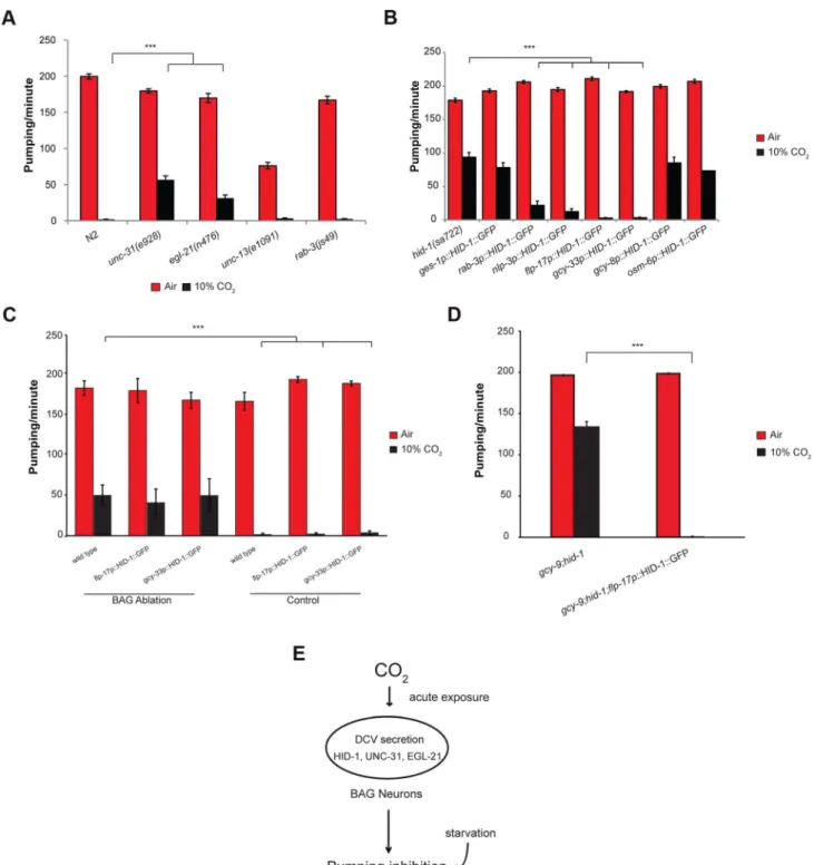
C. elegansortholog of CAPS (calcium-dependent activator protein for secretion), an important component of DCV exocytosis [33]. The geneegl-21encodes theC. elegansortholog of carboxypep-tidase E, an important component in neuropeptide maturation [34]. Following exposure of unc-31(e928) or egl-21(n476)
deletion strains to 10% CO2, the pumping rate of the pharynx
was significantly higher compared with that in wild-type animals exposed to the same concentration of CO2(Figure 4A). We also
tested the role of synaptic vesicle secretion on the pumping response to 10% CO2. The unc-13gene is involved in synaptic
vesicle secretion of neurotransmitters [35,36]. Therab-3gene is a Rab GTPase that affects the distribution of synaptic vesicle populations [37]. Exposure of unc-13(e1091) or rab-3(js49)
mutant strains to 10% CO2showed pumping behavior similar to
that of wild-type strains (Figure 4A). These data suggest that DCVs play an important role in mediating the response of the pharynx to high CO2 levels and that compromising DCV
secretion probably impairs the pumping response to high CO2
levels.
Expression of HID-1 in the BAG neurons is sufficient to restore wild-type CO2response in hid-1mutant strains
HID-1 is expressed in all neuron and gut cells ofC. elegans[30]. To test whether inhibition of pharynx pumping in response to 10% CO2 requires expression of hid-1 in the gut, neurons, or
both, we used transgenic lines that express HID-1 fused to GFP driven by either the pan-neuronal promoter rab-3 or the gut-specific promoter ges-1. Expression of HID-1 under the rab-3
promoter in neurons ofhid-1(sa722) background was sufficient to restore inhibition of the pharynx pumping almost to the levels shown by wild-type animals (Figure 4B). In contrast, expression of HID-1 under theges-1promoter in the gut ofhid-1(sa722) had no significant effect on the response of the pharynx to 10% CO2.
We next asked which subset of neurons is required for mediating the effect of high CO2 levels on the pharynx. The
nlp-3 gene is expressed in sensory neurons (ADF, ASE, ASH, AWB, ASJ, and BAG) as well as in pharyngeal neurons (I1, I2, I3, I4, M1, M3, and NSMR) (Figure S3) [38]. Transgenic expression of HID-1::GFP under the nlp-3 promoter in hid-1(sa722) background was sufficient to restore pharynx pumping inhibition after exposure to 10% CO2 (Figure 4B). High levels of CO2
activate the AFD neurons [23]. Surprisingly, transgenic expression of HID-1::GFP driven by agcy-8promoter in the thermosensory AFD of hid-1(sa722) background did not restore the CO2
-mediated pumping inhibition (Figure 4B), which suggests that the activation of the AFD neurons by high CO2levels is not sufficient
nlp-3are the BAG and the ASE neurons, which are also known to respond to high CO2levels [23]. Transgenic expression of
HID-1::GFP driven by the osm-6promoter, which was expressed in ASE neurons (undetected in BAG neurons), did not restore the CO2-mediated pumping inhibition (Figure 4B). We next tested the
role of BAG neurons in the CO2-dependent pumping inhibition of
the pharynx. Transgenic lines expressing HID-1::GFP under the promoter offlp-17showed expression in the BAG neurons (Figure S3). This expression was sufficient to fully restore the CO2
-dependent pumping inhibition (Figure 4B). Similarly, the expres-sion of HID-1::GFP under thegcy-33promoter in hid-1(sa722) background was specific to the BAG neurons (Figure S3) [21]. This expression was sufficient to fully restore the CO2-dependent
pumping inhibition (Figure 4B). Next, we ablated the BAG
neurons in transgenic worms expressing HID-1::GFP under
flp-17 and gcy-33 promoters in hid-1(sa722) background. We found that following the removal of the HID-1::GFP-expressing BAG neurons, the pumping in 10% CO2was similar to that of
hid-1(sa722) animals (Figure 4C). These results suggest that the specific expression of HID-1 in the BAG neurons is sufficient to induce the CO2-dependent pumping inhibition. We also ablated
the BAG neurons in wild-type background using GFP driven by
gcy-33promoter as a marker. We found that following ablation of the BAG neurons, the pumping in response to 10% CO2 was
similar to that in HID-1-null animals (Figure 4C). These results suggest that the BAG neurons are required for the pumping inhibition. In addition, to test possible cross talk between the neuropeptide secretion pathway and the guanylyl cyclase receptor
Figure 2. The inhibition of pumping following exposure to high CO2level is independent of molecular pathways that regulate CO2 avoidance and oxygen sensing.One-day-old adultC. elegansstrains containing mutations in genes that regulate CO2avoidance (A, B) or O2
sensing (C) were exposed to 10% CO2. The pumping rate was measured under a dissecting microscope during the first minute of exposure to CO2. In
pathway, which is required for CO2avoidance, we measured the
pharyngeal pumping rate of animals carrying both hid-1(sa722) and gcy-9(tm2816)mutations. The pumping rate was similar to that ofhid-1(sa722) animals (Figure 4D). Moreover, in the same genetic background, transgenic expression of HID-1 in the BAG neurons, using the flp-17promoter, restored the suppression of pumping in the presence of high CO2level (Figure 4D), further
demonstrating that the response to CO2 mediated by hid-1 is
independent of the response to CO2 mediated by gcy-9. We
conclude that proper hid-1 activity in the BAG neurons is important to mediate the pumping inhibition by CO2.
Discussion
In humans, high CO2 levels have diverse effects on the lung
epithelium, immunity, and muscle function. However, the effects of acute exposure of muscle cells to high CO2 levels were
unknown. In addition, recent studies suggest that mammals, likeC. elegans, are able to sense elevated CO2levels, which is of broad
physiologic significance.
CO2avoidance and CO2-dependent reduced pharyngeal
pumping are probably regulated via different pathways
Acute exposure of well-fed adultC. elegansanimals to high CO2
levels quickly reduces the pumping rate of the pharynx. This effect depends in part on the nutritional status of the animal, since starved animals exposed to 10% CO2in air continue to pump,
albeit at a significantly slower rate. Our genetic data suggest that the effect of acute exposure to high CO2 levels on the pumping
rate is independent of the avoidance responses of C. elegans to high CO2levels. First, cGMP signaling is required for mediating
the avoidance response, as mutations in the cGMP gated ion channel encoded by tax-2 and tax-4 completely disrupt the avoidance response [2,4]. In contrast, the same mutation intax-4
does not completely rescue the immediate response of the pumping rate to high CO2 levels. Second, mutation in the
insulin-like receptor encoded bydaf-2also disrupts the avoidance response [2,4]. The pumping rate of daf-2 mutants under exposure to 10% CO2is dramatically reduced, like in the
wild-type animals. The limited recovery of the pumping rate indaf-2
mutants at 10% CO2 could be due to the effect of daf-2 on
starvation regulating pathways [39,40]. Third, interference with proper function of ciliated sensory neurons by mutations inosm-3
and che-10 also significantly changes the avoidance response. Again, in the pumping assay, similar mutations in these genes did not change the response of C. elegans to high CO2 levels. In
addition, mutations in ets-5 and gcy-9, which were previously shown to be required for the calcium response of the BAG neurons to CO2, did not change the response of the pharynx to high CO2
levels. Finally, the rescue of pumping byhid-1in the BAG neurons was not affected bygcy-9mutations.
C. elegansanimals presumably interpret high CO2levels as a
harmful cue that leads to avoidance and pumping inhibition. The ability of the animal to stop eating for several minutes probably allows it to avoid undesirable food. Surprisingly, although both the avoidance and the pumping responses to the same stressful cue are immediate, our genetic data suggest that different molecular pathways mediate the two responses to high CO2.
The potential role of neuropeptides in the response of the pharynx to high levels of CO2
Our genetic screen identified hid-1 as a regulator of the pumping response to high CO2levels, as mutations in the hid-1
gene blunted the response of the pharynx to high CO2 levels.
HID-1 is required for the neuropeptide secretion pathway [30,32]. Indeed, mutations in other known genes in peptidergic signaling,
unc-31and egl-21, could also partially suppress the pharyngeal pumping suppression upon exposure to 10% CO2. Neuropeptides
are important signaling molecules in many physiological responses both inC. elegansand in other organisms. InC. elegansthere are more than 250 neuropeptides that play a role in feeding and metabolism, and most neurons inC. eleganssecrete neuropeptides [41]. Neuropeptides are also secreted from the intestine [38], and
hid-1, an important peptidergic signaling gene, is expressed both in the nervous system and in the intestine [30,32]. Neuropeptide signaling was previously shown to regulate pumping inhibition in
Figure 3. HID-1 is required for sensing CO2 level in the pharynx. (A) One-day-old adult hid-1(yg316) and N2 worms were exposed to 5%, 10%, or 20% CO2balanced with 21% O2and N2. The
pumping rate was measured under a dissecting microscope while the animals were exposed to the different gas mixtures. A gas mixture of 21% O2and 79% N2was used as a normal air control. (B) The inhibition
of the pumping rate of the pharynx after exposure to high CO2level in
hid-1(yg316) allele mutants is significantly reduced. Similarly, the inhibition of the pumping rate of the pharynx after exposure to high CO2level is reduced in otherhid-1allele mutants (sa772andsa1058).
Transgenic expression of HID-1 fused to eGFP in thesa722or yg316 background (hid-1(sa722);HID-1::GFP or hid-1(yg316);HID-1::GFP) is sufficient to restore the effect of high CO2level on the pumping rate
back to the wild-type phenotype. In all experiments N$30 animals. Different groups were compared by one-way ANOVA followed byttest. ***P,.001. Error bars indicate SEM.
Figure 4. The effect of high CO2level on the pharynx requires HID-1 activity in the BAG neurons.(A) One-day-old adultC. elegansstrains
containing mutations inunc-31oregl-21genes, which are important for proper neuropeptide secretion and maturation, show a significant rescue of the pumping rate after exposure to 10% CO2. In contrast, strains with mutations inunc-13orrab-3, which promote synaptic vesicle secretion, do not
show a changed pharynx response to 10% CO2. (B) Transgenic expression of HID-1 in the gut using the gut-specificges-1promoter (ges-1p
-HID-1::GFP) was not sufficient to restore pumping phenotype to wild type after exposure to 10% CO2. In contrast, transgenic expression of HID-1 in
neurons using therab-3promoter (rab-3p-HID-1::GFP) was sufficient to restore pumping rate after exposure to 10% CO2almost back to wild-type
levels. Cell-specific expression of HID-1 in the AFD neurons (gcy-8p-HID-1::GFP) or in the amphid and tail ciliated neurons, including ASE neurons (osm-6p-HID-1::GFP), did not restore the CO2effect on the pumping back to wild-type levels, whereas cell-specific expression of HID-1 in sensory and
pharyngeal neurons (nlp-3p-HID-1::GFP) or in BAG neurons (flp-17p-HID-1::GFP andgcy-33p-HID-1::GFP) was sufficient to restore the effect of high CO2
level back to the wild-type phenotype. (C) The BAG neurons of wild-typeC. elegansexpressinggcy-33::GFP were laser ablated and the pharyngeal pumping rate was measured in normal air and 10% CO2. Similarly, the BAG neurons offlp-17p::HID-1::GFP andgcy-33p::HID-1::GFP strains were laser
the absence of food [42]. Specifically, unc-31 mutants demon-strate continuous pumping in the absence of food, unlike wild-type animals [42]. Sincehid-1also partially suppresses the inhibition of pumping in the absence of food (data not shown), we cannot completely rule out the possibility that hid-1 generally inhibits pumping and acts in parallel with CO2.
The pharynx response to 10% CO2 is probably mediated by
several different neuropeptides, since pumping inhibition could not be inhibited by deletion of individual neuropeptide genes known to be overexpressed in the BAG neurons, includingflp-10,
flp-16,flp-27,nlp-1,flp-17, andnlp-14(Figure S4). Neuropep-tide secretion can only partially explain the response of the pharynx to high levels of CO2, since none of the peptidergic
signaling mutants we examined at 10% CO2(hid-1,unc-31, and
egl-21) exhibited the pumping rate seen at normal air levels (Figure 4). Thus we cannot completely rule out the possibility that the effects ofunc-31and egl-21are due to the other pathway(s) that must be acting in parallel with hid-1. This implies the existence of other, HID-1-independent mechanisms that must regulate the response of the pharynx to CO2levels. For example, it
is possible that high CO2 levels trigger other presynaptic inputs
that mediate the effect on the pharynx in parallel with the peptidergic signaling, or that CO2has also a direct postsynaptic
effect on the pharyngeal muscles that inhibits their normal function. Interestingly, such parallel pathways depend on the CO2levels, sincehid-1completely rescues the pumping inhibition
at 5% CO2 and fails to rescue the pumping at 20% CO2
(Figure 3A).
The presence of HID-1 is specifically required in the BAG neurons
Using transgenic lines that express HID-1 either in the gut or in the nervous system we have determined that thehid-1activity is specifically required in neurons to mediate the effect of high CO2
levels on the pharynx. We used an AFD-specific promoter to show that hid-1 activity in the AFD neurons, which are activated by high CO2levels [23], is not sufficient to mediate the effect of high
CO2levels on the pharynx. In contrast, transgenic expression of
HID-1::GFP under the nlp-3 promoter is sufficient to restore CO2-mediated pharynx pumping inhibition. Using the
BAG-specific promoters flp-17 and gcy-33 and performing ablation experiments on the BAG neurons, we have further narrowed the CO2effect on the pharynx to the BAG neurons. Paradoxically,
our genetic data (Figures 2 and 4) suggest the existence of different molecular pathways for the avoidance and the pharynx responses. However, the BAG neurons in the pharynx response are the same neurons that control the avoidance response. Interestingly, mutant animals that block CO2-mediated calcium response in the BAG
neurons still show normal pumping inhibition. The existence of such a pathway is especially surprising given that DCV secretion is expected to depend on an increase in calcium levels.
The physiological and molecular effects of high CO2levels, in
both vertebrates and invertebrates, have been the focus of several recent studies [2,4,6,7,12,14,22,24,43]. However, the sensing mechanism of cells to high CO2 levels is yet largely unknown.
Soluble adenylyl cyclases are bicarbonate sensors in several organisms including mammals [15,44,45]. In C. elegans, which do not have soluble adenylyl cyclases, the soluble guanylyl cyclases GCY-31 and GCY-33 are important for eliciting CO2avoidance
in the BAG neurons [23]. However, it is yet unknown whether the
gcygenes are directly activated by either CO2or HCO32.Our
results show that neither GCY-31 nor GCY-33 are required for mediating the effect of high CO2 levels on the pharynx
(Figure 2C).
Our study sheds new light on the response ofC. elegansto high CO2 levels. It also shows that the CO2-induced response is
differentially regulated across different tissues. Furthermore, different levels of CO2 lead to various outcomes in the same
tissue. Deciphering the mechanisms underlying these fundamental pathways will hopefully help us to better understand the CO2
-induced responses that are activated in human diseases.
Materials and Methods
Strains
Worms were handled as described elsewhere [46]. The following strains were used in this study: N2 (wild type); CF1041, daf-2(e1370); CB3329,che-10(e1809); CX2948,tax-4(p678); PR802,
osm-3(n1540); DA609, npr-1(ad609); CZ3714, gcy-31(ok296); CZ3715,gcy-33(ok232); CX6448,gcy-35(ok769); AX1296, gcy-36(db42); JT722, hid-1(sa722); JT1058, hid-1(sa1058); YG316,
hid-1(yg316); YG2310, hid-1(yg316); jsEx896 [hid-1p ::HID-1::GFP]; NM3017,hid-1(sa722)and lin-15(n765); jsEx896[ hid-1p::HID-1::GFP]; NM3053, hid-1(sa722) and lin-15(n765); jsEx897[rab-3p::HID-1::GFP]; NM3139, hid-1(sa722) and lin-15(n765); jsEx909[ges-1p::HID-1::GFP]; YG2313,hid-1(sa722); ygEx317 [gcy-8p::HID-1::GFP]; YG2318,hid-1(sa722); ygEx318 [nlp-3p::HID-1::GFP]; YG2319, hid-1(sa722); ygEx319 [ flp-17-p::HID-1::GFP]; YG2340,hid-1(sa722); ygEx320 [gcy-33-p ::HID-1::GFP]; DA509,unc-31(e928); KP2018,egl-21(n476); YG2302,
unc-13(e1091); YG2320,ets-5(tm1734); YG2321,ets-5 (tm1755); YG2322, gcy-9(tm2816); YG2323, gcy-9(tm2816) and hid-1(sa722); YG2324, gcy-9(tm2816) and hid-1(sa722); ygEx321 [flp-17-p::HID-1::GFP]; RB1340, nlp-1(ok1469); RB2575, flp-19(ok3587); VC2012,flp-27(gk3331); VC1108,nlp-14(ok1517)/ szT1 X; RB1989,flp-10(ok2624); RB2275, flp-16(ok3085). All strains were obtained from theC. elegansGenome Center (CGC) or the National BioResourse Project (NBRP), except for CX2948, which was kindly provided by the De-Bono laboratory, and NM3017, NM3053, and NM3139, which were kindly provided by the Nonet laboratory [2,30].
Measurement of pumping rate
A standard NGM plate covered with a lid-shaped chamber with inlet and outlet holes to allow gas flow was used to measure the pumping rate under different concentrations of CO2in air. The
chamber was connected to a mechanical valve that controlled the humidified gas mixture entering the chamber. For all pumping assays, NGM plates were seeded with 20mL of OP50 5 h before the experiment to allow normal feeding and to keep worms in a restricted area. A single 1-day-old adult worm was seeded on a plate just before the start of the experiment. Initially, normal air mixture (21% O2, 79% N2) flowed into the chamber and worms
were allowed to adjust for 1 min. The number of pharynx muscle contractions was subsequently measured for 1 min under normal air conditions. Then the airflow was switched to a high-CO2gas
mixture, and after 10 s the pharynx muscle contraction rate was measured again. To measure pumping rate after starvation,
pharynx is mediated by neuropeptide secretion via dense core vesicles (DCVs) in BAG neurons. The CO2response is decreased after starvation. In all
experimentsN$30 animals, except in panel C inflp-17p::HID-1::GFP (N= 5) andgcy-33p::HID-1::GFP (N= 10). Different groups were compared by one-way ANOVA followed byttest. ***P,.001. Error bars indicate SEM.
well-fed wild-type 1-day-old adult worms were collected using M9 buffer and washed four or five times in M9 buffer. Worms were then seeded on either NGM plates with no bacteria or NGM plates seeded with OP50, for 4 h prior to measurements. All pumping assays were performed at 22uC.
EMS screen and SNP mapping
The EMS mutagenesis was performed essentially as described elsewhere [46]. Briefly, wild-type (N2) worms in the L4 stage were exposed to 50 mM EMS in M9 buffer for 4 h and then transferred to fresh plates for 2–3 h (P0) for recovery. After
recovery, five P0animals were transferred again to an NGM plate
and allowed to lay F1 progeny. Adult F1 animals were cloned onto individual NGM plates and their L4-adult F2 progeny where exposed to 10% CO2. F2 worms that continued the
pumping of the pharynx even after exposure to 10% CO2were
isolated. In total, we scored the progeny of ,1200 F1 animals.
The isolated strains were outcrossed three times. The mutation was mapped as described elsewhere [27]. Mutant worms were crossed with the Hawaiian strain and F1 progeny were isolated. Then 44 F2 recombinants that continued the pumping after exposure to 10% CO2 were isolated, and the DNA of their F3
and F4 offspring was extracted using a Gentra Puregene kit (Qiagen, cat. no. 158667). Whole genome sequencing was performed using the Applied Biosystems SOLiD 3 deep sequencing apparatus. The positions of the Hawaiian SNPs were mapped on the DNA of theyg316 strain. A 1.2-MB region in chromosome X that did not contain any Hawaiian SNP was found. Within this region a premature stop codon (W625X) in the coding sequence ofhid-1 was found to cause the phenotype as described in the text.
Plasmid constructs, transgenes, and laser ablation
The NM1699 construct, which contains the hid-1 promoter driving the genomichid-1coding region fused to eGFP, was a kind gift from the Nonet laboratory [30]. The NM1699 construct was digested with KpnI and AatII to replace the nativehid-1promoter with various neuron-specific promoters. To drive AFD-specific expression an 800-bp fragment upstream of thegcy-8start codon was amplified and subsequently digested with KpnI-AatII to generate pKS10. Similarly, to drive sensory and pharyngeal specific expression, a 700-bp fragment upstream of thenlp-3start codon was amplified and digested with KpnI-AatII to generate pKS20. To drive BAG-specific expression a 3.4-kb fragment upstream of the flp-17 start codon was amplified and fused by PCR to HID-1::GFP from NM1699. In addition, to drive BAG-specific expression a 980-bp fragment upstream of thegcy-33start codon was amplified and subsequently digested with KpnI-AatII to generate pKS30. All plasmids were verified by sequencing and microinjected to either JT722 or YG316 with an elt-2::GFP marker as described elsewhere [47].
Laser ablation was performed using an Andor Revolution XD confocal spinning disk system with a Nikon TiE inverted microscope equipped with a nitrogen pulsed laser and a 365-nm Micropoint dye cell. The microscope and laser were controlled by means of IQ software and the Micropoint Mosaic I System 85-75, respectively. The region of interest was set according to the size of the neuron cell body as revealed by the GFP marker. We used a frequency of 15 Hz, an energy range of 80%–90%, and 3–5 repeats in order to completely ablate the GFP marker in the neuron cell body.
Supporting Information
Figure S1 Pumping inhibition is not rescued by either 30 min of exposure to 10% CO2, pH of 5.0 or 7.0, or mutations in the
carbonic anhydrase genes. (A) One-day-old wild-type (N2) adultC. eleganswere continuously exposed to 10% CO2for 30 min and
pumping rate was measured at different time points. (B) One-day-old wild-type (N2) adult C. elegans were transferred to NGM plates buffered at pH of 5.0, 6.0, or 7.0 followed by exposure to 10% CO2 and measurements of the pharyngeal pumping. (C)
One-day-old adult worms with mutations incah-2,cah-5, orcah-6
genes exposed to 10% CO2 showed pharyngeal pumping rate
similar to that of wild-type animals. (PDF)
Figure S2 The egg-laying rate ofhid-1(yg316)animals exposed to 10% CO2is similar to that of wild-type animals. Gravid animals
were exposed to either normal air conditions or air containing 19% or 10% CO2for 6 h. The number of embryos laid during this
period was measured. (PDF)
Figure S3 Transgenic expression of HID-1::GFP. HID-1 fused to eGFP was expressed under its own promoter in the background of yg316 or under gcy-8, nlp-3, osm-6, flp-17, or gcy-33
promoters in the background of sa722. Arrows indicate the AFD neurons (gcy-8p) and BAG neurons (flp-17p and gcy-33p).The expression ofhid-1p,nlp-3pandosm-6pwas detected in several neurons. Scale bar, 10mm.
(PDF)
Figure S4 Animals with deletions in neuropeptide genes expressed in the BAG neurons still show strong CO2-mediated
pumping inhibition. One-day-old animals with mutations in neuropeptide genes, which are known to be overexpressed in the BAG neurons, were exposed to 10% CO2and pumping rate was
measured. The pumping rate ofnlp-1,nlp-14, andflp-16mutants in 10% CO2 (but not in normal air conditions) was significantly
different from that of the wild-type (N2) animals and showed small but significant rescue. *P,.01. Error bars indicate SEM. (PDF)
Movie S1 Pumping of wild-type C. elegans exposed to 10% CO2. Pumping rate of wild-type animal under dissecting
microscope is presented. Worms are first exposed to normal air and then exposed to 10% CO2.
(AVI)
Movie S2 Pumping of hid-1(yg316) mutant exposed to 10% CO2. Pumping rate of hid-1(yg316) mutant animal under
dissecting microscope is presented. Worms are first exposed to normal air and then exposed to 10% CO2.
(AVI)
Acknowledgments
We thank Tamar Gattegno, Veronika Kravtsov, and Benjamin Podbile-wicz for their help with the ablation experiments, Valery Zayat for helping to create the gcy-33-HID-1::GFP strains, Jennifer Davis for editing the manuscript, and the Nonet and DeBono laboratories for providing strains and constructs.
Author Contributions
References
1. Bowen MF (1991) The Sensory Physiology of Host-Seeking Behavior in Mosquitoes. Annual Review of Entomology 36: 139–158.
2. Bretscher AJ, Busch KE, de Bono M (2008) A carbon dioxide avoidance behavior is integrated with responses to ambient oxygen and food in
Caenorhabditis elegans. Proceedings of the National Academy of Sciences 105: 8044–8049.
3. Carrillo M, Guillermin M, Rengarajan S, Okubo R, Hallem E (2013) O2-Sensing Neurons Control CO2 Response in C. elegans. The Journal of neuroscience: the official journal of the Society for Neuroscience 33: 9675–9683. 4. Hallem EA, Sternberg PW (2008) Acute carbon dioxide avoidance in
Caenorhabditis elegans. Proceedings of the National Academy of Sciences 105: 8038–8043.
5. Suh GSB, Wong AM, Hergarden AC, Wang JW, Simon AF, et al. (2004) A single population of olfactory sensory neurons mediates an innate avoidance behaviour in Drosophila. Nature 431: 854–859.
6. Briva A, Vada´sz I, Lecuona E, Welch LC, Chen J, et al. (2007) High CO2Levels
Impair Alveolar Epithelial Function Independently of pH. PLoS ONE 2: e1238. 7. Vada´sz I, Dada LA, Briva A, Trejo HE, Welch LC, et al. (2008) AMP-activated protein kinase regulates CO2-induced alveolar epithelial dysfunction in rats and
human cells by promoting Na,K-ATPase endocytosis. The Journal of Clinical Investigation 118: 752–762.
8. Vohwinkel CU, Lecuona E, Sun H, Sommer N, Vada´sz I, et al. (2011) Elevated CO2Levels Cause Mitochondrial Dysfunction and Impair Cell Proliferation.
Journal of Biological Chemistry 286: 37067–37076.
9. Lecuona E, Sun H, Chen J, Trejo HE, Baker MA, et al. (2013) Protein kinase A-Ialpha regulates Na,K-ATPase endocytosis in alveolar epithelial cells exposed to high CO(2) concentrations. Am J Respir Cell Mol Biol 48: 626–634. 10. Vadasz I, Dada LA, Briva A, Helenius IT, Sharabi K, et al. (2012) Evolutionary
conserved role of c-Jun-N-terminal kinase in CO2-induced epithelial dysfunc-tion. PLoS ONE 7: e46696.
11. Cummins EP, Oliver KM, Lenihan CR, Fitzpatrick SF, Bruning U, et al. (2010) NF-kB Links CO2 Sensing to Innate Immunity and Inflammation in
Mammalian Cells. The Journal of Immunology 185: 4439–4445.
12. Helenius IT, Krupinski T, Turnbull DW, Gruenbaum Y, Silverman N, et al. (2009) Elevated CO2suppresses specific Drosophila innate immune responses
and resistance to bacterial infection. Proceedings of the National Academy of Sciences 106: 18710–18715.
13. Oliver KM, Lenihan CR, Bruning U, Cheong A, Laffey JG, et al. (2012) Hypercapnia Induces Cleavage and Nuclear Localization of RelB Protein, Giving Insight into CO2Sensing and Signaling. Journal of Biological Chemistry
287: 14004–14011.
14. Wang N, Gates KL, Trejo H, Favoreto S, Schleimer RP, et al. (2010) Elevated CO2selectively inhibits interleukin-6 and tumor necrosis factor expression and
decreases phagocytosis in the macrophage. The FASEB Journal 24: 2178–2190. 15. Chen Y, Cann MJ, Litvin TN, Iourgenko V, Sinclair ML, et al. (2000) Soluble adenylyl cyclase as an evolutionarily conserved bicarbonate sensor. Science 289: 625–628.
16. Cook ZC, Gray MA, Cann MJ (2012) Elevated Carbon Dioxide Blunts Mammalian cAMP Signaling Dependent on Inositol 1,4,5-Triphosphate Receptor-mediated Ca2+
Release. Journal of Biological Chemistry 287: 26291–26301.
17. Townsend PD, Holliday PM, Fenyk S, Hess KC, Gray MA, et al. (2009) Stimulation of Mammalian G-protein-responsive Adenylyl Cyclases by Carbon Dioxide. Journal of Biological Chemistry 284: 784–791.
18. Gates KL, Howell HA, Nair A, Vohwinkel CU, Welch LC, et al. (2013) Hypercapnia impairs lung neutrophil function and increases mortality in murine pseudomonas pneumonia. Am J Respir Cell Mol Biol 49: 821–828. 19. Sharabi K, Lecuona E, Helenius IT, Beitel GJ, Sznajder JI, et al. (2009) Sensing,
physiological effects and molecular response to elevated CO2 levels in
eukaryotes. Journal of Cellular and Molecular Medicine 13: 4304–4318. 20. Brandt JP, Aziz-Zaman S, Juozaityte V, Martinez-Velazquez LA, Petersen JG,
et al. (2012) A Single Gene Target of an ETS-Family Transcription Factor Determines Neuronal CO2Chemosensitivity. PLoS ONE 7: e34014.
21. Guillermin ML, Castelletto ML, Hallem EA (2011) Differentiation of Carbon Dioxide-Sensing Neurons in Caenorhabditis elegans Requires the ETS-5 Transcription Factor. Genetics 189: 1327–1339.
22. Hallem EA, Spencer WC, McWhirter RD, Zeller G, Henz SR, et al. (2011) Receptor-type guanylate cyclase is required for carbon dioxide sensation by
Caenorhabditis elegans. Proceedings of the National Academy of Sciences 108: 254–259.
23. Bretscher AJ, Kodama-Namba E, Busch KE, Murphy RJ, Soltesz Z, et al. (2011) Temperature, oxygen, and salt-sensing neurons inC. elegansare carbon dioxide sensors that control avoidance behavior. Neuron 69: 1099–1113.
24. Sharabi K, Hurwitz A, Simon AJ, Beitel GJ, Morimoto RI, et al. (2009) Elevated CO2levels affect development, motility, and fertility and extend life span in
Caenorhabditis elegans. Proceedings of the National Academy of Sciences 106: 4024–4029.
25. Smith ES, Martinez-Velazquez L, Ringstad N (2013) A chemoreceptor that detects molecular carbon dioxide. J Biol Chem 288: 37071–37081.
26. Zimmer M, Gray JM, Pokala N, Chang AJ, Karow DS, et al. (2009) Neurons Detect Increases and Decreases in Oxygen Levels Using Distinct Guanylate Cyclases. Neuron 61: 865–879.
27. Doitsidou M, Poole RJ, Sarin S, Bigelow H, Hobert O (2010)C. elegansMutant Identification with a One-Step Whole-Genome-Sequencing and SNP Mapping Strategy. PLoS ONE 5: e15435.
28. Ailion M, Thomas JH (2003) Isolation and Characterization of High-Temperature-Induced Dauer Formation Mutants inCaenorhabditis elegans. Genetics 165: 127–144.
29. Burgoyne RD, Morgan A (2003) Secretory Granule Exocytosis. Physiological Reviews 83: 581–632.
30. Mesa R, Luo S, Hoover CM, Miller K, Minniti A, et al. (2011) HID-1, a new component of the peptidergic signaling pathway. Genetics 187: 467–483. 31. Wang L, Zhan Y, Song E, Yu Y, Jiu Y, et al. (2011) HID-1 is a peripheral
membrane protein primarily associated with the medial- and trans- Golgi apparatus. Protein Cell 2: 74–85.
32. Yu Y, Wang L, Jiu Y, Zhan Y, Liu L, et al. (2011) HID-1 is a novel player in the regulation of neuropeptide sorting. Biochem J 434: 383–390.
33. Hammarlund M, Watanabe S, Schuske K, Jorgensen EM (2008) CAPS and syntaxin dock dense core vesicles to the plasma membrane in neurons. The Journal of Cell Biology 180: 483–491.
34. Jacob TC, Kaplan JM (2003) The EGL-21 Carboxypeptidase E Facilitates Acetylcholine Release atCaenorhabditis elegansNeuromuscular Junctions. The Journal of Neuroscience 23: 2122–2130.
35. Richmond JE, Davis WS, Jorgensen EM (1999) UNC-13 is required for synaptic vesicle fusion inC. elegans. Nat Neurosci 2: 959–964.
36. Richmond JE, Weimer RM, Jorgensen EM (2001) An open form of syntaxin bypasses the requirement for UNC-13 in vesicle priming. Nature 412: 338–341. 37. Gracheva EO, Hadwiger G, Nonet ML, Richmond JE (2008) Direct interactions between C. elegans RAB-3 and Rim provide a mechanism to target vesicles to the presynaptic density. Neurosci Lett 444: 137–142.
38. Nathoo AN, Moeller RA, Westlund BA, Hart AC (2001) Identification of neuropeptide-like protein gene families in Caenorhabditis elegans and other species. Proceedings of the National Academy of Sciences 98: 14000–14005. 39. Henderson ST BM, Johnson TE (2006) daf-16 protects the nematode
Caenorhabditis elegans during food deprivation. J Gerontol A Biol Sci Med Sci 61: 444–460.
40. Kimura KD, Riddle DL, Ruvkun G (2011) The C. elegans DAF-2 insulin-like receptor is abundantly expressed in the nervous system and regulated by nutritional status. Cold Spring Harb Symp Quant Biol 76: 113–120. 41. Holden-Dye L, Walker RJ (2013) The roles of neuropeptides in Caenorhabditis
elegans including their importance in the regulation of feeding and metabolism. Protein Pept Lett 20: 636–646.
42. Avery L, Bargmann CI, Horvitz HR (1993) The Caenorhabditis elegans unc-31 gene affects multiple nervous system-controlled functions. Genetics 134: 455– 464.
43. Sun L, Wang H, Hu J, Han J, Matsunami H, et al. (2009) Guanylyl cyclase-D in the olfactory CO2neurons is activated by bicarbonate. Proceedings of the
National Academy of Sciences 106: 2041–2046.
44. Klengel T, Liang WJ, Chaloupka J, Ruoff C, Schroppel K, et al. (2005) Fungal adenylyl cyclase integrates CO2 sensing with cAMP signaling and virulence. Curr Biol 15: 2021–2026.
45. Mogensen EG, Janbon G, Chaloupka J, Steegborn C, Fu MS, et al. (2006)
Cryptococcus neoformansSenses CO2through the Carbonic Anhydrase Can2
and the Adenylyl Cyclase Cac1. Eukaryotic Cell 5: 103–111.