Targeted Echocardiographic Screening for
Latent Rheumatic Heart Disease in Northern
Uganda: Evaluating Familial Risk Following
Identification of an Index Case
Twalib Aliku1, Craig Sable2, Amy Scheel2, Alison Tompsett2, Peter Lwabi3, Emmy Okello3,4, Robert McCarter5, Marshall Summar6, Andrea Beaton2*
1School of Medicine, Gulu University, Gulu, Uganda,2Division of Cardiology, Children’s National Health System, Washington, District of Columbia, United States of America,3Uganda Heart Institute, Kampala, Uganda,4School of Medicine, Makerere University, Kampala, Uganda,5Division of Biostatistics and Informatics, Children’s National Health System, Washington, District of Columbia, United States of America,
6Division of Genetics and Metabolism, Children’s National Health System, Washington, District of Columbia, United States of America
*abeaton@childrensnational.org
Abstract
Background
Echocardiographic screening for detection of latent RHD has shown potential as a strategy to decrease the burden of disease. However, further research is needed to determine opti-mal implementation strategies. RHD results from a complex interplay between environment and host susceptibility. Family members share both and relatives of children with latent RHD may represent a high-risk group. The objective of this study was to use echocardio-graphic family screening to determine the relative risk of RHD among first-degree relatives of children with latent RHD compared to the risk in first-degree relatives of healthy peers.
Methodology/Principal Findings
Previous school-based screening data were used to identify RHD positive children and RHD negative peers. All first-degree relatives5 years were invited for echocardiography screening (2012 World Heart Federation Criteria). Sixty RHD positive cases (30 borderline/ 30 definite RHD) and 67 RHD negative cases were recruited. A total of 455/667 (68%) fam-ily members were screened. Definite RHD was more common in childhood siblings of RHD positive compared to RHD negative (p = 0.05). Children with any RHD were 4.5 times as likely to have a sibling with definite RHD, a risk that increased to 5.6 times when considering only cases with definite RHD. Mothers of RHD positive and RHD negative cases had an unexpectedly high rate of latent RHD (9.3%).
Conclusions/Significance
Siblings of RHD positive cases with RHD are more likely to have definite RHD and the rela-tive risk is highest if the index case has definite RHD. Future screening programs should a11111
OPEN ACCESS
Citation:Aliku T, Sable C, Scheel A, Tompsett A, Lwabi P, Okello E, et al. (2016) Targeted Echocardiographic Screening for Latent Rheumatic Heart Disease in Northern Uganda: Evaluating Familial Risk Following Identification of an Index Case. PLoS Negl Trop Dis 10(6): e0004727. doi:10.1371/journal.pntd.0004727
Editor:Joseph M. Vinetz, University of California San Diego School of Medicine, UNITED STATES
Received:January 11, 2016
Accepted:May 2, 2016
Published:June 13, 2016
Copyright:© 2016 Aliku et al. This is an open access article distributed under the terms of the
Creative Commons Attribution License, which permits unrestricted use, distribution, and reproduction in any medium, provided the original author and source are credited.
Data Availability Statement:All relevant data are within the paper and its Supporting Information files.
consider implementation of sibling screening following detection of an RHD positive child. Larger screening studies of adults are needed, as data on prevalence of latent RHD outside of childhood are sparse. Future studies should prioritize implementation research to answer questions of how RHD screening can best be integrated into existing healthcare structures, ensuring practical and sustainable screening programs.
Author Summary
Rheumatic heart disease (RHD) affects at least 33 million people, most of who live in low-resource environments. RHD is a cumulative process and there exists a latent period between early valve damage and presentation with symptoms. Echocardiographic screen-ing (ultrasound of the heart) has proven highly sensitive for latent RHD detection, but implementation research is needed to effectively develop sustainable public health strate-gies. Critical to this research is determining whom to screen. As family members have both a shared environment and shared genetic susceptibility, they may represent a high-risk group that could be targeted once a case of RHD is identified. We conducted an echo-cardiographic family screening study to determine the risk of RHD in families with and without an RHD positive child and found that siblings of children with latent RHD are more likely to have latent RHD themselves. Our data suggest that siblings may represent a particularly high-risk group that could be targeted for echocardiographic screening. Future studies are needed to answer questions of how RHD screening can best be integrated into existing healthcare structures, ensuring practical and sustainable RHD screening
programs.
Introduction
Rheumatic heart disease (RHD), the long-term consequence of acute rheumatic fever (ARF), is the result of a complex interplay between host and environment. Endemic areas are consis-tently marked by poverty, poor sanitation, and limited access to primary healthcare [1]. These factors increase the incidence of group Aβ-hemolytic streptococcal (GAS) carriage, infection, and transmission. Repeated, untreated GAS infections create the substrate for development of ARF, a systemic immune system over-reaction that results, for many, in RHD [2].
However, environmental exposure is only one component of RHD susceptibility. Even in the presence of endemic GAS and poor primary prevention (penicillin for acute streptococcal pharyngitis) not all children are equally at risk. ARF follows only 3–6% of cases of GAS and only 40% of children with ARF develop chronic RHD [3]. Historically, RHD was noted to clus-ter in families, and a meta-analysis of twin studies showed a pooled concordance risk for ARF of 44% in monozygotic twins and 12% in dizygotic twins, giving an estimated heritability of 60%[4]. The majority of these data were captured from observational studies of ARF, and pre-dated routine echocardiography [5].
In many low-resource settings today, presentation with ARF has become rare even as echo-cardiographic screening of school-aged children has revealed a large burden of latent RHD (RHD apparent on echocardiography that has not previously come to clinical attention). Given what is known about genetic susceptibility and a shared environment, it is reasonable to assume that family members of children with latent RHD may themselves be at greater risk of latent RHD. However contemporary echocardiographic screening of families living in RHD
CS) and General Electric who provided the echocardiography equipment used in this study (AB, CS). The funders played no role in the study design, data collection and analysis, decision to publish, or preparation of the manuscript.
Competing Interests:The authors have declared
endemic areas has not been reported. The objective of this study was to use echocardiographic family screening to determine the relative risk of RHD among first-degree relatives of children with latent RHD compared to the risk in first-degree relatives of healthy peers.
Materials and Methods
General Design
We utilized a cross-sectional family design to compare the risk of RHD among first-degree family members of primary school children previously identified with latent RHD compared to the first-degree family members of age/gender matched children with normal echocardio-grams. The study occurred over a 3-month period from February-April, 2015. Informed con-sent was obtained from all participants at least 18 years of age, and informed ascon-sent and parental permission was obtained for those between 5–17 years. Approval for this study was granted from the Institutional Review Boards at Children’s National Health System, Washing-ton DC, Makerere University School of Medicine, Kampala, Uganda, and the Ugandan National Council of Science and Technology.
Study Population
RHD positive index cases included children with borderline or definite RHD (2012 WHF crite-ria), identified through previous echocardiographic school screening programs in the Gulu District of Northern Uganda in 2014.[6] These children are followed clinically at the Gulu Regional Referral Hospital, Gulu, Uganda. RHD negative index cases were recruited from screen-negative peers who were similar in age and gender and attending the same schools (reflecting the same general socioeconomic status). All RHD positive and RHD negative index children underwent repeated echocardiographic evaluation at time of study enrollment to ensure their RHD status had not changed since first screen in 2014.
The parents/guardians of RHD positive and RHD negative index children were approached to invite them, and all first-degree relatives (5 years of age) in the family, to undergo echocar-diographic screening to evaluate for the presence of latent RHD. Children without at least one parent alive/available were excluded.
Clinical Data
Following family recruitment, a list of all first-degree family members (at least 5 years of age)–
living or deceased was captured. For living family members, age, gender, and known history of ARF/RHD were recorded. For family members who were deceased, attempts were made to understand the cause of death. For those family members who were alive but unavailable for screening, the reason for absence was recorded.
Echocardiographic Protocol
other for subjects20 years of age and normal, definite RHD, or other for subjects>20 years
of age)[7]. All positive studies were confirmed by a second reviewer and, in cases of disagree-ment, a third reviewer determined the final classification.
Statistical Analysis
Demographic information is presented by number and percentage, and where applicable with stan-dard deviation. Continuous variables were compared using Student’s t-test. Fisher’s exact tests were used to evaluate the exact probability under the null hypothesis of observing results as or more extreme based on comparisons of differences in categorical variables between study groups. Poisson Regression was used to estimate the average risk and relative risk of RHD positivity in first-degree family members of RHD positive and RHD negative index children. The method of Poisson Regres-sion was chosen because it appropriately handles outcomes based on counts, including low fre-quency counts, that generally do not meet criteria for tests requiring the data to meet the parametric assumption and appropriately accounts for the natural clustering of family data. Prevalence rates of RHD among mothers vs. fathers vs. siblings are presented as percentages and compared using model z-statistics. Relative risk was also presented according to the presence of borderline vs. defi-nite RHD in index children. Agreement between reviewers was calculated with the Kappa statistic. Greatest emphasis was placed on results that achieved statistical significance at the p<0.05 level, but
substantive differences that achieved borderline significance were also described.
Results
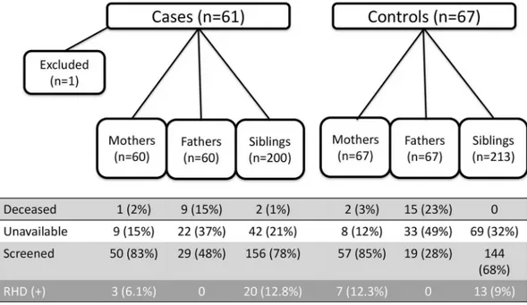
The index group consisted of 61 RHD positive children (30 with definite RHD and 31 with borderline RHD) and 67 RHD negative children (Table 1), generating a complete list of 320 (5.3/child) and 347 (5.2/child) first-degree family members of RHD positive and RHD negative index subjects, respectively, p = 0.22. During enrollment, 1 child (borderline RHD) was excluded from participation when no biological parent could attend screening–leaving 60 RHD positive index cases and 67 RHD negative index cases.
Of the 667 identified first-degree relatives, 455 (68.2%) attended screening including 107 mothers (83.5%), 48 fathers (37.8%), and 300 siblings (72.6%) (Table 2). Fathers (24/127, 19%) were more likely to be deceased than mothers (3/127, 2.4%, p<0.01), with the reasons for
paternal death including HIV/AIDS (6), accidental trauma (5), other illness (5), the LRA con-flict (3), and other/unknown (5). Fathers who were alive were also less likely to be available for screening (55/103, 53% unavailable) than mothers (17/124, 14% unavailable, p<0.01). Siblings
of RHD negative cases were less available to participate in screening (p = 0.03). A breakdown of reasons for all family members who were alive but unavailable and those who are deceased are listed (Table 3). No absent family members were reported to have cardiovascular symptoms and no causes of death were known to be attributable to cardiac disease.
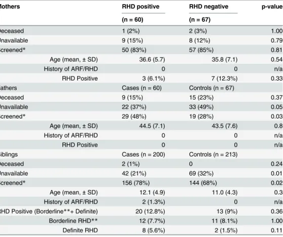
Table 2. Demographics of First-Degree Relatives.
Mothers RHD positive RHD negative p-value
(n = 60) (n = 67)
Deceased 1 (2%) 2 (3%) 1.00
Unavailable 9 (15%) 8 (12%) 0.79
Screened* 50 (83%) 57 (85%) 0.81
Age (mean,±SD) 36.6 (5.7) 35.8 (7.1) 0.54
History of ARF/RHD 0 0 n/a
RHD Positive 3 (6.1%) 7 (12.3%) 0.33
Fathers Cases (n = 60) Controls (n = 67)
Deceased 9 (15%) 15 (23%) 0.37
Unavailable 22 (37%) 33 (49%) 0.05
Screened* 29 (48%) 19 (28%) 0.03
Age (mean,±SD) 44.5 (7.1) 43.5 (7.6) 0.8
History of ARF/RHD 0 0 n/a
RHD Positive 0 0 n/a
Siblings Cases (n = 200) Controls (n = 213)
Deceased 2 (1%) 0 0.24
Unavailable 42 (21%) 69 (32%) 0.01
Screened* 156 (78%) 144 (68%) 0.02
Age (mean,±SD) 12.1 (4.9) 11.0 (4.3) 0.3
History of ARF/RHD 2 (1.3%) 0 n/a
RHD Positive (Borderline**+ Definite) 20 (12.8%) 13 (9%) 0.36
Borderline RHD** 12 (7.7%) 11 (8.1%) 1.00
Definite RHD 8 (5.6%) 2 (1.5%) 0.11
*Details only forfirst-degree relatives who were screened
**Only in siblings18 years ARF: Acute Rheumatic Fever RHD: Rheumatic Heart Disease
doi:10.1371/journal.pntd.0004727.t002
Table 1. Demographics of Index Cases.
Index Cases RHD positive*(n = 60) RHD negative (n = 67) p-value
Age (mean,±SD) 12.5 (2.4) 12.4 (2.4) 1.00
Gender (n, % Female) 60% (n = 36) 59.7% (n = 40) 0.29
# First Degree Relatives (n, mean) 320 (5.3) 347 (5.2) 1.00
*Not including 1 case of borderline RHD excluded for no biological parents.
The prevalence of all latent RHD was similar in first-degree relatives of RHD positive and RHD negative cases 9.8% vs. 9.0% (23/235 screened vs. 20/220 screened, p = 0.87). Similarly there was no difference between prevalence of definite latent RHD between groups (Cases: 11/235, 4.3% vs. Controls: 9/220, 4.1%, p = 1.00). Definite RHD was more likely to be found in mothers, with 9.3% (10/107 screened) having echocardiographic evidence of definite RHD, compared to fathers 0% (0/48 screened, p = 0.03), and siblings 3.3% (10/300 screened, p = 0.02). Borderline RHD, a category reserved only for those20 years of age, was similar prevalence between siblings of RHD positive vs. RHD negative (7.7% vs. 8.1%, p = 1.00). How-ever, definite RHD was more common among siblings of RHD positive cases (5.2% vs. 1.4%, p = 0.11), but only reached borderline significance. There were 7 families (4 cases, 3 controls) where 2 or more first-degree relatives were found to be RHD positive (Fig 2).
There was no increased familial, or sibling risk of RHD in the first-degree relatives of RHD positive cases (borderline & definite RHD) vs. RHD negative cases. However, RHD positive cases had a 4.5 times greater chance of having a sibling with definite RHD (p = 0.05) and this risk increased to 5.6 times greater chance if you limited the comparison to RHD positive cases with definite RHD (n = 30, p = 0.03) (Table 4).
There was 97% agreement between reviewers 1 and 2 (κ= 0.86, 95% CI 0.78–0.93), with 13 cases of non-agreement adjudicated by the third reviewer. All cases of non-agreement were between the diagnoses of“borderline RHD”or“normal”with 100% agreement on the diagno-sis of definite RHD.
Discussion
This is the first study to assess the utility of echocardiography screening of first-degree relatives of children with latent RHD. Siblings of RHD positive cases with any RHD are more likely to have definite RHD and the relative risk goes from 4.5 to 5.6 if the index case has definite RHD. Additionally, we found that nearly 10% of mothers had latent RHD by echocardiography while no fathers were positive. Unlike our sibling results, the likelihood of a mother being positive for RHD has no association with a positive index case.
Echocardiographic screening has shown potential as a public health strategy to decrease the global burden of RHD. Population studies, mostly involving schoolchildren, have revealed a weighted pooled prevalence of 1.3% of children living in endemic areas show evidence of latent RHD [8]. Early detection of these children provides the opportunity for secondary prophylaxis,
Table 3. Breakdown of Reasons for Unavailability or Death.
Death
HIV/AIDS Accident LRA Conflict Other Illness Other/
Unknown
Total
Father 6 5 3 5 5 24
Mother 2 1 3
Sibling 1 1 2
Alive but Unavailable
Distance (lives/works far from home)
Divorced/Not in Contact with Family
Could not get off work
Boarding School/School in another city
Other/ Unknown
Total
Father 20 7 22 0 6 55
Mother 0 4 6 0 7 17
Sibling 9 0 7 56 39 111
LRA: Lord’s Resistance Army
monthly penicillin injections that prevent recurrent streptococcal infections, rheumatic fever, and further valve damage. This is of particular importance in areas such as sub-Saharan Africa where RHD remains endemic, ARF rarely comes to clinical attention [9], and RHD patients most commonly present late, with advanced disease and resulting complications [10].
Optimal implementation strategies, the who, when, in what setting, and how often to screen, have received little study to date, yet these details are critical to developing cost-effective and sustainable screening programs. Our study suggests that siblings of children identified with latent RHD are a high-risk group, and should be prioritized for screening. Siblings of index controls showed a 1.4% prevalence of definite RHD, which is comparable to previously pub-lished data from the pediatric population in Gulu, Uganda [6]. In contrast, siblings of RHD positive cases were found to have a 5.2% prevalence of latent, but definite RHD–and to be at 4.5–5.6 times risk, depending if you included index cases with borderline and definite RHD or
Table 4. Relative Risk of Rheumatic Heart Disease.
Relative Risk
p-value
If any RHD–risk of having another affectedfirst-degree relative (23/235 vs. 20/ 220)
1.05 (0.6– 1.8)
0.85
If any RHD–risk of having a family member with definite RHD (11/255 vs. 9/232) 1.2 (0.54– 2.8)
0.61
If any RHD–risk of having a sibling with definite RHD (8/60 vs. 2/67) 4.5 (1–20.2) 0.052 If definite RHD–risk of having a sibling with definite RHD (5/30 vs. 2/67) 5.6 (1.1–
27.2)
0.033
RHD: Rheumatic Heart Disease
doi:10.1371/journal.pntd.0004727.t004
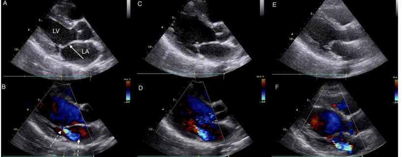
Fig 2. Images from a family with 3 RHD positive members: Index Case, Sibling and Mother.Parasternal long axis echocardiographic still frames in early systole in black and white (panels A, C, and E) and color Doppler (panels B, D, and F) of RHD positive index case (panels A and B), sibling (panels C and D) and mother panels (E and F). The black and white panels all show a thickened anterior mitral valve leaflet (solid arrow) and prolapse (excessive tip motion) of tip of anterior mitral valve leaflet. The mother has the most extreme thickening of the valve along with thickened chordae and immobile valve motion (not seen on these frames). The color Doppler panels show similar mitral regurgitation jets (dotted arrows) measuring approximately 3 cm in each subject. Both RHD positive index case and sibling also had aortic insufficiency jet measuring>1 cm (not shown). Abbreviations: LA–left atrium, LV–left
ventricle.
only those with definite RHD respectively. In a resource-constrained setting, identification of this increased risk could translate into strategic targeting of siblings for single or repeated echo-cardiographic screening and for education on primary prevention. Strategies such as this would need formal evaluation, but hold promise to save financial and human resources.
There is a similar precedent from the World Health Organization for prioritizing household contacts when an index case of tuberculosis is identified [11]. In tuberculosis, household screening has been shown to dramatically improve prevention, early diagnosis, and outcomes [12,13]. Similar to tuberculosis, RHD has a strong environmental component. RHD originates from group A Streptococcus (GAS), which is endemic in areas of poverty and overcrowding. While the risk of invasive GAS infection among household contacts is only mildly increased when an index case is identified [14], it is likely that family members are exposed to the same streptococcal strains at a similar frequency over time. Thus, while familial chemoprophylaxis is not recommended for individual invasive streptococcal infections [15], our data suggest famil-ial echocardiographic screening, once RHD is identified, may be worthwhile.
In addition to a shared environment, first-degree relatives also share genetic susceptibility, which is thought to play a crucial role in RHD development [2,16]. Engel et al. reported a meta-analysis of twin-studies that included 435 twin pairs between 1933 and 1964[4]. The pooled concordance risk for ARF was 44% in monozygotic twins and 12% in dizygotic twins (OR 6.39, p<0.001), with an estimated heritability of 60%. To date, targeted genomic
investiga-tions have examined select genes involved in immune regulation including human leukocyte antigens (HLA), transforming growth factor-beta1, toll-like receptor 5, angiotensin I-convert-ing enzyme gene, PTPN22, and signal transducers and activators of transcription (STATs) gene polymorphisms [17–23]. The most robust data comes from studies of the major histo-compatibility complex human leukocyte antigens [3,24], with many finding polymorphisms within the HLA-DR locus, including a study from Uganda [25]. However, most of these inves-tigations have been small and no single or combination haplotype has consistently emerged [3,
24]. In this study, we cannot separate the influence of host susceptibility from that of the shared environment; our phenotypic data supports the concept of genetic predisposition in RHD. Future genome-wide association studies are needed to link phenotype to genotype and eluci-date the drivers of familial susceptibility.
While not the primary objective of this investigation, our study design captured some of the first data on the community burden of RHD in adults. We found an unanticipated high preva-lence of latent RHD among adult females (10/107 or 9.3% of those screened), while no cases were identified in adult males (0/43 screened). These findings are similar to those by Paar et al, who reported a preponderance of female adults both available for screening and having RHD in Nicaragua (91% of all adult cases of definite RHD)[26].
Our study has several limitations. While we were able to capture most mothers and over three-quarters of siblings for screening, we were able to screen less than half of the fathers. This was due to both higher levels of paternal mortality and for those fathers who were alive, high rates of living and working outside of Gulu. Absentee rates for fathers were comparable between cases and controls, but we cannot accurately determine prevalence of latent RHD among fathers. Additionally, a greater number of siblings were captured from RHD positive compared to RHD negative cases. While there was no difference in death rates between siblings and no evidence of poorer cardiovascular health captured among reasons for sibling non-attendance, the impact of this difference cannot be known. We included as RHD positive index cases both children identi-fied with borderline and with definite RHD. While our most significant findings were associated with a sub-analysis of siblings of definite RHD cases, it is possible that the inclusion of borderline cases weakened our overall phenotype, and study power, which could result in underestimation of risk. It is also important to remember that the total number of RHD cases in our families was small, leading to wide confidence intervals and less certainty in our findings, making them more hypothesis-generating than definitive. Finally, we did not specially control for variations in socio-economic status level between families. However, index controls were recruited from the same schools as cases to indirectly equilibrate socio-economic conditions between groups, and the number of people per household did not differ between cases and controls.
It is also necessary to point out that many important practical and logistical questions remain before a public health strategy that includes echocardiographic screening can be broadly recommended. Our study demonstrates that, given the right resources, large-scale echocardiographic screening is feasible in low-income impoverished areas of the world. How-ever, healthcare resources in most endemic areas are highly constrained, and lack of human and financial resources commonplace. Studies examining optimal training strategies and use of less-expensive handheld echocardiography in the hands of non-experts are beginning to address these barriers [31–34]. The natural history of latent RHD, in particular the category of borderline RHD, is unknown. Natural history studies of children with latent RHD are ongoing and may provide answers [32,35,36,37].
Conclusions
In conclusion, siblings of RHD positive cases with any RHD are more likely to have definite RHD and the relative risk is highest if the index case has definite RHD. Future screening pro-grams should consider implementation of sibling screening following detection of an RHD posi-tive index case. Follow-up of this cohort is needed to determine if latent RHD that exists in more than one family member is more likely to persist and progress. Future studies should prioritize implementation research to answer questions of how RHD screening can best be integrated into existing healthcare structures, ensuring practical and sustainable screening programs.
Supporting Information
S1 Table. World Heart Federation Criteria for the Echocardiographic Diagnosis of Latent Rheumatic Heart Disease (RHD).
(DOCX)
Acknowledgments
Author Contributions
Conceived and designed the experiments: TA CS AS PL EO MS AB. Performed the experi-ments: TA AS AT AB. Analyzed the data: TA CS RM AB. Wrote the paper: TA CS AS AT PL EO MS RM AB.
References
1. Carapetis JR, Steer AC, Mulholland EK, Weber M. The global burden of group A streptococcal dis-eases. Lancet Infect Dis. 2005; 5(11):685–94. PMID:16253886
2. Carapetis JR, McDonald M, Wilson NJ. Acute rheumatic fever. Lancet. 2005; 366(9480):155–68. PMID:16005340
3. Bryant PA, Robins-Browne R, Carapetis JR, Curtis N. Some of the people, some of the time: suscepti-bility to acute rheumatic fever. Circulation. 2009; 119(5):742–53. doi:10.1161/CIRCULATIONAHA. 108.792135PMID:19204317
4. Engel ME, Stander R, Vogel J, Adeyemo AA, Mayosi BM. Genetic susceptibility to acute rheumatic fever: a systematic review and meta-analysis of twin studies. PLoS One. 2011; 6(9):e25326. doi:10. 1371/journal.pone.0025326PMID:21980428
5. Davies AM, Lazarov E. Heredity, infection and chemoprophylaxis in rheumatic carditis: an epidemiolog-ical study of a communal settlement. J Hyg (Lond). 1960; 58(3):263–76.
6. Beaton A, Lu JC, Aliku T, Dean P, Gaur L, Weinberg J, et al. The utility of handheld echocardiography for early rheumatic heart disease diagnosis: a field study. Eur Heart J Cardiovasc Imaging. 2015; 16 (5):475–82. doi:10.1093/ehjci/jeu296PMID:25564396
7. Remenyi B, Wilson N, Steer A, Ferreira B, Kado J, Kumar K, et al. World Heart Federation criteria for echocardiographic diagnosis of rheumatic heart disease—an evidence-based guideline. Nat Rev Car-diol. 2012; 9(5):297–309. doi:10.1038/nrcardio.2012.7PMID:22371105
8. Rothenbuhler M, O'Sullivan CJ, Stortecky S, Stefanini GG, Spitzer E, Estill J, et al. Active surveillance for rheumatic heart disease in endemic regions: a systematic review and meta-analysis of prevalence among children and adolescents. Lancet Glob Health. 2014; 2(12):e717–26. doi:10.1016/S2214-109X (14)70310-9PMID:25433627
9. Zhang W, Mondo C, Okello E, Musoke C, Kakande B, Nyakoojo W, et al. Presenting features of newly diagnosed rheumatic heart disease patients in Mulago Hospital: a pilot study. Cardiovasc J Afr. 2013; 24(2):28–33. doi:10.5830/CVJA-2012-076PMID:23612950
10. Okello E, Wanzhu Z, Musoke C, Twalib A, Kakande B, Lwabi P, et al. Cardiovascular complications in newly diagnosed rheumatic heart disease patients at Mulago Hospital, Uganda. Cardiovasc J Afr. 2013; 24(3):80–5. doi:10.5830/CVJA-2013-004PMID:23736132
11. World Health Organization. Systematic screening for active tuberculosis: an operational guide.http:// apps.who.int/iris/bitstream/10665/181164/1/9789241549172_eng.pdf?ua=12015.
12. Khaparde K, Jethani P, Dewan PK, Nair SA, Deshpande MR, Satyanarayana S, et al. Evaluation of TB Case Finding through Systematic Contact Investigation, Chhattisgarh, India. Tuberc Res Treat. 2015; 2015:670167. doi:10.1155/2015/670167PMID:26236503
13. Pothukuchi M, Nagaraja SB, Kelamane S, Satyanarayana S, Shashidhar, Babu S, et al. Tuberculosis con-tact screening and isoniazid preventive therapy in a South Indian district: operational issues for program-matic consideration. PLoS One. 2011; 6(7):e22500. doi:10.1371/journal.pone.0022500PMID:21799875 14. Davies HD, McGeer A, Schwartz B, Green K, Cann D, Simor AE, et al. Invasive group A streptococcal infections in Ontario, Canada. Ontario Group A Streptococcal Study Group. N Engl J Med. 1996; 335 (8):547–54. PMID:8684408
15. Robinson KA, Rothrock G, Phan Q, Sayler B, Stefonek K, Van Beneden C, et al. Risk for severe group A streptococcal disease among patients' household contacts. Emerg Infect Dis. 2003; 9(4):443–7. PMID:12702224
16. Azevedo PM, Pereira RR, Guilherme L. Understanding rheumatic fever. Rheumatol Int. 2012; 32 (5):1113–20. doi:10.1007/s00296-011-2152-zPMID:21953302
17. Gupta U, Mir SS, Chauhan T, Garg N, Agarwal SK, Pande S, et al. Influence of protein tyrosine phos-phatase gene (PTPN22) polymorphisms on rheumatic heart disease susceptibility in North Indian popu-lation. Tissue Antigens. 2014; 84(5):492–6. doi:10.1111/tan.12440PMID:25273327
19. Gupta U, Mishra A, Rathore SS, Agarwal SK, Pande S, Garg N, et al. Association of angiotensin I-con-verting enzyme gene insertion/deletion polymorphism with rheumatic heart disease in Indian population and meta-analysis. Mol Cell Biochem. 2013; 382(1–2):75–82. doi:10.1007/s11010-013-1719-2PMID: 23749169
20. Mohamed AA, Rashed LA, Shaker SM, Ammar RI. Association of tumor necrosis factor-alpha polymor-phisms with susceptibility and clinical outcomes of rheumatic heart disease. Saudi Med J. 2010; 31 (6):644–9. PMID:20563362
21. Kamal H, Hussein G, Hassoba H, Mosaad N, Gad A, Ismail M. Transforming growth factor-beta1 gene C-509T and T869C polymorphisms as possible risk factors in rheumatic heart disease in Egypt. Acta Cardiol. 2010; 65(2):177–83. PMID:20458825
22. Chou HT, Tsai CH, Tsai FJ. Association between angiotensin I-converting enzyme gene insertion/dele-tion polymorphism and risk of rheumatic heart disease. Jpn Heart J. 2004; 45(6):949–57. PMID: 15655270
23. Zhu L, Zou LJ, Hua R, Li B. Association of single-nucleotide polymorphisms in toll-like receptor 5 gene with rheumatic heart disease in Chinese Han population. Int J Cardiol. 2010; 145(1):129–30. doi:10. 1016/j.ijcard.2009.06.046PMID:19616328
24. Gerbase-DeLima M, Scala LC, Temin J, Santos DV, Otto PA. Rheumatic fever and the HLA complex. A cosegregation study. Circulation. 1994; 89(1):138–41. PMID:8281640
25. Okello E, Beaton A, Mondo CK, Kruszka P, Kiwanuka N, Odoi-Adome R, et al. Rheumatic heart dis-ease in Uganda: the association between MHC class II HLA DR alleles and disdis-ease: a case control study. BMC Cardiovasc Disord. 2014; 14:28. doi:10.1186/1471-2261-14-28PMID:24581333 26. Paar JA, Berrios NM, Rose JD, Caceres M, Pena R, Perez W, et al. Prevalence of rheumatic heart
dis-ease in children and young adults in Nicaragua. Am J Cardiol. 2010; 105(12):1809–14. doi:10.1016/j. amjcard.2010.01.364PMID:20538135
27. Patel S, Schechter MT, Sewankambo NK, Atim S, Lakor S, Kiwanuka N, et al. War and HIV: sex and gender differences in risk behaviour among young men and women in post-conflict Gulu District, North-ern Uganda. Glob Public Health. 2014; 9(3):325–41. doi:10.1080/17441692.2014.887136PMID: 24580099
28. Accorsi S, Fabiani M, Nattabi B, Corrado B, Iriso R, Ayella EO, et al. The disease profile of poverty: mor-bidity and mortality in northern Uganda in the context of war, population displacement and HIV/AIDS. Trans R Soc Trop Med Hyg. 2005; 99(3):226–33. PMID:15653126
29. Diao M, Kane A, Ndiaye MB, Mbaye A, Bodian M, Dia MM, et al. Pregnancy in women with heart dis-ease in sub-Saharan Africa. Arch Cardiovasc Dis. 2011; 104(6–7):370–4. doi:10.1016/j.acvd.2011.04. 001PMID:21798468
30. Hameed A, Karaalp IS, Tummala PP, Wani OR, Canetti M, Akhter MW, et al. The effect of valvular heart disease on maternal and fetal outcome of pregnancy. J Am Coll Cardiol. 2001; 37(3):893–9. PMID:11693767
31. Lu JC, Sable C, Ensing GJ, Webb C, Scheel J, Aliku T, et al. Simplified rheumatic heart disease screen-ing criteria for handheld echocardiography. J Am Soc Echocardiogr. 2015; 28(4):463–9. doi:10.1016/j. echo.2015.01.001PMID:25660669
32. Beaton A, Okello E, Aliku T, Lubega S, Lwabi P, Mondo C, et al. Latent rheumatic heart disease: out-comes 2 years after echocardiographic detection. Pediatr Cardiol. 2014; 35(7):1259–67. doi:10.1007/ s00246-014-0925-3PMID:24827080
33. Mirabel M, Bacquelin R, Tafflet M, Robillard C, Huon B, Corsenac P, et al. Screening for rheumatic heart disease: evaluation of a focused cardiac ultrasound approach. Circ Cardiovasc Imaging. 2015; 8 (1).
34. Engelman D, Kado JH, Remenyi B, Colquhoun SM, Watson C, Rayasidamu SC, et al. Teaching focused echocardiography for rheumatic heart disease screening. Ann Pediatr Cardiol. 2015; 8 (2):118–21. doi:10.4103/0974-2069.157024PMID:26085762
35. Bhaya M, Beniwal R, Panwar S, Panwar RB. Two years of follow-up validates the echocardiographic criteria for the diagnosis and screening of rheumatic heart disease in asymptomatic populations. Echo-cardiography. 2011; 28(9):929–33. doi:10.1111/j.1540-8175.2011.01487.xPMID:21854437
36. Saxena A, Ramakrishnan S, Roy A, Seth S, Krishnan A, Misra P, et al. Prevalence and outcome of sub-clinical rheumatic heart disease in India: the RHEUMATIC (Rheumatic Heart Echo Utilisation and Moni-toring Actuarial Trends in Indian Children) study. Heart. 2011; 97(24):2018–22. doi: 10.1136/heartjnl-2011-300792PMID:22076022