SELECTION OF BIOSURFACTAN/BIOEMULSIFIER-PRODUCING BACTERIA FROM HYDROCARBON-CONTAMINATED SOIL
Sabina Viramontes-Ramos, Martha Cristina Portillo-Ruiz, María de Lourdes Ballinas-Casarrubias, José Vinicio Torres-Muñoz, Blanca Estela Rivera-Chavira, Guadalupe Virginia Nevárez-Moorillón*
Facultad de Ciencias Químicas, Universidad Autónoma de Chihuahua. P.O. Box 1542- C Chihuahua, Chih. Mexico.
Submitted: April 19, 2009; Returned to authors for corrections: October 08, 2009; Approved: February 18, 2010.
ABSTRACT
Petroleum-derived hydrocarbons are among the most persistent soil contaminants, and some
hydrocarbon-degrading microorganisms can produce biosurfactants to increase bioavailability and degradation. The aim
of this work was to identify biosurfactant-producing bacterial strains isolated from
hydrocarbon-contaminated sites, and to evaluate their biosurfactant properties. The drop-collapse method and minimal
agar added with a layer of combustoleo were used for screening, and positive strains were grown in liquid
medium, and surface tension and emulsification index were determined in cell-free supernantant and cell
suspension. A total of 324 bacterial strains were tested, and 17 were positive for the drop-collapse and
hydrocarbon-layer agar methods. Most of the strains were Pseudomonas, except for three strains (Acinetobacter, Bacillus, Rhodococcus). Surface tension was similar in cell-free and cell suspension measurements, with values in the range of 58 to 26 (mN/m), and all formed stable emulsions with motor
oil (76-93% E24). Considering the variety of molecular structures among microbial biosurfactants, they
have different chemical properties that can be exploited commercially, for applications as diverse as
bioremediation or degradable detergents.
Key words: biosurfactants, bioemulsifiers, soil microbiota, hydrocarbon contamination.
INTRODUCTION
The release of contaminants to the environment, including
petroleum and petroleum-derived products, is one of the main
causes of global contamination (29). It is also a risk for human
and animal health, since many of these contaminants have
demonstrated to be toxic and carcinogenic (27). Hydrocarbon
molecules that are released into the environment are hard to
remove, since they adsorb to surfaces and are trapped by
capillarity in a water–immiscible phase. Bioremediation has
proven to be an alternative to diminish the effects caused by
hydrocarbon contamination of soil and water, using the
metabolic capacities of microorganisms that can use
hydrocarbons as source of carbon and energy, or that can
modify them by cometabolism. The efficiency of removal is
directly related to the compounds’ chemical structure, to its
bioavailabity (concentration, toxicity, mobility and access) and
to the physicochemical conditions present in the environment
(10).
Biosurfactants are produced by many organisms, in order
to metabolize water-immiscible substrates, allowing its
adsorption, emulsification or dispersion. For the
microorganisms, production of biosurfactants is an advantage
in soil, giving them advantages in specific conditions (30).
Microbial biosurfactants are mainly produced by aerobic
microorganisms, using as carbon sources carbohydrates,
hydrocarbons, animal or vegetal oils or a mixture of them (6,
14). Biosurfactants can be intracellular (remain attached to the
cell wall) and/or can be excreted to the media (1). When the
biosurfactants are intracellular, their structure include
membrane lipids, and promote the transport of insoluble
substrates through the membrane; when they are extracellular,
the biosurfactants help on the substrate solubilization and are
usually a complex structure of lipids, proteins and
carbohydrates (27). The main difference in the chemical nature
of the different biosurfactant molecules is in the hydrophilic
head; allowing for a wide range of variation in their physical
and biological properties (20).
In the literature, the terms biosurfactant and bioemulsifier
are considered interchangeable, but although all bioemulsifiers
are considered biosurfactants, not all the biosurfactants produce
stable emulsions. Biosurfactants can reduce the surface tension
between two liquids, and bioemulsifiers induce a dispersion of
undissolved material throughout the liquid, by formation and
stabilization of droplets of the dispersed phase (13, 14).
Microbial biosurfactants are classified by its chemical
composition and its microbial origin (13). Low molecular
weight (glycolipidis or lypopeptides) can diminish surface
tension, but does not form stable emulsions (10). On the other
hand, biopolymers are less effective on lowering the surface
tension, but are highly effective on the production of
emulsions, and have a considerable specificity for the substrate
(30).
The search for biosurfactant-producing microbial strains is
still an interesting area of research (3), because of the diversity
of the molecules and the wide variety of uses that those
molecules have. There are several methods used for screening
and isolation of interesting bacterial strains; an easy and
common method is the drop-collapse test (5), and it has been
described that the carbon source used to grow the
microorganisms is important on the identification of
biosurfactat producing strains (6). Therefore, the aim of this
work was to identify biosurfactant and/or bioemulsifier
producing bacteria from a collection of soil bacterial isolates
obtained from hydrocarbon-contaminated sites.
METHODS
Bacterial strains
Bacterial strains were isolated from soil contaminated with
either combustoleo (Fuel Oil No. 6) or used mobile oil, which
was used on laboratory-scale bioreactors to study different
bioremediation strategies. Remediation strategies studied
included biostimulation (fertilizer solution N 0.03%, P 0.01%)
and bioaugmentation (bacterial consortium obtained by
selective enrichment with gasoline vapors), as well as a control
bioreactor where only water was added (24). Microbial activity
in the bioreactors was followed for six months in the mobile oil
bioreactors and for one year in the combustoleo reactors. From
the bioreactors, soil samples were taken periodically (every two
weeks) and screened for hydrocarbon-degrading bacteria by
inoculation into M9 mineral salt agar (Na2HPO4 6g, KH2PO4
3g, NaCl 0.5g, NH4Cl .1 g, MgSO4·7H2O 0.24g and CaCl2
0.01g, Bacto-Agar 15 g, distilled water 1 l) without carbon
source added to the plates, and were incubated at 25°C in a
chamber saturated with gasoline vapors from 5-7 days.
Colonies with different colonial morphology were selected and
inoculated into Tryptic Soy Agar (TSA) (Bioxon, Mexico) to
obtain pure cultures, and strains were maintained by periodical
transfer into TSA. Selected strains were characterized by Gram
stain and biochemical tests, including carbohydrate
fermentation (sucrose, trehalose, glucose, arabinose, xylose,
galactose, maltose, ribose, mannose, lactose, fructose) and
catalase and oxidase reactions (17). All inorganic salts were
JTBaker (Mexico City, Mexico) and carbohydrates for
biochemical test were purchased form Sigma (Mexico City,
Growth conditions
Pure cultures were grown on M9 minimal salt broth added
with glucose (2% w/v) olive oil, paraffin or sucrose (1% w/v),
and incubated at 120 rpm for five days, according to Bodour
and Miller (4). After growth, biosurfactant production was
tested. Cells were recovered by centrifugation (12,000 x g, 5 min) and resuspended in 1 ml of M9 broth. Cell suspension and
supernatant were used in the biosurfactant activity assay.
Qualitative evaluation of biosurfactant production
The drop-collapse method was used for initial
identification of biosurfactant-producing bacteria (4). Briefly,
1.8 l of 10W 40 Pennzoil (Pennsylvania, USA) oil was added
to each well of a 96 well microtiter plate lid. The lid was
equilibrated for 24h at room temperature, and then 5 l of the
culture were added to the surface of oil. The shape of the drop
was inspected after 1 min; if the drop remained beaded, the
result was scored as negative. If the drop collapsed, the result
was scored as positive. Tests were carried by triplicate, using
culture supernatant and cell suspensions. Also, biosurfactant
production was monitored in M9 Agar plates added with a
layer of combustoleo (fuel oil No. 6), according to the method
proposed by Kiyohara et al. (16). Also, a set of M9 Agar plates added with 10mM of glucose were used to test the effect of an
additional carbon source on biosurfactant production. Plates
were inoculated by aseptically transferring a bacterial colony
with sterile toothpicks onto the M9 Agar plate. Plates were
incubated at 28°C for 7 days.
Determination of Surface Tension
Bacterial strains that were positive in the drop-collapse
test were also evaluated for surface tension and for stable
emulsion formation. Strains were grown in M9 broth added
with glucose (2% w/v for Pseudomonas) or sucrose (1% w/v for Bacillus, Acinetobacter, Rhodococcus) and incubated at 120 rpm for five days. For surface tension measurements, 5 ml of
broth supernatant were transferred to a glass tube that was
submerged in a water bath at a constant temperature (28°C).
Surface tension was calculated by measuring the height
reached by the liquid when freely ascended trough a capillary
tube (23). As control, non-inoculated broth was used, and the
surface tension was calculated according to the following
formula:
2
g
rh
=
γ
(1)γ = Surface tension (mN/m); δ = Density (g/mL); g = gravity
(980 cm/s2); r = capillary radius (0.05 cm); h = height of the
liquid column (cm).
Determination of emulsification index
For determination of the emulsification index, 2 ml of
supernatant or cell suspension and 3 ml of a selected
hydrocarbon were mixed in a test tube and vortexed for 2 min.
The test tubes were maintained at 25°C and the height of
emulsion layer was measured after 24 h to determine the
emulsification index (11). The equation used to determine the
emulsification index (E24) is as follows:
%
100
24
X
solution
total
of
height
layer
emulsion
of
height
E
=
(2)Statistical analysis
The data analysis was carried out with Minitab Inc.
software (version14.13). One-way ANOVA was used to
determine whether significant (p < 0.05) variation occurred
among emulsification index and surface tension measurements
of bacterial isolates. Tukey’s-test was used to perform multiple
comparisons between means.
RESULTS AND DISCUSSION
Screening of biosurfactant producing bacteria
Bacterial strains were isolated from two different soils
contaminated with hydrocarbons (fuel oil No. 6 and used motor
oil) that were subjected to different bioremediation procedures
in laboratory-scale bioreactors and were isolated during the
period that the bioreactors were followed (24). A total of 324
microbial isolates were selected for test of biosurfactant
production based on their growth on M9 agar plates without
atmosphere as well as on the basis of their colonial
morphologies. Of all isolates, 102 were obtained from a site
contaminated with used mobile oil and 222 from a site
contaminated with fuel oil No. 6 (combustoleo). According to
the initial characterization (colonial morphology, Gram stain
and biochemical tests), bacterial isolates belonged to the genera
Pseudomonas, Rhodococcus, Bacillus, Micrococcus, Staphylococcus, Acinetobacter and Serratia; also, some colonies resembled Actinomycetes. Besides being typical soil
microorganisms, all the genera have been reported to be
present in hydrocarbon-contaminated sites, and have also been
reported as hydrocarbon degraders (7, 29).
One of the factors that is important in biosurfactant
production is the carbon source present, since some
microorganisms produce biosurfactants in water insoluble
substrates, such as vegetable or mineral oils, and other
microorganisms can produce this metabolites when
carbohydrates are present as carbon sources (6). Therefore, all
324 bacterial strains were grown in M9 added with glucose,
sucrose or olive oil, and the supernatant was used to select for
biosurfactant-production bacteria by the drop-collapse test.
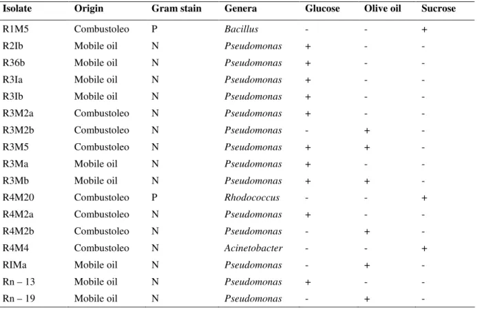
Seventeen isolates were positive and of those, 11 strains were
growing in glucose, 6 in olive oil and only 3 in sucrose; of
those, 3 strains produced biosurfactant in olive oil as well as in
glucose as substrates (Table 1). The substrates used have been
reported to promote biosurfactant production (4, 26, 32).
Although the biodegradability capacity of the 324 strains has
not been tested by reduction of hydrocarbons in pure culture,
many are likely to degrade hydrocarbons since they were
isolated in M9 Agar plates, incubated in a
hydrocarbon-saturated atmosphere as carbon source. However, only a small
proportion were biosurfactant producers, which support the
concept of bacterial community, whereas the biosurfactant
producers will liberate the surface-tension-active molecules,
that will help other microorganisms to degrade the wide variety
of hydrocarbons found at the site (28, 30).
Table 1. Biosurfactant production and preliminary identification of isolates growing in glucose, sucrose and olive oil as the carbon source (Gram stain: P, Gram-positive; N, Gram-negative. Biosurfactant production: +, positive response; -, negative response)
Isolate Origin Gram stain Genera Glucose Olive oil Sucrose
R1M5 Combustoleo P Bacillus - - +
R2Ib Mobile oil N Pseudomonas + - -
R36b Mobile oil N Pseudomonas + - -
R3Ia Mobile oil N Pseudomonas + - -
R3Ib Mobile oil N Pseudomonas + - -
R3M2a Combustoleo N Pseudomonas + - -
R3M2b Combustoleo N Pseudomonas - + -
R3M5 Combustoleo N Pseudomonas + + -
R3Ma Mobile oil N Pseudomonas + - -
R3Mb Mobile oil N Pseudomonas + + -
R4M20 Combustoleo P Rhodococcus - - +
R4M2a Combustoleo N Pseudomonas + - -
R4M2b Combustoleo N Pseudomonas - + -
R4M4 Combustoleo N Acinetobacter - - +
RIMa Mobile oil N Pseudomonas - + -
Rn – 13 Mobile oil N Pseudomonas + - -
The drop-collapse test was considered positive when
within one minute, the drop expanded on the oily surface of the
microplate lid. Besides the 17 positive isolates, another 21
strains (18 in olive oil, 1 in maize starch, 1 in sucrose and 1 in
paraffin) showed a partially collapsed drop after one minute,
suggesting that those microorganisms produced only a small
amount of biosurfactant, or that it remained intracellular (4).
Biosurfactant-producing strains had a turbid and pigmented
growth in broth when growing in glucose, with a foamy aspect;
those that grew in olive oil had a milky aspect, with growth
attached to the substrate; while the strains positive in sucrose
did not had a characteristic growth. Most of the positive strains
were identified as Pseudomonas spp., but identification could not be obtained up to species level. The Pseudomonas genera is one of the most reported for biosurfactant production, and it
has also been reported that the most known biosurfactant that
they produce, is a rhamnolipid (22, 33). Although most of the
reports on rhamnolipid production are attributed to
Pseudomonas aeruginosa strains, there are reports of other Pseudomonas related microorganisms, such as Burholderia plantarii (2) and Ps. chlororaphis (15) that produce biosurfactants.
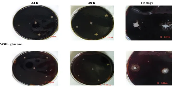
When the 17 isolates described in Table 1 were inoculated
in an M9 agar plate covered with a layer of combustoleo, a
clear zone around the colonies was observed in the glucose-free
plates after 24 h of incubation. After 4 days of incubation,
emulsification zones were observed and increased after longer
incubation times (Figure 1). It has been suggested that the
biosurfactant remains attached to the cell surface until a
saturation point is reached; then the biosurfactant is liberated to
the surroundings and emulsification occurs (8) as the main
mechanism to introduce water-insoluble substrates to the cell
interior (7). Southam et al. (31) demonstrated by transmission electron microscopy, the interphase between
hydrocarbon-degrading bacteria and small oil droplets that were
encapsulated by the biosurfactant. This phenomenon can
explain the clear area and further emulsion observed in
glucose-free medium. On the other hand, Das and Mukherjee
(12) found that supplementing the medium with glucose as a
co-carbon source enhanced the rate of PAH degradation in
selected bacteria. With the use of glucose, in the M9 Agar, the
microorganisms only increased the clear zone around the
colonies, but no emulsion was observed (Figure 1). The
presence of a water-soluble substrate such as glucose, can lead
to a hydrophobic cell membrane, and the production of a
biosurfactant with no emulsification activity (27).
Figure 1. Growth of bacterial strain R2Ib in M9 Agar plates with a top layer of combustoleo and with or without glucose (10 mM)
Tensioactive properties of biosurfactants
Surface tension measurement showed that in order to give
a positive drop collapse test, a surface tension lower than 45
mN/m was necessary. Surface tension of M9 minimal broth
was 69.97 mN/m. No differences were observed in the surface
tension of cell-free supernantant and cell suspensions for all
bacterial strains tested (p>0.05, data no shown), opposing
results obtained by Batista et al. (3). Glycolipids produced by Pseudomonas are low molecular weight compounds, which can lower the medium surface tension below 30 mN/m (25). The
lower surface tension values, both with or without cells, was
reached by Pseudomonas strains (26.7 mN/m), and were closely followed by the Bacillus strain (33.32 mN/m) (Table 2). Even though lypopeptides produced by Bacillus are known as one of the most powerful microbial biosurfactants (20),
Pseudomonas rhamnolipids are also effective, and both of them are extracellular (21). Isolates that liberate biosurfactants into
the culture medium are interesting from an industrial point of
view, because the product can be easily removed from the
culture media (19).
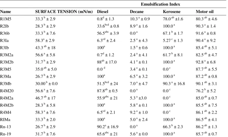
Table 2. Surface tension and emulsification index of bacterial strains isolated from hydrocarbon-contaminated soils
Emulsification Index
Name SURFACE TENSION (mN/m) Diesel Decane Kerosene Motor oil
R1M5 33.3 b ± 2.9 0.8d ± 1.3 10.3 a ± 0.9 78.0 ab ±1.6 80.3 ab ± 4.6
R2Ib 28.3 b ± 2.9 33.6bcd ± 0.8 8.9 a ± 1.6 100.0 a 90.3 a ± 1.4
R36b 33.3 b ± 7.6 56.5abc ± 3.9 0.0 a 67.1 b ± 1.7 91.6 a ± 0.8
R3Ia 58.3a ± 2.9 6.3cd ± 2.4 2.5 a ± 4.3 5.27 c ± 1.3 90.4 a ± 9.2
R3Ib 43.3 ab ± 18 100a 1.5 a ± 0.6 100.0 a 85.4 ab ± 5.1
R3M2a 56.6 a ± 5.8 0.7d ± 1.2 2.4 a ± 4.1 61.7 b ± 8.1 82.5 ab ± 4.7
R3M2b 31.7 b ± 2.9 88ab ± 17.0 4.1 a ± 0.1 100.0 a 92.8 a ± 6.8
R3M5 35.0 ab ± 5.0 0.0 d 3.4 a ± 0.1 0.0 c 87.7 ab ± 5.5
R3Ma 26.7 b ± 2.9 100a 6.5 a ± 3.2 100.0 a 87.2 ab ± 0.8
R3Mb 30.00 b ± 0.0 51.5bcd ± 24 7.0 a ± 4.7 90.3 a ± 16.8 90.1 ab ± 3.1
R4M20 56.6 a ± 7.6 87.8ab ± 0.5 0.0 a 0.0 c 76.2 b ± 5.2
R4M2a 46.7 ab ± 17 55.9abc ± 21 5.3 a ±3.0 0.0 c 85.0 ab ± 0.7
R4M2b 28.3 b ± 5.8 100a 5.8 a ± 0.1 100.0 a 85.5 ab ± 7.5
R4M4 58.3 a ± 7.6 6.5cd ± 2.1 9.2 a ± 1.0 0.0 c 86.1 ab ± 2.2
RIMa 33.3 b ± 2.0 100a 5.0 a ± 2.4 100.0 a 86.5 ab ± 4.1
Rn-13 26.7 b ± 2.9 90.2a ± 16.9 0.0 a 66.3 b ± 2.3 86.2 ab ± 1.3
Rn-19 31.7 b ± 7.6 65.6abc ± 21 5.6 a ± 0.0 100.0 a 85.7 ab ± 0.7
Results are shown as average and standard deviation of three replicates. In a column, different letters represent different statistical groups (p<0.05).
The terms biosurfactant and bioemulsifier are used as
synonyms in scientific literature. However, while the molecular
structure of the surfactant is well defined (a surfactant has both
hydrophilic and hydrophobic moieties present within the same
molecule), the term emulsifier is often used in an
application-oriented manner to describe the combination of all the surface
active compounds that constitute the emulsion secreted by the
cell to facilitate the assimilation of an insoluble substrate (14).
The debate is if a surfactant that reduces the surface tension of
water, can form stable emulsions (3, 9). As described in Table
2, most of the strains that had the lower surface tension values
emulsions. Also, there was no significant difference between
the emulsions formed by the released biosurfactant or the cell
suspension (p>0.05). Negative control was the M9 minimal
medium mixed with the different hydrophobic substrates. For
all water-insoluble compounds tested, emulsions were more
stable in the hydrocarbonated portion of the oil-water mixture.
When a supernatant without cells was used, emulsification
index (after 24 hours) ranged from 0 to 100% for diesel, from
0.0 to 100% for kerosene, and from 76.2 to 92.8% for motor oil
(Table 2), with high statistical differences between strains
within each compound (F=15.55 for diesel, F=100.60 for
kerosene and F=2.35 for motor oil respectively; p<0.01). For
decane, emulsion index was in the range of 0-10% and there
were no differences among strains (p>0.05).
Emulsification index can vary with bacterial growth phase,
bacterial interactions and hydrophobic compound tested (18).
The highest emulsification index values of diesel, kerosene and
motor oil were detected for Pseudomonas strains. Monteiro et al. (22) reported an emulsification index of 70% after 30 days of incubation, demonstrating that emulsions produced by P. aeruginosa rhamnolipids are stable, and can be used in the control of environmental contamination. Only Bacillus and Acinetobacter formed stable emulsions with decane. Emulsions formed by Acinetobacter were small, but optically clear, probably due to vesicles rich in phosphatidiletanolamine that
are formed, as observed by Desai & Banat (13), and the
emulsion formed by Rhodococcus cells incorporated air in the emulsion, giving a column height higher than the controls.
Hydrocarbon contaminated sites can be considered as
enrichment environments for selection of
hydrocarbon-degrading and/or biosurfactant producing microbial strains.
Production of biosurfactants and bioemulsifiers by soil
microorganisms provide them with an advantage in
contaminated sited, since they can use water insoluble carbon
sources for growth. Identification and selection of microbial
strains with those capacities, can lead to the identification and
functional characterization of their biosurfactants. Considering
that there is a wide variety of molecular structures among
microbial biosurfactants, they also have different chemical
properties that can be exploited commercially, for applications
as diverse as bioremediation or degradable detergents.
ACKNOWLEDGEMENT
This project was partially funded by grant
UACH-CA-073-2007 (Universidad Autónoma de Chihuahua). Authors
Viramontes-Ramos and Portillo-Ruiz held a scholarship from
CONACYT (Consejo Nacional de Ciencia y Tecnología,
México) for graduate studies.
REFERENCES
1. Adamczak, M.; Bednarski, W. (2000). Influence of medium composition and aeration on the synthesis of biosurfactants produced by Candida antarctica. Biotechnol. Lett. 22, 313-316.
2. Andrä, J.; Rademann, J.; Howe, J.; Koch, M.H.J.; Heine, H.; Zähringer, U.; Brandenburg, K. (2006). Endotoxin-like properties of a rhamnolipid exotoxin from Burkholderia (Pseudomonas) plantarii: immune cell stimulation and biophysical characterization. Biol. Chem. 387, 301-310. 3. Batista, S.B.; Mounteer, A.H.; Amorim, F.R.; Tótola, M.R. (2006).
Isolation and characterization of biosurfactant/bioemulsifier-producing bacteria from petroleum contaminated sites. Bioresour. Technol. 97, 868-875.
4. Bodour, A.; Miller–Maier, R. (1998). Application of a modified drop-collapse technique for surfactant quantitation and screening of biosurfactant–producing microorganisms. J. Microbiol. Methods 32, 273-280.
5. Bodour, A.A.; Drees, K.P.; Maier, R.M. (2003). Distribution of biosurfactant-producting bacteria in undisturbed and contaminated arid Southwestern soils. Appl. Environm. Microbiol. 69, 3280-3287. 6. Bognolo, G. (1999). Biosurfactants as emulsifying agents for
hydrocarbons. Colloids Surfaces A: Physicochem. Engineering Aspects
152, 41-52.
7. Bouchez, M.; Rakatozafy, H.; Marchal, R.; Leveau, J.; Vandecasteele, J. (1999). Diversity of bacterial strains degrading hexadecane in relation to the mode of substrate uptake. J. Appl. Microbiol. 86, 421-428.
8. Cassidy, D.; Hudak, A.; Dale, D.; Atekwana, E.; Rossbach, S.; Duris, J.; Attekwana, E.; Sauck, W. (2002). In situ rhamnolipid production at an abandoned petroleum refinery. Soil Sediment Contamination 11, 769-787. 9. Chen, C.Y.; Baker, S.C.; Darton, R.C. (2007). The application of a high throughput analysis method for the screening of potential biosurfactants from natural sources. J. Microbiol. Methods 70, 503-510.
10. Christofi, N.; Ivshina, I. (2002). Microbial surfactants and their use in field studies of soil remediation. J. Appl. Microbiol. 93, 915-929. 11. Cooper, D.G.; Gondenberg, B.G. (1987). Surface-active agents from two
12. Das, K.; Mukherjee, A. (2007). Crude petroleum-oil biodegradation efficiency of Bacillus subtilis and Pseudomonas aeruginosa strains isolated from a petroleum-oil contaminated soil from North-East India.
Bioresour. Technol. 98, 1339-1345.
13. Desai, J.D.; Banat, I.M. (1997). Microbial production of surfactants and their commercial potential. Microbiol. Mol. Biol. Rev. 61, 47-64. 14. Fiechter, A. (1992). Biosurfactants: moving towards industrial
application. Tibtech 10, 208-217.
15. Gunther IV, N.W.; Nuñez, A.; Fett, W.; Solaiman, D.Y.K. (2005). Production of rhamnolipids by Pseudomonas chlororaphis, a nonpathogenic bacterium. Appl. Environm. Microbiol. 71, 2288-2293. 16. Kiyohara, H.; Nagao, K.; Yano, K. (1982). Rapid screen for bacteria
degrading water–insoluble, solid hydrocarbons on agar plates. Appl. Environm. Microbiol. 43, 454–457.
17. Koneman, E. (2005). Color Atlas and Textbook of Diagnostic Microbiology. 6th Edition. Lippincott Williams & Wilkins, USA. 18. Krepsky, N.; Da Silva, F.S.; Fontana, L.F.; Crapez, M.A.C. (2007).
Alternative methodology for isolation of biosurfactant producing bacteria.
Braz. J. Biol. 67, 117-124.
19. Kuyukina, M.; Ivshina, I.; Philp, J.; Christofi, N.; Dunbar, S.; Ritchkova, M. (2001). Recovery of Rhodococcus biosurfactants using methyl tertiary – butyl ether extraction. J. Microbiol. Methods 46, 149-156.
20. Lu, J.R.; Zhao, X.B.; Yaseen, M. (2007). Biomimetic amphiphiles: biosurfactants. Current Opinion Colloid Interface Sci. 12, 60-67. 21. Mata, J.; Karns, J.; Torrents, A. (1999). High – performance liquid
chromatography method for the characterization of rhamnolipid mixtures produced by Pseudomonas aeruginosa UG2 on corn oil. J. Chromatogr.
864, 211-220.
22. Monteiro, S.A.; Sassaki, G.L.; Souza, L.M.; Meira, J.A.; Araújo, J.M.; Mitchell, D.A.; Ramos, L.P.; Krieger, N. (2007). Molecular and structural characterization of the biosurfactant produced by Pseudomonas aeruginosa DAUPE614. Chem. Phys. Lipids 147, 1-13.
23. Munguia, T.; Smith, C.A. (2001). Surface tension determination through capillary rise and laser difraction patterns. J. Chem. Educ. 78, 343-344. 24. Nevárez-Moorillón, G.V.; Piñón-Castillo, H.; Torres-Muñoz, V.;
Muñoz-Castellanos, L.N.; Vélez-Sánchez Verín, C.; Espinoza, J.V.; Riojas-
González, H.H.; Hernández-Castillo, D. (2004) Soil contaminated with combustoleo (residual fuel no. 6). Remediation strategies. In Magar, V.I., Kelley, M.E. (eds). In Situ and On-site Bioremediation-2003. Battelle Press, Columbia OH. USA. Paper E-22
25. Nitschke, M.; Costa, S.G.V.A.O.; Haddad, R.; Goncalves, L.A.G.; Eberlin, M.N.; Contiero, J. (2005). Oil wastes as unconventional substrates for rhamnolipid biosurfactant production by Pseudomonas aeruginosa LBI. Biotechnol. Prog. 21, 1562-1566.
26. Olivera, N.L.; Commendatore, M.G.; Morán, A.C.; Esteves, J.L. (2000). Biosurfactant-enhanced degradation of residual hydrocarbons from ship bilge wastes. J. Ind. Microbiol. Biotechnol. 25:70-73.
27. Prabhu, Y.; Phale, P. (2003). Biodegradation of phenanthrene by
Pseudomonas sp. Strain PP2: novel metabolic pathway, role of biosurfactant and cell surface hydrophobicity in hydrocarbon assimilation. Appl. Microbiol. Biotechnol. 61, 342-351.
28. Rahman, K,; Banat, I.; Thahira, J.; Thayumamavan, T.; Lakshmanaperumalsamy, P. (2002). Bioremediation of gasoline contaminated soil by a bacterial consortium amended with poultry litter, coir pith and ramnolipid biosurfactant. Bioresour. Technol. 81, 25-32. 29. Rahman, K.; Rahman, T.; Korkoutas, Y.; Petsas, I.; Marchant, R.; Banat,
I. (2003). Enhanced bioremediation of n – alcane in petroleum sludge using bacterial consortium amended with rhamnolipid and micronutrients.
Bioresour. Technol. 90, 159-168.
30. Ron, E.; Rosenberg, E. (2002). Biosurfactants and oil bioremediation.
Current Opinion Biotechnol. 13, 249-252.
31. Southam, G.; Whitney, M.; Knickerbocker, S. (2001). Structural characterization of the hydrocarbon degrading bacteria – oil interface: implications for bioremediation. Internat. Biodeterior. Biodeg. 47, 190-201.
32. Vipulanandan, C.; Ren, X. (2000). Enhanced solubility and biodegradation of naphthalene with biosurfactant. J. Environm. Eng. Geophys. 126, 629-633.