SC
IEN
TIARUM POLONOR U
M
ACTA
Acta Sci. Pol., Technol. Aliment. 7(4) 2008, 61-72
Corresponding author – Adres do korespondencji: Dr in . Anna Gramza-Michałowska,
Depart-ANTIOXIDANT POTENTIAL OF HERBS EXTRACTS
AND IMPACT ON HEPG2 CELLS VIABILITY
Anna Gramza-Michałowska
1, Zyta Abramowski
2, Eduardo Jovel
2,
Marzanna H
11
Pozna University of Life Sciences, Poland
2
University of British Columbia Vancouver, Canada
Abstract. Mercury poisoning is responsible for inducing serious adverse effects in living organisms. One of protection factors could be substances proven to possess high antioxi-dant and metal chelating activity – plant polyphenols. There are many sources of poly-phenols in plant kingdom but the most interesting for food industry could be widely con-sumed herbs. Aim of the research was to evaluate antioxidative potential of selected plant extracts and its influence on HepG2 cells in different conditions. Ethanolic herbs extracts were characterised by total polyphenol content. Antioxidant activity was estimated with
use of DPPH• and ABTS+• radicals scavenging methods and FRAP. Research included
cells viability estimation by the MTT assay and cells exposition to HgCl2, chemical agent
inducing cell death. Analysis of herbs extracts antioxidative activity showed best potential represented thyme and marjoram, highest FRAP was evaluated in samples with mint and marjoram extracts. On the basis of received results it was found that examined plant ex-tracts showed weak protection against Hg presence in examined cells environment.
Key words: herbs, plant extracts, cytotoxicity, HepG2 cells, FRAP, radicals
INTRODUCTION
Williams et al. 2005, Suhaj 2006]. Antioxidants present in food are very important for human health since the reactive oxygen species are recognised as aging and carcino-genesis factor. Plant components as antioxidants play important role in foods and living organisms because of the radicals scavenging ability and reducing cells degradation in human body [Madsen and Bertelsen 1995, Jin et al. 2004, Matkowski and Piotrowska 2006, Yoo et al. 2008]. Polyphenols are recognised as great scavengers of free radicals, hydroxyl radicals and superoxide anion radicals [Hanasaki et al. 1994, Cao and Cao 1999, Kahkonen et al. 1999].
Environment is widely polluted with heavy metals like mercury or lead. Mercury poisoning is responsible for inducing serious adverse effects in living organisms. Evi-dence indicates that cellular damage mediated by reactive oxygen species may be in-volved with heavy metals intoxication [Chen et al. 2002, Hermes-Lima et al. 1991]. Substances that are well proven to possess high antioxidant and metal chelating activity are polyphenols, also present in herbs. There are many results suggesting antioxidants role in the treatment of heavy metals poisoning as metal ions chelators and scavengers of free radicals [Matsingou et al. 2001]. Although plant extracts might protect cell from oxidative stress the mechanism remains unclear.
In present work the antioxidative activity as radical scavengers of herbs extracts had been evaluated. The aim of this study was to estimate total polyphenol content and cor-relation with antiradical activity. Second aim was the introductory evaluation of herbs extracts activity as cells protectors from oxidative damage in presence of mercury.
MATERIALS AND METHODS
Chemicals. The following chemicals were used: [3-(4,5-dimethylthiazol-2-yl)-2,5- -diphenyl-tetrazolium bromide] (MTT); HgCl2 (Sigma); ethanol; HCl; ddH2O;
phos-phate-buffered saline – PBS, Ca2+ and Mg2+ free, purchased from Gibco BRL (Gaithersburg, MD, USA); (+)-sodium L-ascorbate (Sigma); EDTA (Sigma); 6-hydroxy-2,5,7,8-tetramethylchromane-2-carboxylic acid – Trolox (Aldrich); iron (III) chloride FeCl3 (Aldrich); 2,4,6 Tri (2-pirydyl)-s-triazine (Fluka).
2,2’-azinobis-(3-ethyl-benzothiazoline-6-sulphonic acid) diammonium salt (ABTS) (Fluka); 2,2-diphenyl-1-picrylhydrazyl (DPPH) (Sigma-Aldrich); ethanol POCH (Poland). For the extracts ster-ilization Syringe filters were used (Nalgene 25 mm). All chemicals were of the highest analytical grade and purchased from common sources. Measurements were taken on Flu-oStar Galaxy (BMG Labtechnologies Ltd.) – multifunctional microplate reader. Cell cultures were grown on Falcon microtestTM tissue culture plates, 96 well (Becton Dick-inson Labware).
Antiradical activity assays. Plant polyphenols antioxidative activity was estimated using DPPH• [Sanchez-Moreno et al. 1998] and ABTS+• [Re et al. 1999] radicals
scav-enging ability of examined extracts. Results were expressed as mg Trolox per 1 g of extract’s dry weight (mg T/g).
FRAP assay. FRAP assay was performed as previously described by Benzie and Strain [1996] with modifications of Griffin and Bhagooli [2004]. After addition of FRAP reagent next readings were performed after 4 min ( = 600 nm). The changes in absorbance were compared to that of a standard that was run simultaneously. Final results were expressed as M Trolox equivalents ( MT) per sample concentration.
Cell culture. HepG2 cells (human hepatocellular carcinoma cells – liver tumor) were cultured in 75-cm2 cell culture flasks to confluence and harvested using a solution containing 0.05% (v/v) trypsin and 0.02% (w/v) EDTA in PBS. For experiments, cells were grown in 96-well plates, and inspected under an inverted system microscope, car-ried out before starting the experiments.
Cell viability assay. Cell viability was estimated by the MTT assay, which is based on the cleavage of a tetrazolium salt by mitochondrial dehydrogenases in viable cells [Loikkanen et al. 1998]. Data were mean percentages of viable cells versus the respec-tive controls.
Cells exposure to chemical agents and plant extracts. Cells were exposed to chemical agent inducing cell death – HgCl2. The possible protective effect of extracts,
when added immediately before toxic compound, was assessed according to Fallarero et al. [2003]. All extracts were dissolved, sterilized by filtering through 0.2- m filters, and then added to cells. Extract concentrations in all experiments were selected considering that extracts up to 0.1 mg·mL-1 are not toxic to HepG2 cells during exposure. Cells were exposed to HgCl2 for 24 hours in darkness at 37°C. Final concentration of ethanol was
in all cases lower than or equal to 3%, concentrations which have no effect on cell vi-ability in this cell line.
Statistical analysis. Data were expressed as mean values of three independent ex-periments (each in triplicate) and analysed by the analysis of variance (p ≤ 0.05) to estimate the differences between values of compounds tested.
RESULTS AND DISCUSSION
Evaluations of total polyphenols content with Folin-Ciocalteu method showed that examined herbs extracts differed significantly. Highest polyphenols content was evalu-ated in mint (221.6 mg GAE/g) and thyme extracts (217.1 mg GAE/g). Nearly 20% less polyphenols content was evaluated in samples of cilantro (183.8 mg GAE/g) and rose-mary (171.0 mg GAE/g). Significantly lower amount was evaluated in marjoram extract (119.8 mg GAE/g). Garlic extract was found to contain the lowest polyphenol amount among examined extracts (41.1 mg GAE/g).
Fig. 1. Total polyphenols content in herbs extracts, mg gallic acid/g dry weight
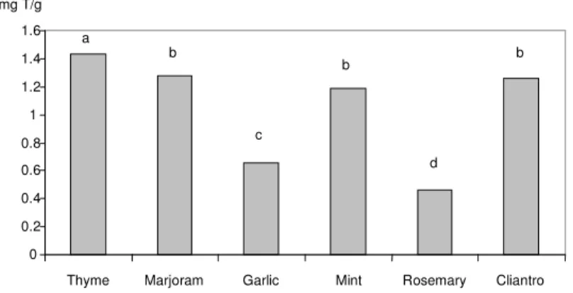
Fig. 2. DPPH radical scavenging activity of selected herbs extracts, mg Trolox/g dry weight
Statistical analysis of received data showed that herbs extracts possessed radical scavenging ability, depending on their concentration. It was evaluated that with higher extracts concentration, also antiradical activity increases, reaching highest at concentra-tion of 0.1%. Analysis of DPPH• scavenging activity calculated on equal Trolox con-centration showed that all herbs extracts exhibited activity significantly higher than of garlic and rosemary extracts. As the result of the DPPH• analysis herbs extracts were ranked as follows: rosemary < garlic < mint < marjoram < cilantro < thyme. Statistical analysis evaluations did not confirm the correlations between antiradical activity and total polyphenols in examined herbs extracts (r = 0.86, p < 0.05).
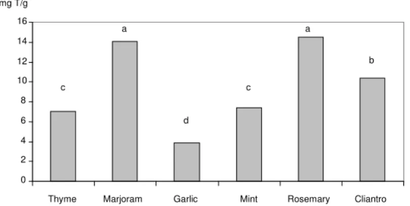
Second method for the evaluation of antiradical activity of herbs extracts was method using the ABTS+• radical (Fig. 3). On the basis of present research it was evalu-ated that highest radical scavenging activity possessed equally rosemary (14.53 mg T/g) and marjoram (14.09 mg T/g) extract. Weaker antiradical activity was evaluated in
s a m p l e s
0 0.2 0.4 0.6 0.8 1 1.2 1.4 1.6
Thyme Marjoram Garlic Mint Rosemary Cliantro mg T/g
a
b
c
b
d b
0 50 100 150 200 250
Thyme Marjoram Garlic Mint Rosemary Cliantro mg GAE/g
a a
b c
d
Fig. 3. ABTS radical scavenging activity of selected herbs extracts, mg Trolox/g dry weight
with cilantro (10.34 mg T/g), mint (7.38 mg T/g) and thyme extracts (7.00 mg T/g). Among examined extracts garlic possessed lowest antiradical activity (3.91 mg T/g). Thyme and mint extract did not scavenge the radicals as well as in presence of DPPH• radical, rosemary however, was a better scavenger of ABTS+• radical.
Statistical analysis of received data showed that antiradical activity was dependent from its concentration, higher extracts concentration resulted in higher antiradical activ-ity, reaching highest at concentration of 0.1%. Analysis of ABTS+• scavenging activity calculated on equal Trolox concentration allowed to rank the extracts as follows: garlic < thyme < mint < cilantro < marjoram < rosemary. On the basis of statistical analysis results it was stated that ABTS+• scavenging activity was poorly correlated with total polyphenol content (r = 0.72, p < 0.05).
Antioxidative activity of examined plant extracts was also measured with Ferric Re-ducing Antioxidant Power, FRAP method (Fig. 4). Results of the evaluations presented as µM Trolox per dry extract’s concentration, showed increasing activity with extracts concentration. Highest FRAP values were evaluated in samples of mint (1665.5 uMT) and marjoram (1614.9 uMT), similarly high activity showed rosemary extract (1586.4 uMT). Other extracts exhibited significantly lower activity, lowest however was found in garlic sample (35.4 uMT).
As the result of the FRAP analysis herbs extracts were ranked as follows: garlic < cilantro < thyme < rosemary < mint < marjoram. Statistical analysis of relationships between ferric reducing antioxidant power of ethanol herbs extracts and total polyphe-nol content showed high correlations (r = 0.93, p < 0.05).
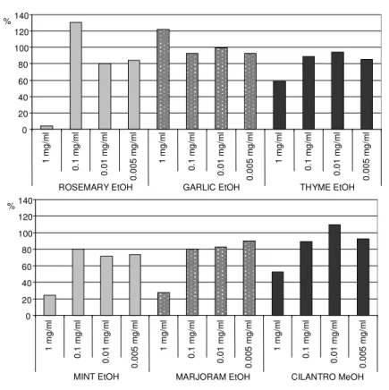
Exposure of HepG2 cells to different herb extracts significantly decreased cell vi-ability and in some cases stimulated its apoptosis. It was found that with increasing extracts concentration also the percentage of cells viability changed (Fig. 5). Highest ex-tracts concentration (1 mg/mL) however significantly decreased cell’s viability. It was evaluated that rosemary, mint and marjoram extracts strongly influenced cells viability, ranging 27.5 – 3.8%. Thyme and cilantro extracts decreased cell’s viability for nearly 60% at highest concentration. Surprisingly rosemary, garlic and cilantro extract addition resulted in cells proliferation, reaching 130.4; 122.2 and 109.7% respectively.
0 2 4 6 8 10 12 14 16
Thyme Marjoram Garlic Mint Rosemary Cliantro mg T/g
c c
d
a a
Fig. 4. FRAP assay for selected herbs extracts, uM T
Further research on Hg influence was conducted on the extracts concentration which did not cause the cells apoptosis or proliferation (0.005 mg/mL). No correlations be-tween total polyphenols content and cell viability were evaluated (r = 0.21, p < 0.05).
Using the MTT method, the toxicity of Hg on HepG2 cells was evaluated (Fig. 6). There was no statistical difference in cells viability when the mercury concentration was 58 ( g Hg/mL) or exceeded to 60 ( g Hg/mL). As seen on Figure 6, it was found that all examined extracts at 0.005 mg/mL concentration caused cells proliferation, except cilantro sample. Results showed that plant extracts did not or weakly protected HepG2 cells from Hg influence. It also must be noticed that HepG2 cells are very unstable and sensitive to reaction conditions. Among herbs extracts best protection offered addition of garlic (3.56%) and rosemary extract (3.39%). Cells apoptosis was evaluated in sam-ple with mint extract. This suggests that no chelating activity was evaluated during cells exposure to Hg in chosen concentration.
0 200 400 600 800 1000 1200 1400 1600 1800
a b c d e a b c d e a b c d e R O S E M A R Y E tO H G A R L IC E tO H T H Y M E E tO H uM Trolox a – 1.25%; b – 0.63%; c – 0.31%; d – 0.21%; e – 0.11%
0 200 400 600 800 1000 1200 1400 1600 1800
Fig. 5. Viability of HepG2 cells exposed for 24 h to various concentrations of plant extracts (MTT assay; 20 000 cells/well)
Fig. 6. Viability of HepG2 cells exposed for 24 h to plant extracts and mercury Hg60 (MTT assay; 20 000 cells/well)
-5 15 35 55 75 95 115 135 155 175 195 215 235 255 275 295 315 ROSEMARY EtOH+60 GARLIC EtOH+60 THYME EtOH+60 MINT EtOH+60 MARJORAM EtOH+60 CILANTRO MeOH+60 DMSA+60 ROSEMARY EtOH GARLIC EtOH THYME EtOH MINT EtOH MARJORAM EtOH CILANTRO MeOH DMSA % 0 20 40 60 80 100 120 140 1 m g /m l 0 .1 m g /m l 0 .0 1 m g /m l 0 .0 0 5 m g /m l 1 m g /m l 0 .1 m g /m l 0 .0 1 m g /m l 0 .0 0 5 m g /m l 1 m g /m l 0 .1 m g /m l 0 .0 1 m g /m l 0 .0 0 5 m g /m l %
MINT EtOH MARJORAM EtOH CILANTRO MeOH
0 20 40 60 80 100 120 140 1 m g /m l 0 .1 m g /m l 0 .0 1 m g /m l 0 .0 0 5 m g /m l 1 m g /m l 0 .1 m g /m l 0 .0 1 m g /m l 0 .0 0 5 m g /m l 1 m g /m l 0 .1 m g /m l 0 .0 1 m g /m l 0 .0 0 5 m g /m l
ROSEMARY EtOH GARLIC EtOH THYME EtOH
Summarizing it was found that HepG2 cells exposure to Hg significantly decreased cell viability. The results pre-suggest that supplementation could play an important role in modulating oxidative stress in HepG2 cells exposed for a mercury influence. Accord-ing to results of Yoo et al. [2008] it was suggested that antioxidant effect of herbs on cell viability could be explained by two mechanisms. One is direct action as reactive oxygen species ROS scavengers, second is indirect action through induction of antioxi-dative enzymes and intracellular communications protection.
DISCUSSION
Herbs could be very promising antioxidants sources. Of the six selected herbs used for the research, it is quite difficult to show the best source of natural antioxidants. As the results showed herbs extracts exhibited different antiradical and antioxidant activity, depending on the evaluation method used, as a result of methods complexity of the involved antioxidative mechanism. Example of that could be the cilantro extract that exhibited strong antioxidative activity and low FRAP values.
Wojdyło et al. [2007] examined selected herbs for their total polyphenol content and showed that rosemary possessed higher amount that thyme, what was in agreement with results of Cosio et al. [2006] and Parejo et al. [2002]. There are researches showing extremely high differences in polyphenols content of garlic extracts. Leelarungrayub et al. [2006] evaluated nearly 500 (mg GAE/g) of garlic extract, Nencini et al. [2007] however, is in agreement with the present research, with rage from 0.32-0.64 (mg GAE/g). Results of other research showed that herbs are relatively high in polyphenols, depending genotypic, environmental and sampling differences [Parejo et al. 2002, Shan et al. 2005]. Total polyphenols content also differs with method used. Folin-Ciocalteu procedure does not give the exact picture of the polyphenols quality and quantity in tested extract. Main polyphenols evaluated in herbs were phenolic acids, like hydroxyn-namic acid, caffeic acid, rosmarinic, p-coumaric and ferulic acids [Zheng and Wang 2001, Kim and Lee 2004, Wojdyło et al. 2007, Fecka and Turek 2008]. Other flavon-oids found in herbs were quercetin, kaempferol, apigenin and luteolin [Justesen and Knuthsen 2001, Shan et al. 2005].
Present research showed that rosemary extract exhibited best FRAP values, marjo-ram and thyme extracts however possessed also strong antioxidant activity measured with FRAP. Other authors results showed similar FRAP values for rosemary and thyme extracts [Wojdyło et al. 2007]. In present research all examined herbs extracts reduced the ferric ion. In contrast the weakest ability to reduce the ferric ion exhibited cilantro extract, as in previous results of DPPH• and ABTS+• methods. Rosemary which exhib-ited the highest scavenging of ABTS+• and ferric ion reducing ability did not reveal the same activity as DPPH• radical scavenger. The weakest activity however, in all evalua-tions was found in garlic sample.
All examined extracts possessed high polyphenols content but there was no simple correlation between antioxidant capacity and total polyphenol content that would be confirmed in assays used for the research. The present research results are in agreement with other showing poor correlation between total polyphenols content and antioxidant activity [Czapecka et al. 2005, Wong et al. 2005].
Results of Chen et al. [2002] showed that cell’s exposure to heavy metals induce oxidative stress and stimulates lipid peroxidation of lipid membrane. As a result of the process radicals and other lipid degradation products (like aldehydes) are formed, being extremely toxic for the cells. Strong chelating activity of herbs extracts made herbs polyphenols good candidates for treatment of mercury toxicity. There are however, results showing prooxidant activity of natural products. It was found that copper and tea catechins presence induced lipid peroxidation and DNA cleavage, resulting cells death [Hayakawa et al. 1997]. Chen et al. [2002] suggested that plant polyphenols could be toxic; so much consideration for safety should be required if used as therapeutic agents or nutrition supplements.
The results of the above analysis suggest a complexity of the antioxidative mecha-nism involved in different herbs extracts.
CONCLUSIONS
On the basis of the received results the following statements were formed: – plant extracts differed with total polyphenols content;
– according to antioxidative activity it was found that best radical scavenging activ-ity represented thyme, marjoram and rosemary extracts;
– highest FRAP was evaluated in samples of mint, marjoram and rosemary extracts; – no significant correlations between total polyphenols content and antiradical
ac-tivity of examined extracts was evaluated;
– higher extracts concentrations resulted in low HepG2 cells viability in presence of mercury;
– research showed weak herbs extracts protection against Hg presence in examined HepG2 cells environment.
Acknowledgments
REFERENCES
Aoshima H., Ayabe S., 2007. Prevention of the deterioration of polyphenol-rich beverages. Food Chem. 100, 350-355.
Benzie I.F.F., Strain J.J., 1996. The ferric reducing ability of plasma (FRAP) as a measure of antioxidant power: the FRAP assay. Anal. Biochem. 239, 70-76.
Cao Y.H., Cao R.H., 1999. Angiogenesis inhibited by drinking tea. Nature 398, 381.
Chen L., Yang X., Jiao H., Zhao B., 2002. Tea catechins protect against lead-induced cytotoxic-ity, lipid peroxidation and membrane fluidity in HepG2 cells. Toxicol. Sci. 69, 149-156. Cosio M.S., Buratti S., Mannino S., Benedetti S., 2006. Use of an electrochemical method to
evaluate the antioxidant activity of herb extracts from the Labiatae family. Food Chem. 97, 725-731.
Czapecka E., Mareczek A., Leja M., 2005. Antioxidant activity of fresh and dry herbs of some Lamiaceae species. Food Chem. 93, 223-226.
Fallarero A., Loikkanen J.J., Mannisto P.T., Castaneda O., Vidal A., 2003. Effects of aqueous extracts of Halimeda incrassata (Ellis) Lamouroux and Bryothamnion triquetrum (S.G.Gmelim) Howe on hydrogen peroxide and methyl mercury induced oxidative stress in GT1-7 mouse hypothalamic immortalized cells. Phytomedicine 10, 39-47.
Fecka I., Turek S., 2008. Determination of polyphenolic compounds in commercial herbal drugs and spices from Lamiaceae: thyme, wild thyme and sweet marjoram by chromatographic techniques. Food Chem. 108, 1039-1053.
Gramza A., Khokhar S., Yoko S., Gliszczynska-Swiglo A., Hes M., Korczak J., 2006. Antioxi-dant activity of tea extracts in lipids and correlation with polyphenol content. Eur. J. Lipid Sci. Tech. 108/4, 351-362.
Griffin S.P., Bhagooli R., 2004. Measuring antioxidant potential in corals using the FRAP assay. J. Exp. Marine Biol. Ecol. 302, 201-211.
Hanasaki Y., Ogawa S., Fukuki S., 1994. The correlation between active oxygens scavenging and antioxidative effects on flavonoids. Free Rad. Biol. Med. 16, 845-850.
Hayakawa F., Kimura T., Maeda T., Fujita M., Sohmiya H., Fujii M., Ando T., 1997. DNA cleav-age reaction and linoleic acid peroxidation induced by tea catechins in the presence of cupric ion. Biochim. Biophys. Acta 1336, 123-131.
Hermes-Lima M., Pereira B., Bechara E.J.H., 1991. Are free radicals involved in lead poisoning? Xenobiotica 21, 1085-1090.
Horwitz W., 1970. Official methods of analysis of the association of Official Analytical Chemists (AOAC). Washington 15.049-15.055, 10 Ausgabe.
Jin D., Hakamata H., Takahashi K., Ktani A., Kusu F., 2004. Determination of quercetin in hu-man plasma after ingestion of commercial canned green tea by semi-micro HPLC with elec-trochemical detection. Biomed. Chromat. 18, 876-882.
Justesen U., Knuthsen P., 2001. Composition of flavonoids in fresh herbs and calculation of flavonoid intake by use of herbs in traditional Danish dishes. Food Chem. 73, 245-250. Kahkonen M.P., Hopia A.I., Vuorela H.J., Rauha J.P., Pihlaja K., Kujala T.S., Heinonen M.,
1999. Antioxidant activity of plant extracts containing phenolic compounds. J. Agric. Food Chem. 47, 3954-3962.
Kim D.O., Lee C.Y., 2004. Comprehensive study on vitamin C equivalent antioxidant capacity (VCEAC) of various polyphenolics in scavenging a free radical and its structural relation-ships. Crit. Rev. Food Sci. Nutr. 44, 4, 253-273.
Leelarungrayub N., Rattanapanone V., Chanarat N., Gebicki J.M., 2006. Quantitative evaluation of the antioxidant properties of garlic and shallot preparations. Nutrition 22, 266-274. Loikkanen J.J., Naarala J., Savolainen K.M., 1998. Modification of glutamate-induced oxidative
Matkowski A., Piotrowska M., 2006. Antioxidant and free radical scavenging activities of some medicinal plants from the Lamiaceae. Fititerapia 77, 346-353.
Matsingou T.C., Kapsokefaloi M., Salifoglou A., 2001. Aqueous infusions of Mediterranean herbs exhibit antioxidant activity towards iron promoted oxidation of phospholipids, linoleic acid and deoxyribose. Free Rad. Res. 35, 593-605.
Nencini C., Cavallo F., Capasso A., Franchi G.G., Giorgio G., Micheli L., 2007. Evaluation of antioxidative properties of Allium species growing wild in Italy. Phytotherapy Res. 21, 874-878.
Parejo I., Viladomat F., Bastida J., Rosas-Romeo A., Flerlage N., Burillo J., Codina C., 2002. Comparison between the radical scavenging activity and antioxidant activity of six distilled and nondistilled mediterranean herbs and aromatic plants. J. Agric. Food Chem. 50, 6882- -6890.
Park E.J., Pezzuto J.M., 2002. Botanicals in cancer chemoprevention. Cancer Metast. Rev. 21, 231-255.
Re R., Pellegrini N., Proteggente A., Pannala A., Yang M., Rice-Evans C., 1999. Antioxidant activity applying an improved ABTS radical cation decolorization assay. Free Rad. Biol. Med. 26, 1231-1237.
Sánchez-Moreno C., Larrauri J.A., Saura-Calixto F., 1998. A procedure to measure the antiradical efficiency of polyphenols. J. Sci. Food Agric. 76, 270-276.
Shan B., Cai Y.Z., Sun M., Corke H., 2005. Antioxidant capacity of 26 spice extracts and charac-terization of their phenolic constituents. J. Agric. Food Chem. 53, 7749-7759.
Suhaj M., 2006. Spice antioxidants isolation and their antiradical activity: a review. J. Food Comp. Anal. 19, 531-537.
Williams M.J.A., Sutherland W.H.F., McCormick M.P., Yeoman D.J., de Jong S.A., 2005. Aged garlic extract improves endotherial function in men with coronary artery disease. Phytother. Res. 19, 314-319.
Wojdyło A., Oszmia ski J, Czemerys R., 2007. Antioxidant activity and phenolic compounds in 32 selected herbs. Food Chem. 105, 940-949.
Wong C.C., Li H.B., Cheng K.W., Chen F., 2006. A systematic survey of antioxidant activity of 30 Chinese medicinal plants using the ferric reducing antioxidant power asay. Food Chem. 97, 705-711.
Yoo K.M., Lee C.H., Lee H., Moon B., Lee C.Y., 2008. Relative antioxidant and cytoprotective activities of common herbs. Food Chem. 106, 929-936.
Zheng W., Wang Y., 2001. Antioxidant activity and phenolic compounds in selected herbs. J. Agric. Food Chem. 49, 5165-5170.
POTENCJAŁ PRZECIWUTLENIAJ CY EKSTRAKTÓW ZIOŁOWYCH
ORAZ WPŁYW NA PRZE YWALNO KOMÓREK HEPG2
Streszczenie. Zatrucia rt ci prowadz do powstania wielu niepo danych skutków w or-ganizmach ywych. Jednym z czynników ochronnych mog by polifenole ro linne, któ-rych wysoki potencjał przeciwutleniaj cy oraz zdolno ci wi zania jonów metali potwier-dziło wiele bada . Spo ród ró nych ródeł tych zwi zków powszechnie spo ywane zioła mog by grup najbardziej interesuj c dla przemysłu spo ywczego. Celem bada było okre lenie potencjału przeciwutleniaj cego wybranych ekstraktów ro linnych oraz ich wpływu na komórki HepG2 w ró nych warunkach. Badania obejmowały okre lenie
stop-nia prze ywalno ci komórek w metodzie MTT oraz poddanych działaniu HgCl2, zwi zku
wyko-rzystaniem rodników DPPH• and ABTS+• oraz FRAP. Na podstawie uzyskanych wyni-ków stwierdzono, e badane ekstrakty słabo chroniły komórki przed działaniem Hg. Ana-liza aktywno ci przeciwutleniaj cej ekstraktów ziołowych wskazała najwy szy potencjał tymianku i majeranku, natomiast w metodzie FRAP najwi ksz aktywno stwierdzono dla ekstraktów mi ty, majeranku i rozmarynu.
Słowa kluczowe: zioła, ekstrakty ro linne, cytotoksyczno , komórki HepG2, FRAP, rodniki
Accepted for print – Zaakceptowano do druku: 27.10.2008