The Effects of Experimental Sleep Apnea on
Cardiac and Respiratory Functions in 6 and
18 Month Old Dystrophic (
mdx
) Mice
Milind R. Chaudhari1,2,3☯¤, James A. Fallavollita2,4,5, Gaspar A. Farkas1☯
*
1Department of Exercise and Nutrition Sciences, University at Buffalo, Buffalo, New York, United States of America,2Department of Medicine, University at Buffalo, Buffalo, New York, United States of America, 3Department of Epidemiology and Environmental Health, University at Buffalo, Buffalo, New York, United States of America,4Center for Research in Cardiovascular Medicine, University at Buffalo, Buffalo, New York, United States of America,5VA Western New York Healthcare System at Buffalo, Buffalo, New York, United States of America
☯These authors contributed equally to this work.
¤ Current address: Department of Medicine, and Department of Epidemiology and Environmental Health, University at Buffalo, Buffalo, New York, United States of America
Abstract
Duchenne muscular dystrophy (DMD) is a fatal disease where over 90% of patients suc-cumb to respiratory or cardiac failure. Sleep apnea and sleep disordered breathing (SDB) are noted in a plurality of DMD patients, and the resulting nocturnal episodic hypoxia (EH) cannot be ruled out as a contributing factor to cardiac and respiratory dysfunction. In this study, we investigated the impact of long-term episodic hypoxia, which mimics the cyclic hypoxia seen in sleep apnea, on cardiac and respiratory function in a murine model of DMD (mdxmice). Since the severity and prevalence of sleep apnea in DMD increases with age,
we studied the impact of EH on young (6-month) and on older (18-month)mdxmice. Mice
were either exposed for 12 weeks to EH (8 hours/day, 5 days/week) or to room air. We noted a significant increase in left ventricular (LV) dilatation (transthoracic echocardiogra-phy) on EH exposure in both age groups, but reduced LV contractility was seen only in 6-month old mice. With EH exposure, an increased fibrosis (hydroxyproline) was noted in both cardiac and diaphragm muscle in 18-month but not 6-month old mice. No significant change in relative diaphragm strength (in-vitro) was noted on EH exposure in 18-month old mice. In contrast, EH exposed 6-month old mice showed a significant increase in relative diaphragm strength. EH exposure did not result in any significant change in ventilatory parameters (barometric plethysmography) in awake 6-month oldmdxmice. In contrast,
18-month oldmdxmice showed considerable ventilatory dysfunction, consistent with reduced
ventilatory reserve. Our findings highlight that sleep apnea impacts respiratory and cardiac function in muscular dystrophy, and that EH can have divergent effects on both systems. To our knowledge, this is the first comprehensive study to investigate the impact of EH on car-diac and respiratory function inmdxmice.
OPEN ACCESS
Citation:Chaudhari MR, Fallavollita JA, Farkas GA (2016) The Effects of Experimental Sleep Apnea on Cardiac and Respiratory Functions in 6 and 18 Month Old Dystrophic (mdx) Mice. PLoS ONE 11(1): e0147640. doi:10.1371/journal.pone.0147640
Editor:Diego Fraidenraich, Rutgers University -New Jersey Medical School, UNITED STATES
Received:August 17, 2015
Accepted:January 5, 2016
Published:January 25, 2016
Copyright:© 2016 Chaudhari et al. This is an open access article distributed under the terms of the Creative Commons Attribution License, which permits unrestricted use, distribution, and reproduction in any medium, provided the original author and source are credited.
Data Availability Statement:All data files are available from the Dryad database (DOI:10.5061/ dryad.k6n5k).
Funding:This work was supported by the National Institutes of Health R21AR53901 - (NIH.Gov) GAF.
Introduction
Duchenne muscular dystrophy (DMD) is an X-linked recessive neuromuscular disorder that
affects approximately 1 out of 3500 male births [1]. DMD is a lethal disease that is
character-ized by the absence of dystrophin, a structural protein in skeletal and cardiac muscle, that links
the cytoskeleton to the extracellular matrix [2,3]. Dystrophin deficient skeletal and cardiac
muscles, are susceptible to a progressive myopathy. Indeed, respiratory and cardiac muscle
dys-function together contributes to the death of over 90% of DMD patients [3,4].
Ventilatory abnormalities and related respiratory symptoms are noted early in the course of DMD. Respiratory muscle weakness, especially that of the diaphragm, leads to respiratory
insufficiency and 2/3 of DMD patients die in their third decade from respiratory failure [4]. By
18 years of age, cardiac manifestations are also detected in about 90% of DMD patients. The most prevalent cardiac defect in DMD is dilated cardiomyopathy, and eventually leads to
car-diac failure in 10–20% of DMD patients [4].
Sleep is an especially vulnerable period for patients with DMD, and many are diagnosed
with sleep disordered breathing (SDB). It has been reported that>80% DMD patients have
SDB with overt signs of alveolar hypoventilation [5,6]. Smith et al. [7] reported that patients
with DMD endure bouts of episodic hypoxia (EH) during sleep, during which SaO2levels fall
from baseline values of 95.4 ± 0.6% to a mean nadir of 74.2 ± 3.9%. These nocturnal episodes of hypoxemia were reported to be more severe and more frequent in older DMD patients. Indeed, sleep apnea in non-DMD patients is known to contribute to cardiovascular
complica-tions such as systemic hypertension, coronary heart diseases and stroke [8–10]. Due to the
high incidence of SDB in DMD patients, we hypothesized that EH may be a contributing factor to their cardiac and respiratory dysfunction.
Themdxmouse, a murine model of DMD presenting with a deficiency of dystrophin,
results in a progressive myopathy of the diaphragm [11,12]. Cardiomyopathy is also observed
inmdxmice, but at a later age. Signs of cardiomyopathy are detected inmdxmice at 9–10
months of age and become more prominent with age [13,14].
Although cardiac and respiratory dysfunctions consistent with DMD are indeed detected in
mdxmice, they do not have spontaneous SDB. In order to better define the possible
conse-quences of SDB that is so prevalent in DMD, the current study assessed the putative impact of
EH on cardiac and respiratory function inmdxmice. EH is a standard and well-studied
tech-nique for experimental sleep apnea in mice [15]. Since SDB is more severe and prevalent in
older DMD patients, we investigated whether the effects of EH inmdxmice are compounded
by age. The purpose of this study, therefore, was to assess the impact of EH on cardiac and
respiratory function in 6 and 18-month oldmdxmice. To our knowledge, this is the first
com-prehensive study to investigate the consequences of EH simultaneously on cardiac and
respira-tory function in young and oldermdxmice.
Methods
The study was performed on 80 malemdxmice (C57BL/10ScSn-Dmdmdx/J) purchased from
Jackson Laboratories (Maine, USA). The animals were purchased at 3–4 weeks of age and
housed in the Laboratory Animal Facility at the University at Buffalo. Mice of similar age were
housed five to six per cage with food and water availablead libitum. Throughout the study,
mice were maintained on a 12 hour light/dark cycles (6:00 am-6:00 pm).
Ethics Statement
collection was performed under anesthesia (ketamine/xylazine) in order to minimize pain and distress.
Episodic Hypoxia (EH)
Starting at 3-mo of age and at 15-mo of age, 40mdxmice from each age group were randomly
assigned either: 1) to a control group (n = 20) exposed continuously to room air (RA) or 2) to an EH group (n = 20) exposed 5 days a week, 8 hours per day for 12 weeks to EH. Mice were weighed weekly throughout the experimental period. Studies in young and old mice were run concurrently. Mice assigned to the EH protocol were slowly habituated over a 2 weeks period to progressively longer and more severe levels of hypoxia. This initial habituation period was not included in the 12 week experimental period. Mice of both age groups assigned to the EH group were placed 8 hours a day during the diurnal period in sealed chambers (8:00 am and
4:00 pm, the normal sleep period for mice). During EH exposure, the O2levels in the chamber
were altered repeatedly in cycles (90-second cycle length) between 21% and 6% by alternating
the flow of N2(99.5%, medical grade) and room air. The O2levels in the cages were
continu-ously measured with an oxygen analyzer (S-3A, AEI technology Inc. Pittsburgh, PA). Mice housed within a given cage were all treated to the same exposure (EH or RA). Mice were returned to their home cages at the conclusion of the daily EH exposure. RA and EH mice were housed in a common room, and were thus exposed to the same environmental conditions
(sounds from valves opening/closing). Food and water were availablead libitumduring all
phases of the study.
At the end of the 12 week experimental period, mice from all four groups were studied in a random order. The younger mice were designated as 6-mo RA or 6-mo EH, while the older mice were designated as 18-mo RA or 18-mo EH.
Respiratory function analysis
In the 2 weeks prior to the terminal procedures, and over the weekend as not to interfere with the daily exposure to EH, breathing patterns in awake mice were measured using a
modifica-tion of the whole body plethysmography technique [16]. Mice were placed inside a chamber
that prevented backward rotation but did not cause chest wall compression. Mice were habitu-ated on numerous occasions to a restrainer as well as to plethysmography in order to minimize any effects of stress related changes in ventilation. The apparatus was connected to a flow meter (Dwyer Instruments Inc., Michigan City, IN), a pressure transducer (Model #7, Hewlett Packard, Palo Alto, CA) and a polygraph machine (Model 7 polygraph, Grass, Quincy, MA) for the purpose of maintaining a constant air flow, detecting pressure changes due to breathing
and recording the transducer signal, respectively. The inflowing and outflowing O2
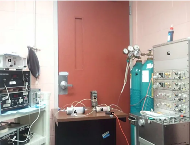
concentra-tions were measured by O2analyzer (S-3A, AEI technology Inc. Pittsburgh, PA) (Fig 1).
Ventilation was measured at rest and during hypercapnic respiratory stimuli (4% CO2, 26%
O2, and Bal N2). Resting ventilation was measured 15 min after placing the animal in the
cham-ber and 5 minutes after administering the hypercapnic mixture. To record ventilation, airflow was momentarily stopped and the chamber was sealed after which pressure changes due to breathing were recorded. An amount of 0.1 mL was injected and withdrawn via a 1 cc syringe into the chamber for the purpose of calibration.
Body temperatures of all mice were assumed to be 37˚C and to remain constant during the
ventilation protocol. Chamber temperature was monitored continuously via a probe mounted inside the chamber. Barometric pressure measurements were recorded every day. Breathing fre-quency (f) was calculated directly from the ventilation-induced pressure swings. Tidal volume
by Drorbaugh and Fenn [17] modified by Bartlett and Tenney [18]. Respiratory ventilation (VE)
was calculated (VE= VT× f) and values were expressed per body weight (ml/kg/min).
Echocardiography
Transthoracic echocardiography was performed on anesthetized [ketamine (100mg/kg) -xyla-zine (5 mg/kg) IP] mice using Vivid 7 echo machine (GE medical systems) with a 13-MHz lin-ear transducer (i13L). Mice were held in the supine position, the anterior chest wall was shaved, warm ultrasound gel applied to the chest, and the transducer probe was placed over the left hemithorax. Parasternal and short-axis two-dimensional images of the left ventricle (LV) were obtained to determine correct M-mode cursor positioning. Multiple short axis M-mode images of the left ventricle were obtained and these images were analyzed for LV functional parameters in triplicate. Heart rate (HR) was determined from at least three consecutive RR intervals. The other LV indices measured included LV internal dimension at end-diastole (LVIDd), and end-systole (LVIDs), septal wall thickness (SWT) and posterior wall thickness
(PWT) as recommended by the American Society of Echocardiography[19]. Left ventricular
end-systolic volume (ESV) and end-diastolic volume (EDV) were estimated assuming spherical geometry. The LV percent fractional shortening (%FS) was calculated as follows: %FS = 100 x
(LVIDd–LVIDs) / LVIDd. Estimated LV mass (in diastole) was calculated from following
Fig 1. Modified Barometric Plethysmography.Photograph of plethysmography set up for ventilatory function analysis. (A) Animal chamber; (B) Reference chamber; (C) Transducer; (D) Thermometer; (E) Polygraph machine; (F) Gas mixture; (G) Flow meter.
formula: LV mass (g) = 0.80 x [1.04 (IVSd + LVPWd + LVIDd)3- (LVIDd)3] + 0.6. All the LV dimension data are presented as the average of measurements from at least 3 selected beats on the image.
Tissue removal and blood collection
Following completion of the echocardiography, blood was collected from the orbital vein in a capillary tube for hematocrit (Hct) analysis.
The diaphragm (costal and crural) muscle was then quickly removed and placed in cooled
(4°C) Kreb’s solution aerated with a 95% O2, 5% CO2gas. The diaphragm was cut into two
halves, each containing both crural and costal parts. One half was used for measurement of
contractile properties while the other half was either frozen in liquid N2for biochemical
prop-erties or mounted on thick cork sheet, supported with O.C.T. compound and snap-frozen in
isopentane (2-methylbutane) cooled to its melting point with liquid N2for histological
analy-ses. Muscle tissue samples were then stored at -80°C for subsequent histological or hydroxy-proline analysis.
The whole heart was then removed, blotted dry on filter paper and weighed. The heart was then either cut horizontally through the ventricles and mounted on thick cork sheet, supported with O.C.T. compound, and snap-frozen in isopentane (2-methylbutane) cooled to its melting
point with liquid N2for histology or frozen in liquid N2and stored at -80°C for subsequent
hydroxyproline analysis.
Contractile properties of diaphragm
Details of the technique employed for measuringin vitromuscle contractile properties have
been described previously [20]. In brief, portions of the costal diaphragm muscle were
removed, cut into small bundles, and placed in a muscle bath. The muscle bath was filled with
Krebs solution composed of the following (mM): NaCl, 137; KCl, 4; MgCl2, 1; KH2PO4, 1;
NaHCO3, 12; CaCl2, 2; and glucose, 6.5. The diaphragm segments were vertically mounted
between large platinum stimulating electrodes and immersed in a Krebs solution maintained at
36°C perfused with 95% O2-5% CO2. The muscle bundles were stimulated supra-maximally
with monophasic pulses of 0.2 ms duration.
Isometric force measurements were recorded with a Grass FT-03C force transducer (Grass Instruments, Quincy, Massachusetts). Muscle force signals were displayed on an oscilloscope and measured directly from the oscilloscope tracings. Muscle length was changed by raising or lowering the force transducer to a desired level. After a 5-min thermo-equilibration period, the
bundles were adjusted to Lo, defined as the length at which peak twitch force were recorded. Lo
was measured in triplicate using a micrometer. The following procedures were performed in the following order: (1) twitch characteristics, (2) force-frequency response.
Twitch Characteristics. In each muscle bundle, five twitches were recorded at Loand the
average value was used to determine time-to-peak tension (TPT), half-relaxation time (½RT),
and twitch force (Pt). TPT is defined as the time from the start of the contraction to the point
where peak tension was achieved. ½RT is defined as the time from Ptto the point where twitch
tension dropped by 50%.
Force-Frequency Curve. Each muscle bundle was sequentially stimulated at frequencies of 10, 20, 35, 50, 70, 100, 120, and 150 Hz. The stimulation was maintained until plateau in
force was attained, usually requiring 350–500 ms. Approximately 30 sec elapses between each
stimulus train. Maximum tetanic force (Po) was recorded and the twitch-to-tetanus ratio (Pt/
On completion of the measurements, the bundle was removed from the apparatus, blotted dry, and weighed. Muscle cross-sectional area (CSA) was approximated by dividing the muscle
mass by its length and muscle density (1.056 g/cm3) [21]. Specific force was expressed as N/cm2.
Histological analysis
Tissue samples previously mounted on cork for histological analysis were removed from the
freezer and equilibrated to -20°C for 15–20 min. The cork was mounted on a Microtome
cryo-stat block with O.C.T. compound. Serial sections (10μthick) of the tissue were cut, mounted
on glass cover slides and stained by H&E method [22].
Images of the tissues (10x, 20x) were obtained with a color coolSNAP CCD camera and ana-lyzed with Metamorph imaging software (Molecular Devices, CA, USA). Tissue slides and images were coded and the individual performing the analysis was blinded to the code. For each muscle tissue, at least 200 myofibers were analyzed for fiber size, number of central nuclei,
tissue thickness and percent area of interstitial space [23].
Hydroxyproline analysis
To determine collagen content, a whole heart and a total hemi-diaphragm were assayed for hydroxyproline. A modified colorimetric hydroxyproline assay adapted from Prockop and
Udenfriend [24] and Switzer and Summer [25] was used. For diaphragm, the tissue was dry
blotted, weighed and then hydrolyzed using 6 N HCl (1ml HCl per 10mg of tissue) at 110°C for 16 hours. For determination of heart hydroxyproline content, whole heart tissue was dry blot-ted, weighed and then hydrolyzed using 6N HCl (0.5ml HCl per 10mg of tissue) at 120°C for 48 hours. Samples of hydrolysate from either tissue were then diluted with deionized water and oxidized using chloramine-T solution for 25 minutes. Toluene and KCl was added to remove the impurities. The remaining solution was heated in boiling water for 30 minutes. Toluene
extraction was repeated again and Ehrlich’s reagent (p-dimethylamino-benzaldehyde) was
mixed with the toluene layer. After 30 minutes of incubation, the diaphragm and heart tissue samples were read using a BioTek microplate reader (BioTek Instruments, Winooski, VT)
against hydroxyproline standards (0.75, 1.5, 3.0, 6.0μg) at 562nm or 558nm, respectively.
Hydroxyproline content was expressed asμg hydroxyproline/mg muscle wet weight.
Cardiomyocyte size determination
Hematoxylin-eosin (H&E) stained short-axis tissue sections of 10-μm thickness were digitized
using light microscopy (x600 magnification; Nikon Eclipse E600). Myocytes with myofilaments surrounding the nucleus were identified separately from the left ventricular free wall and
inter-ventricular septum (minimum 50 myocytes; mean ± SEM—60.8±9.3). Myocyte diameter was
determined as the minimum transverse diameter at the level of the nucleus (Image Pro Plus
analysis software; Media Cybernetics) [26].
Statistical analysis
group, paired t-test was used. Statistical significance was set at p<0.05. All the values in the
text, tables and figures are presented as Mean ± SEM.
Results
Body weight, Heart weight and Hematocrit
Body weights at baseline and at the end of experimental period are shown inTable 1. At
base-line, no significant difference was noted in body weights ofmdxmice across all the study
groups. At the end of the 12 weeks experimental period, EH exposed mice from both the 6-mo and the 18-mo groups weighed significantly less as compared to their RA age matched controls.
Consistent with previous studies, over the course of study, EH exposedmdxmice in both age
groups did not exhibit weight gain. Instead, EH exposure led to decreases in body weight in both 6-mo and 18-mo old mice (3% and 17% respectively). As stated previously, food and water were available ad libitum during all phases of the study, even during EH exposure.
As noted previously in non-DMD mice [27,28], Hct levels were significantly higher in EH
exposed 6-mo oldmdxmice compared to their age matched RA controls (Table 1). In contrast,
Hct levels in EH exposed 18-mo oldmdxmice were similar to levels noted in age matched RA
mice (Table 1).
As shown inTable 1, 18-mo old RA mice showed significantly higher absolute as well as
rela-tive (heart weight/body weight) heart weight compared to 6-mo old RA mice. Consistent with
previous findings [27], relative heart weights (heart weight/body weight) were noticed to be
sig-nificantly higher in EH exposed mice as compared to their RA counterparts in both age groups.
Cardiac function
Echocardiography. Echocardiographic analysis of cardiac function, especially LV
func-tion, is represented inFig 2, and the data are shown inTable 2. RA exposedmdxmice in 6 and
18-mo old groups did not show any significant difference in HR at rest under anesthesia.
How-ever, EH exposedmdxmice in both age groups exhibited significantly lower HR than their age
matched RA controls.
There were no detectable differences in left ventricular dimensions (LVIDs, LVIDd), vol-umes (EDV, ESV) and contractility (EF and FS) between 6-mo and 18-mo old RA exposed
mdxmice. However, 18-mo old RAmdxmice showed significantly thicker septal wall (SWT)
and higher LV mass (LVM) compared to 6-mo old RA mice.
In both age groups, chronic EH exposure led to significant changes in LV dimension and function. A significant increase in LV dimension (LVIDs and LVIDd) was noted in EH
Table 1. Effect of 12 weeks of episodic hypoxia (EH) on body weight, hematocrit and heart weight in both 6-mo and 18-mo oldmdxmice.
6-mo RA 6-mo EH 18-mo RA 18-mo EH
(n = 22) (n = 18) (n = 23) (n = 12)
Initial Body Weight, g 34.9±0.6 35.5±0.7 36.9±0.6 35.5±0.5 Final Body Weight, g 38.1±0.8 34.5±0.6b 35.9±0.8 29.4±0.5b
Hematocrit, % 44.2±0.5 46.1±0.4b 42.6±0.7 43.9±0.7
Heart Weight, mg 149.2±2.6 142.0±3.0 157.7±3.5a 148.3±5.6
Heart weight/Final Body Weight, mg/g 3.9±0.1 4.1±0.1b 4.4±0.1a 5.1±0.2b
Values represent Mean±SEM; n, number of mice.
asignificant difference (p<0.05) 6-mo vs 18-mo old RAmdxmice. bsignificant difference (p<0.05) RA vs EH exposed mice of same age.
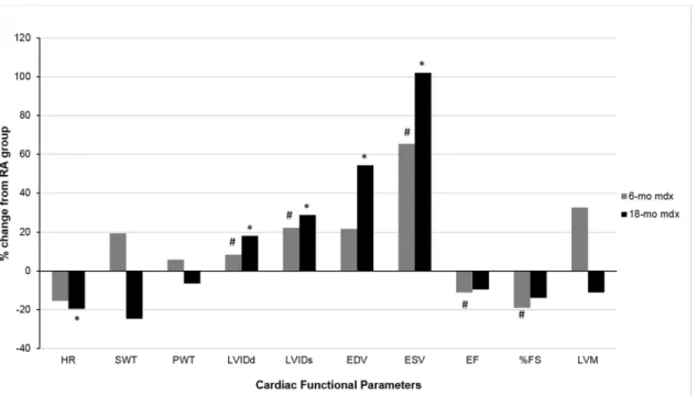
exposed 6-mo and 18-mo oldmdxmice compared to their age matched RA counterparts. In 18-mo EH mice, LV volume (ESV, EDV) were significantly higher than their RA controls. In 6-mo EH group, though ESV was significantly greater than 6-mo RA mice, EDV failed to show any significant difference. As expected, a significant decrease in LV contractility (EF, FS) was Fig 2. Effect of episodic hypoxia (EH) exposure on left ventricular functional parameters.The values are represented as percent change in LV parameter from RA exposed mice recorded by M-mode echocardiogram in 6-mo and 18-mo oldmdxmice.*, indicates significant change (p<0.05) in cardiac functional parameters of 18-mo oldmdxmice from their respective RA controls. #, indicates significant change (p<0.05) in cardiac functional parameters of 6-mo oldmdxmice from their respective RA controls.
doi:10.1371/journal.pone.0147640.g002
Table 2. Left Ventricular parameters measured by M-mode echocardiogram in 6-mo and 18-mo oldmdxmice exposed to room air (RA) and epi-sodic hypoxia (EH).
6-mo RA 6-mo EH 18-mo RA 18-mo EH
(n = 7) (n = 7) (n = 9) (n = 5)
HR bpm 330±14 279±11b 310±20 250±13b
SWT mm 1.47±0.12 1.75±0.27 2.01±0.20a 1.51±0.18
PWT mm 1.49±0.14 1.58±0.09 1.58±0.08 1.48±0.11
LVIDd mm 2.60±0.07 2.82±0.06b 2.50±0.13 2.95±0.10b
LVIDs mm 1.48±0.12 1.82±0.08b 1.50±0.11 1.94±0.17b
EDV ml 0.047±0.004 0.057±0.005 0.044±0.007 0.068±0.007b
ESV ml 0.010±0.002 0.017±0.002b 0.011±0.002 0.021±0.005b
EF % 79.2±3.3 70.5±2.2b 76.7±2.7 69.5±5.1
%FS % 42.6±3.1 34.6±1.6b 40.1±2.4 34.5±4.1
LVM mg 135±20 179±27 177±15a 157±19
Values represent means±SEM. p<0.05. asigni
ficant difference (p<0.05) 6-mo vs 18-mo old RAmdxmice. bsigni
ficant difference (p<0.05) RA vs EH exposedmdxmice of same age.
HR, heart rate; SWT, septal wall thickness; PWT, posterior wall thickness; LVIDd, left ventricular internal dimension at end-diastole; LVIDs, left ventricular internal dimension at end-systole; EDV, end diastolic volume; ESV, end systolic volume; FS, fractional shortening; LVM, left ventricular mass.
noted in 6-mo EH group compared to their age matched RA controls. However, EH exposed
18-mo oldmdxmice failed to show any significant decrease in LV contractility than to their
age matched RA exposed mice.
Histological Analysis. Representative images of LV sections from each group are shown inFig 3A. Cardiomyocyte diameter from both free LV wall and septal wall was determined (Fig
3C). EH exposure did not result in any significant difference in cardiomyocyte size in both
6-mo and 18-mo oldmdxmice (Table 3).
Hydroxyproline Analysis. The hydroxyproline levels in 18-mo oldmdxmice heart tissue
were significantly higher (~ 34%) as compared to 6-mo oldmdxmice heart in both EH and RA
groups (Fig 3B).
In 6-mo oldmdxmice, EH exposure did not lead to changes in heart hydroxyproline levels
(1.27 ± 0.08 vs 1.42 ± 0.15μg/mg wet weight, RA and EH respectively, NS). In contrast, EH
exposure in 18-mo old mice was associated with an elevation in heart hydroxyproline level
(1.70 ± 0.15 vs 2.42 ± 0.17μg/mg wet weight, P<0.05).
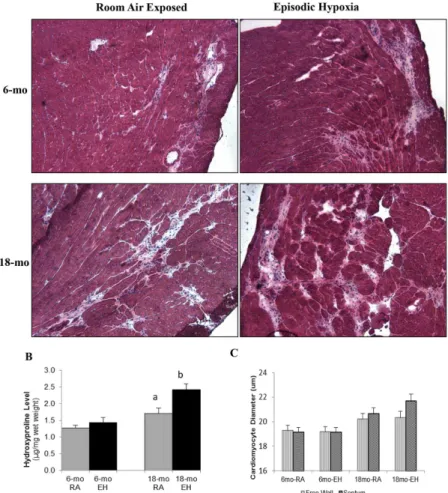
Fig 3. Histology of cardiac muscle and hydroxyproline content.(A) Photomicrograph of cardiac muscle (H&E stained, 10x) from 6-mo and 18-mo old mdx mice exposed to room air (RA) and episodic hypoxia (EH). Note higher amount of fibrosis and interstitial space in 18-mo oldmdxmice as compared to 6-mo oldmdx
mice. EH exposed mice heart showed higher amount of fibrosis and interstitial space as compared to RA controls in 18-mo oldmdxmice. (B) Cardiac hydroxyproline levels (μg/mg wet tissue weight) measured by biochemical analysis. a, significant difference (p<0.05) 6-mo vs 18-mo old RAmdxmice; b, significant difference (p<0.05) RA vs EH group of same age. (C) Cardiomyocyte Size, Effect of aging and episodic hypoxia on cardiomyocyte (LV free wall and septal) size in mdx mice.
Respiratory function
Ventilation. As a comprehensive global assessment of ventilatory function, breathing
pat-terns at rest and in response to hypercapnic challenge (4% CO2) were measured in awake mice.
The data are shown inTable 4. At rest (Table 4), we observed that 18-mo-RA animals had a
significantly different ventilatory profile than 6-mo old mice. Overall, exposure to EH in both 6-mo and 18-mo old mice did not significantly affect resting ventilatory patterns.
In response to hypercapnia, 6-mo oldmdxmice from both RA and EH groups showed
sig-nificant increases in ventilation (f, VT, and VE) over their resting RA values (Table 4).
Although, 18-mo old RA mice increased ventilation in response to hypercapnia over their rest-ing values, the response was much less than that observed in 6-mo old mice. In sharp contrast, hypercapnia failed to elicit any ventilatory response in 18-mo EH mice above their resting
val-ues (Table 4).
Diaphragm Contractility. In order to assess respiratory muscle function, force output of the isolated diaphragm (primary respiratory muscle) bundles were measured. Intrinsic
dia-phragm forces corrected for cross sectional area were significantly reduced in 18-mo oldmdx
Table 3. Histological parameters of diaphragm and cardiac muscle.
6-mo RA 6-mo EH 18-mo RA 18-mo EH
Cardiac Muscle
Free Wall Cardiomyocyte diameter (μm) 19.3±0.4 19.2±0.4 20.2±0.5 20.3±0.5
Septal Wall Cardiomyocyte diameter (μm) 19.1±0.4 19.1±0.4 20.6±0.5 21.7±0.6
Diaphragm Muscle
Thickness,μm 586±25 622±23 415±25a 350±16b
Interstitial Space, % 17.5±0.6 17.1±0.9 32.1±1.1a 35.9±1.4b
Central Nucleated Fibers, % 27±2 30±2 11±1a 13±1
Values represent Mean±SEM; n, number of mice. aindicates signi
ficant difference(p<0.05) 6-mo vs 18-mo old RAmdxmice. bindicates signi
ficant difference (p<0.05) RA vs EH group of same age.
doi:10.1371/journal.pone.0147640.t003
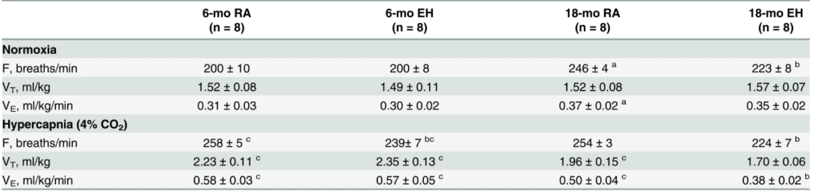
Table 4. Ventilatory parameters at rest and during hypercapnic exposures in 6-mo and 18-mo oldmdxmice exposed to RA and EH.
6-mo RA 6-mo EH 18-mo RA 18-mo EH
(n = 8) (n = 8) (n = 8) (n = 8)
Normoxia
F, breaths/min 200±10 200±8 246±4a 223±8b
VT, ml/kg 1.52±0.08 1.49±0.11 1.52±0.08 1.57±0.07
VE, ml/kg/min 0.31±0.03 0.30±0.02 0.37±0.02a 0.35±0.02
Hypercapnia (4% CO2)
F, breaths/min 258±5c 239±7bc 254±3 224±7b
VT, ml/kg 2.23±0.11c 2.35±0.13c 1.96±0.15c 1.70±0.06
VE, ml/kg/min 0.58±0.03c 0.57±0.05c 0.50±0.04c 0.38±0.02b
Values represent Mean±SEM; n, number of mice.
asignificant difference (p<0.05) 6-mo vs 18-mo old RAmdxmice. bsignificant difference (p<0.05) RA vs EH exposed mice of same age. csignificant difference (p<0.05) normoxia vs hypercapnia in all the groups F, frequency; VT, tidal volume; VE, minute ventilation
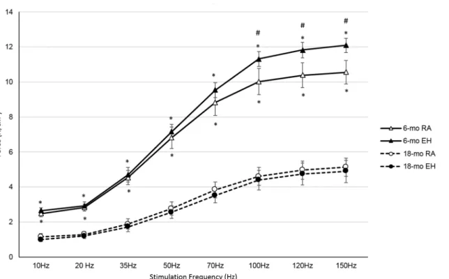
mice as compared to 6-mo oldmdxmice across all the stimulation frequencies (Table 5and Fig 4). On average, maximal tetanic force for 18-mo old RAmdxmice was ~50% lower than that generated by 6-mo old RA mice.
In response to EH exposure, diaphragms from 6-mo mice generated significantly higher
forces (~14%) compared to forces recorded in 6-mo old RA mice (Table 5). In contrast,
dia-phragm force output was similar in RA and EH exposed 18-mo oldmdxmice (Fig 4).
Histological analysis. In order to assess structural changes at the cellular level, we ana-lyzed histological parameters in both diaphragm and cardiac muscles. Representative images of
diaphragm sections from each group are shown inFig 5A. Histological data are shown in
Table 3. The diaphragms from 18-mo oldmdxmice were significantly thinner (~30%), with
increased interstitial space (~80%) as compared to 6-mo oldmdxmice.
In 6-mo old mice, EH exposure had no impact on diaphragm thickness and interstitial space. In contrast, EH exposure in 18-mo old mice led to significant decreases in diaphragm thickness (~16%) and to a concomitant increase in interstitial space (~12%).
Central nucleation, an indicator of regeneration process in muscle fiber after degeneration
or damage [29], was measured (Table 3). Diaphragm muscle fibers of 6-mo oldmdxmice in
both RA and EH group showed significantly higher (245% and 230% respectively) number of
centrally nucleated fibers than those noted in 18-mo oldmdxmice (Table 3). EH exposure had
no impact on central nucleation in either 6-mo old or 18-mo oldmdxmice.
Hydroxyproline Analysis. Hydroxyproline content, an index of collagen content and
fibrosis in muscle, was also measured. Diaphragms from 18-mo old RAmdxmice revealed
sig-nificantly higher hydroxyproline levels (~230%) as compared to 6-mo old RA mice (Fig 5B).
There was no significant difference in diaphragm hydroxyproline levels between RA and EH
exposed 6-mo oldmdxmice (3.3 ± 0.3 vs 4.4 ± 0.4μg/mg wet weights respectively, N. S.), while
EH exposure in18-mo oldmdxmice led to significantly higher hydroxyproline level compared
to age matched RA controls (13.3 ± 0.4 vs 10.9 ± 0.8μg/mg wet weights respectively, p<0.05).
Discussion
To our knowledge, this is the first comprehensive study to investigate the combined impact of
EH and aging on cardiac and respiratory function in the dystrophin-deficientmdxmice. The
mdxmouse has been validated as a model for DMD and has been studied extensively for
sev-eral decades. Although limb muscles ofmdxmice do not show the same extent of pathology or
degeneration as seen in DMD [11], the diaphragm muscle has been shown to closely mirror
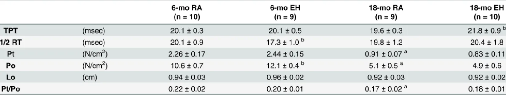
Table 5. In-vitro mechanical characteristics of diaphragm muscle of 6-mo and 18-mo oldmdxmice exposed to RA and EH for 12 weeks.
6-mo RA 6-mo EH 18-mo RA 18-mo EH
(n = 10) (n = 9) (n = 9) (n = 10)
TPT (msec) 20.1±0.3 20.1±0.5 19.6±0.3 21.8±0.9b
1/2 RT (msec) 20.1±0.9 17.3±1.0b 19.8±1.2 20.4±1.8
Pt (N/cm2) 2.26±0.17 2.44±0.15 0.91±0.07a 0.83±0.11
Po (N/cm2) 10.6±0.7 12.1±0.4b 5.1±0.5a 4.9±0.6
Lo (cm) 0.94±0.03 0.96±0.02 0.92±0.03 0.92±0.02
Pt/Po 0.22±0.02 0.20±0.01 0.17±0.02a 0.18±0.01
Values represent Mean±SEM; n, number of mice
aindicates significant difference(p<0.05) 6-mo vs 18-mo old RAmdxmice. bindicates significant difference (p<0.05) RA vs EH group of same age.
TPT, time to peak twitch tension;½RT, half relaxation time; Pt, peak twitch force; Po, maximal isometric force; Lo, optimal muscle length.
the pathology and degeneration-regeneration pattern noted in skeletal muscles of DMD
patients [30]. In addition, the cardiac muscles of themdxmouse also present with a similar
pathological profile as seen in DMD [13].
Dysfunctions of the respiratory and cardiac systems primarily occur at different stages in
the course of DMD. Respiratory abnormalities are usually noted first, at around 8–10 years of
age, while cardiac abnormalities in DMD are usually detected at about 18 years of age [31].
Similarly, inmdxmice the respiratory abnormalities are diagnosed prior to detecting cardiac
dysfunction. The respiratory muscle dysfunction, including that of the diaphragm, begins at
3–4 weeks of age inmdxmice and progresses as the animal age, while cardiomyopathy inmdx
mice is detected usually after 9 months of age [13,32]. In the current study, 3-mo oldmdx
mice were selected to represent a younger stage where respiratory but not cardiac dysfunction
would be expected, and 15-mo oldmdxmice represented an older group, where both cardiac
and respiratory dysfunction would be present simultaneously.
SBD and sleep apnea are common findings in DMD. Indeed, over 80% patients show dis-turbed breathing during sleep with gas exchange abnormalities and episodes of hypopnea and apnea. Smith et al. (1988) reported that patients of DMD undergo repeated bouts of hypoxia during sleep, during which SaO2 levels falls from baseline of 95.4 +/- 0.6% (SEM) to a mean
nadir of 74.2 +/- 3.9% [7]. Since sleep apnea patients experience similar repeated bouts
hypox-emia during sleep, experimentally induced intermittent hypoxia/episodic hypoxia have been used in animal models to simulate sleep apnea. To validate whether our EH model induced apneic events that were akin to those previously reported in DMD, we conducted an initial pilot study to assess SaO2 levels (MouseOx, Starr Life Sciences, Oakmont, PA) during EH in Fig 4. Force-frequency profile of diaphragm from 6-mo and 18-mo old mdx mice exposed to room air (RA) and episodic hypoxia (EH).At all stimulation frequencies diaphragm forces were significantly lower in 18-mo oldmdxmice in both RA and EH groups. 6-mo oldmdxmice exposed to EH showed higher diaphragm forces compared to RA group at higher stimulation frequencies, but in 18-mo oldmdxmice diaphragm forces were not significantly
different in RA and EH groups.*, indicates significant difference between 6-mo RA vs 18-mo RA and 6-mo EH vs 18-mo EH. #, indicates significant difference between 6-mo RA vs 6-mo EH.
awakemdxmice. In five 4-mo oldmdxmice, we determined that SaO2during the EH as
selected for the current study (21% to 6% FiO2), fluctuated between 97.2 ± 1.7% during RA and
reached a nadir of 71.84 ± 1.8% after 90 sec of the hypoxic exposure (unpublished observation). Thus, the EH exposure used in the current study closely mimicked findings as reported in DMD patients.
Effect of EH on body weight and hematocrit
Consistent with previous EH studies in non-DMD rodents [27,28],mdxmice exposed to EH
of in 6-mo old groups similarly responded with changes in body weight and Hct levels (Table 1). However, 18-mo oldmdxmice did not show significant elevations in Hct level on EH exposure, suggesting failed compensation. EH increases hypoxic stress which results in a Fig 5. Histology of diaphragm muscle and hydroxyproline content.(A) Photomicrograph of diaphragm muscle (H&E stained, 20x) from 6-mo and 18-mo old mdx mice exposed to room air (RA) and episodic hypoxia (EH). Note higher amount of fibrosis and interstitial space in 18-mo oldmdxmice as compared to
6-mo oldmdxmice. EH exposed mice diaphragm showed higher amount of fibrosis and interstitial space as
compared to RA controls in both 6-mo and 18-mo oldmdxmice. Difference in diaphragm thickness as well as presence of muscle fibers with central nuclei can also be noticed in this image. (B) Diaphragm hydroxyproline levels (μg/mg wet tissue weight) measured by biochemical analysis. a, significant difference(p<0.05) 6-mo vs 18-mo old RAmdxmice; b, significant difference (p<0.05) RA vs EH group of same age.
decrease in body weight of mice over the experimental period. Long term hypoxia exposure
normally leads to activation of hypoxia inducible factor (HIF) 1-α, which in turn causes
increased production of erythropoietin[33,34]. Erythropoietin subsequently increases red
blood cell production leading to elevated Hct level [35]. The impact on weight and Hct levels
support that EH exposedmdxmice of both age groups received a hypoxic stimulus that was
physiologically challenging.
Cardiac function
In DMD patients, cardiac muscles initially undergo hypertrophy due to muscle damage. With increasing age, damaged cardiac tissue undergoes fibrosis, necrosis and loss of muscle fibers,
ultimately leading to dilated cardiomyopathy [36]. In DMD patients, left ventricular wall
ini-tially shows signs of myocardial fibrosis which then spread to the interventricular septum and
subsequently to the right ventricle [13]. Similar cardiac pathology has been reported to occur
inmdxmice [14]. Chronic exposure to sleep apnea causes an increase in systemic blood
pres-sure and later it can result in left ventricular hypertrophy and cardiomyopathy [10]. To our
knowledge, no study has investigated the impact of EH on cardiac dysfunction inmdxmice.
This is not trivial, as some studies have reported that more than 80% of DMD patients suffer
from sleep disturbed breathing with significant nocturnal desaturation [5].
In this study, we observed decreased heart rate at rest in EH exposed mice as compared to
their RA controls in both 6-mo and 18-mo oldmdxmice. However, as Ketamine/Xylazine
anesthesia is known to have negative ionotropic effect, the results may have been confounded even at similar dose of anesthetic. Although some studies have shown increase in heart rate
after exposure to hypoxia in non-DMD mice due to stimulation of sympathetic system [15,
37], the results are inconsistent, as other studies, consistent with our findings, have reported
decreased heart rate in awake mice after hypoxic exposure [38].
Consistent with pathophysiology of cardiomyopathy in DMD andmdxmice [39–41], we
expected more severe cardiac dysfunction in our 18-mo old group as compared to 6-mo old group. Although older mice had significantly thicker septal wall and higher LV mass than 6-mo old mice, LV contractility was noted not to be significantly different. In contrast to previous
stud-ies, which reported decrease in EF and FS with aging [42,43], we did not see significant difference
in EF and FS in both age groups. One possible explanation is the development of cardiac
dysfunc-tion by 6 month of age inmdxmice, which therefore is not significantly different than older 18
monthmdxmice. With 12 weeks of EH exposure, no significant difference was noted in LVM
and SWT in both age group mice. Even though, not significantly different, once cannot ignore opposite trend in both LVM and SWT parameters as a result of 12 weeks EH exposure in both age groups. These findings suggest that 12 weeks EH exposure results in hypertrophic changes in
6-mo old mice and increased muscle necrosis with muscle fiber loss in older 18-momdxmice.
Contrary to a previous studies [13,39], who failed to detect cardiomyopathy using
non-invasive techniques (echocardiogram) before 7–8 months of age inmdxmice, we found that if
mdxmice are exposed to episodic hypoxia, detectable cardiomyopathy can be detected at an
early age inmdxmice. Danialou et al., 2001 [44] reported thatmdxhearts are susceptible to
damage under mechanical stress and reported abnormal cardiac function using invasive tech-niques. Our findings support that increased stress, in the form of EH exposure, in 6-mo old
mdxmice results in decreased LV contractility. It was interesting to see early signs of dilated
cardiomyopathy (increased LVID) at this age with EH exposure, as dilated cardiomyopathy is
usually seen in later stages of life in bothmdxmice as well as DMD patients [31,45]. EH
exposed 18-mo oldmdxmice also showed signs of dilated cardiomyopathy which is consistent
As shown previously in wild-type andmdxmice [13], we noted increases in hydroxyproline
levels, an indicator of cardiac fibrosis, with aging (Fig 3B). Signs of increased myocardial
fibro-sis with mononuclear infiltrates were also evident on histological examination of heart tissue (Fig 3A). EH exposure in wild-type mice has also been shown to increase cardiac fibrosis [46]. In our study, although EH exposure led to increased hydroxyproline in 18-mo old mice, no
sig-nificant increase in hydroxyproline level was noted in 6-mo oldmdxmice, suggesting increased
susceptibility of older cardiac muscle to damage in response to EH. Increase in fibrosis with
aging has been reported previously in normal wild type as well asmdxmice [39,47]. In older
mdxmice, there are many factors that have been implicated in the increased fibrosis such as
increased muscle injury-repair mechanism and increased TGF-βlevel [39,48].
A previous study[13] has shown cardiomyocyte hypertrophy with aging inmdxmice. In
our study, we did not see significant difference in cardiomyocyte diameter between 6-mo and
18-momdxmice. Cardiomyocyte diameter tended to be larger in 18-momdxmice as
com-pared to 6-mo old mice, but failed to reach significance (Table 3andFig 3C). We also found no
significant effect of EH exposure on cardiomyocyte size inmdxmice. No study has reported
the effect of EH inmdxmice. However, in non-DMD mice, several studies reported
cardio-myocyte hypertrophy in response to EH exposure [49,50], while others observed no
hypertro-phy with EH exposure [51]. No significant change seen in cardiomyocyte size in response to
EH can be due to altered response to stress in these mice due to dystrophin deficiency. There are at least two possible mechanisms, mechanical damage and oxidative stress, which may account for the cardiac dysfunction seen following EH exposure. Exposure to EH in non-DMD rodent models increases the mechanical stress on the heart resulting in hypertension
[27,38], decreased cardiac contractility [46] and ischemic events [52]. The lack of dystrophin
makes cardiac muscle fibers more susceptible to membrane damage during mechanical stress.
As a result, cardiac muscle membrane becomes more permeable to influx of Ca2+[53,54]. Ca2+
overload is associated with a cascade of events such as increased protease activity, impaired mitochondrial oxidative phosphorylation leading to cardiomyocyte necrosis, and ultimately to
cell death [54]. In addition, sleep apnea as well as long term exposure to EH have been shown
to be associated with myocardial ischemia [55–57]. Episodic hypoxia with its resultant
fluctua-tions in SaO2levels causes repeated episodes of hypoxia/reoxygenation, which leads to increase
susceptibility of muscle to ischemia/reperfusion injury on chronic exposure [52]. The role of
reactive oxygen species in ischemia/reperfusion injury has been well documented [58–60].
Thus, synergistic effect of mechanical instability and oxidative stress inmdxmice following EH
may be singularly or partially responsible for the cardiac dysfunction that we observed. Addi-tional studies will be required to investigate which of these two possible mechanisms play the major role in cardiac dysfunction.
Respiratory function
Due to the absence of dystrophin, skeletal muscles inmdxmice are susceptible to injury
induced by increased mechanical stress [2]. The loss of dystrophin makes the sarcolemma
more fragile leading to abnormal calcium influx and recurrent cycles of myofiber
degenera-tion-regeneration, thus leading to progressive muscle weakness [30,53]. Along with increased
susceptibility to muscle injury, these muscles also have impaired repair mechanism. In normal
muscle repair, satellite cells play a major role in regeneration of muscle fibers [61]. In DMD
andmdxskeletal muscle, however there is continuous degeneration-regeneration of muscle
fibers, along with inflammation [62], and satellite cells lose their capacity over time for muscle
repair [23]. Damaged muscle fibers are eventually replaced by fibrotic tissue leading to muscle
diaphragmatic force [63,64] (Fig 4), thickness, interstitial space [65], central nucleation [65],
and hydroxyproline levels (Table 3andFig 5B).
On histological analysis, the higher number of muscle fibers exhibiting central nucleation in
6-mo old RAmdxmice supports elevated degeneration-regeneration process in diaphragm
muscle fibers. In contrast, diaphragms of 18-mo old RAmdxmice exhibited significantly lower
number of central nucleation suggesting satellite cells exhaustion and/or reduced regenerative
capacity with ageing [66,67]. Histological analysis of diaphragm muscle also revealed scattered
areas of fibrosis consistent with our hydroxyproline analysis. Consistent with previous findings
[11], 18-mo old diaphragm tissue demonstrated more areas of fibrosis and mononuclear
infil-trates than noted at 6 months.
Relative force of isolated diaphragm corrected for cross sectional area, as shown inFig 4, in
reality underestimates the effect of age on in-situ maximal force generation. Since the
dia-phragm was not only noted to be thinner in older RA mice (Table 3), but that a larger fraction
of the diaphragm was occupied by non-muscular tissues (Fig 5A), we can estimate that absolute
peak diaphragm forces that could be generated in-situ (assuming equal neural activation)
would be 28% lower in older RAmdxmice as compared to their younger counterparts. In
con-trast, 18-mo old EH exposedmdxmice showed no change in relative diaphragm strength (Fig
4). However, since these mice also demonstrated significantly thinner diaphragms (Table 3)
with larger fraction of cross-section occupied by fibrotic tissue (Table 3), one would predict
that in-situ diaphragm forces in 18-mo EH mice would be further compromised by 20%.
Previously, we reported [68] that 12 weeks of EH exposure in 6-mo oldmdxmice resulted
in a reduction in relative diaphragm force by about 30%. In sharp contrast, relative diaphragm
strength in the current study in 6-month oldmdxmice increased (Table 5) following 12 weeks
of EH exposure (Fig 4).The divergent responses to EH between our current and our former
study is likely attributed to the hypoxic stress levels imposed and the resultant physiological responses on the respiratory system. One would expect that, more severe hypoxia would result in a more robust ventilatory response (diaphragm activation) during EH. The severity of whole body activity levels have been reported to result in contrasting findings on limb muscles
strength. Voluntary activity inmdxmice appears to have a beneficial effect on limb muscle
strength [69–71], whereas forced activity at higher intensities (and muscle recruitment) has
been shown to be detrimental and to enhance weakness of limb muscles [72,73]. Thus, a
simi-lar conclusion can be drawn here for diaphragm strength, in that respiratory muscle recruit-ment at less severe levels can lead to improverecruit-ments in diaphragm strength, whereas more severe recruitment and higher stress can lead to reductions in diaphragm strength.
Ventilatory parameters were recorded using modified barometric plethysmography in all
mdxmice at rest and in response to hypercapnia (4% CO2). Consistent with a previous study
[65], older, 18-mo RAmdxmice showed higher frequency of breathing compared to younger,
6-mo RA mice. Since tidal volume between these groups were similar, ventilation was noted to
be higher in oldermdxmice. Aging, as has been previously reported [65], was associated with
an increase in ventilatory dysfunction, as 18-mo oldmdxmice showed decreased response to
4% CO2exposure as compared to 6-mo oldmdxmice. Consistent with our diaphragm strength
data (Fig 4) and the implications to in-situ diaphragm force discussed above, ventilatory
func-tion in awake mice showed no significant effect in ventilatory parameters due to EH exposure
in 6-mo oldmdxmice (Table 4). In contrast, 18-mo oldmdxmice exposed to EH show
consid-erable ventilatory dysfunction and in addition, failed to increase ventilation above resting room air baseline values in response to the hypercapnic challenge. As this effect appears to be focused on the control of respiration, rather than the diaphragm muscle itself, ventilatory
dys-function in18-mo EHmdxmice is most likely secondary to reduced ventilatory reserve, or to a
chemosensory response and carotid body dysfunction inmdxmice [74]. Additional studies are needed to determine if carotid body dysfunction is exacerbated by sleep apnea in DMD.
There are several limitations of the current study design. One relates to the absence of wild type non-DMD mice. As our intent was to investigate the functional impact of EH on cardiac and respiratory function in dystrophin deficient mice, we purposely restricted our study to
mdxmice. In the absence of normal wild type mice, we cannot address whether EH causes
more or less severe cardiac and respiratory dysfunction inmdxmice. Although not a goal of
our study, additional research is required to study this comparison. Another limitation is related to the use of anesthesia for the assessment of cardiac function. Ketamine/xylazine
anes-thesia is known to exert negative chronotropic and ionotropic effects in mice [75,76]. For this
reason, our original intent was to evaluate cardiac function in awake mice, and initial attempts were successful in all groups with the exception of the 18-mo old EH group. Since the first two 18-mo EH awake mice died during echocardiography and as we could ill afford to lose addi-tional 18-mo EH mice during the procedure, we elected to study all animals under anesthesia. Also, we limited our analysis to LV function. Echocardiographic assessment of RV dimension and function is very challenging, especially in murine models due to their small size, retroster-nal position and irregular shape. As technical limitation preclude accurate measurement of RV function, we decided to focus on LV function.
In summary, this is the first comprehensive study to report the effect of experimentally induced sleep apnea on cardiac and respiratory functions in DMD mice. Our results support
that EH induces cardiac and respiratory dysfunction inmdxmice. In addition, the observed
dysfunctions were more severe in oldermdxmice. Our findings also highlight that EH can
have divergent effects on the cardiac and respiratory systems. Indeed, in younger animals, EH produced improvement in isolated diaphragmatic function, while producing mild cardiac dys-function. In older animals, EH proved to be detrimental to both the cardiac and respiratory systems. The results of the present study have important implications in DMD patients. As pre-viously mentioned, over 80% of DMD patients suffer from sleep disordered breathing and a majority of these patients succumb to respiratory or cardiac failure. Although, nocturnal assis-ted ventilation is often employed in DMD patients, it is generally initiaassis-ted during the late ambulatory or non-ambulatory stages, only after a positive diagnosis of hypoventilation or
hypercapnia [4]. It is still debatable when to initiate nocturnal assisted ventilation to achieve
maximum benefit. Based on the current findings, DMD patients may certainly benefit by ear-lier screening and interventions, especially since nocturnal hypoventilation is often detected in
DMD patients presenting with normal daytime ventilation and oxygen saturation [7]. In
addi-tion, future studies are needed to investigate the putative mechanisms responsible for the respi-ratory and cardiac dysfunction and to study whether these deficits, once detected, can be reversed with effective treatment.
Acknowledgments
The authors acknowledge the technical assistance of Kunjan Shah. The authors also thank Gen Suzuki and Brian Weil for their assistance with cardiac histochemistry.
Author Contributions
on cardiac function: JAF MRC. Data analysis and interpretation: MRC GAF. Manuscript draft-ing and revision: MRC GAF JAF.
References
1. Emery AE. The muscular dystrophies. Lancet. 2002; 359(9307):687–95. doi:10.1016/S0140-6736(02)
07815-7PMID:11879882.
2. Hoffman EP, Brown RH Jr, Kunkel LM. Dystrophin: the protein product of the Duchenne muscular dys-trophy locus. Cell. 1987; 51(6):919–28. PMID:3319190.
3. Finsterer J, Stollberger C. The heart in human dystrophinopathies. Cardiology. 2003; 99(1):1–19.
68446. PMID:12589117.
4. [Reports from the annual meeting of the American Thoracic Society, Orlando, Florida, USA, 21–26 May
2004]. Revue des maladies respiratoires. 2004; 21(5 Pt 2):7S5–190. PMID:15806622.
5. Labanowski M, Schmidt-Nowara W, Guilleminault C. Sleep and neuromuscular disease: frequency of sleep-disordered breathing in a neuromuscular disease clinic population. Neurology. 1996; 47 (5):1173–80. PMID:8909425.
6. Barbe F, Quera-Salva MA, McCann C, Gajdos P, Raphael JC, de Lattre J, et al. Sleep-related respiratory disturbances in patients with Duchenne muscular dystrophy. The European respiratory journal: official journal of the European Society for Clinical Respiratory Physiology. 1994; 7(8):1403–8. PMID:7957826.
7. Smith PE, Calverley PM, Edwards RH. Hypoxemia during sleep in Duchenne muscular dystrophy. The American review of respiratory disease. 1988; 137(4):884–8. doi:10.1164/ajrccm/137.4.884PMID:
3354996.
8. Shamsuzzaman AS, Gersh BJ, Somers VK. Obstructive sleep apnea: implications for cardiac and vas-cular disease. JAMA: the journal of the American Medical Association. 2003; 290(14):1906–14. doi:10.
1001/jama.290.14.1906PMID:14532320.
9. Wolk R, Kara T, Somers VK. Sleep-disordered breathing and cardiovascular disease. Circulation. 2003; 108(1):9–12. doi:10.1161/01.CIR.0000072346.56728.E4PMID:12847053.
10. Bradley TD, Floras JS. Sleep apnea and heart failure: Part I: obstructive sleep apnea. Circulation. 2003; 107(12):1671–8. doi:10.1161/01.CIR.0000061757.12581.15PMID:12668504.
11. Stedman HH, Sweeney HL, Shrager JB, Maguire HC, Panettieri RA, Petrof B, et al. The mdx mouse diaphragm reproduces the degenerative changes of Duchenne muscular dystrophy. Nature. 1991; 352 (6335):536–9. doi:10.1038/352536a0PMID:1865908.
12. Grady RM, Teng H, Nichol MC, Cunningham JC, Wilkinson RS, Sanes JR. Skeletal and cardiac myopa-thies in mice lacking utrophin and dystrophin: a model for Duchenne muscular dystrophy. Cell. 1997; 90(4):729–38. PMID:9288752.
13. Quinlan JG, Hahn HS, Wong BL, Lorenz JN, Wenisch AS, Levin LS. Evolution of the mdx mouse car-diomyopathy: physiological and morphological findings. Neuromuscular disorders: NMD. 2004; 14(8–
9):491–6. doi:10.1016/j.nmd.2004.04.007PMID:15336690.
14. Wehling-Henricks M, Jordan MC, Roos KP, Deng B, Tidball JG. Cardiomyopathy in dystrophin-deficient hearts is prevented by expression of a neuronal nitric oxide synthase transgene in the myocardium. Human molecular genetics. 2005; 14(14):1921–33. doi:10.1093/hmg/ddi197PMID:15917272.
15. Fletcher EC, Lesske J, Behm R, Miller CC 3rd, Stauss H, Unger T. Carotid chemoreceptors, systemic blood pressure, and chronic episodic hypoxia mimicking sleep apnea. Journal of applied physiology. 1992; 72(5):1978–84. PMID:1601808.
16. Lee SD, Nakano H, Gosselin LE, Krasney JA, Schlenker EH, Farkas GA. Endogenous opioids modulate ventilation and peak oxygen consumption in obese Zucker rats. American journal of respiratory and criti-cal care medicine. 2000; 162(3 Pt 1):1009–15. doi:10.1164/ajrccm.162.3.9909058PMID:10988122.
17. Drorbaugh JE, Fenn WO. A barometric method for measuring ventilation in newborn infants. Pediatrics. 1955; 16(1):81–7. PMID:14394741.
18. Bartlett D Jr, Tenney SM. Control of breathing in experimental anemia. Respiration physiology. 1970; 10(3):384–95. PMID:5476156.
20. Farkas GA, Gosselin LE, Zhan WZ, Schlenker EH, Sieck GC. Histochemical and mechanical properties of diaphragm muscle in morbidly obese Zucker rats. Journal of applied physiology. 1994; 77(5):2250–
9. PMID:7868442.
21. Close RI. Dynamic properties of mammalian skeletal muscles. Physiological reviews. 1972; 52(1):129–
97. PMID:4256989.
22. Coulton GR, Morgan JE, Partridge TA, Sloper JC. The mdx mouse skeletal muscle myopathy: I. A his-tological, morphometric and biochemical investigation. Neuropathology and applied neurobiology. 1988; 14(1):53–70. PMID:2967442.
23. Matecki S, Guibinga GH, Petrof BJ. Regenerative capacity of the dystrophic (mdx) diaphragm after induced injury. American journal of physiology Regulatory, integrative and comparative physiology. 2004; 287(4):R961–8. doi:10.1152/ajpregu.00146.2004PMID:15191902.
24. Prockop DJ, Udenfriend S. A specific method for the analysis of hydroxyproline in tissues and urine. Analytical biochemistry. 1960; 1:228–39. PMID:13738134.
25. Switzer BR, Summer GK. Improved method for hydroxyproline analysis in tissue hydrolyzates. Analyti-cal biochemistry. 1971; 39(2):487–91. PMID:4324532.
26. Rosa CM, Xavier NP, Henrique Campos D, Fernandes AA, Cezar MD, Martinez PF, et al. Diabetes mellitus activates fetal gene program and intensifies cardiac remodeling and oxidative stress in aged spontaneously hypertensive rats. Cardiovascular diabetology. 2013; 12(1):152. doi: 10.1186/1475-2840-12-152PMID:24134628.
27. Campen MJ, Shimoda LA, O'Donnell CP. Acute and chronic cardiovascular effects of intermittent hyp-oxia in C57BL/6J mice. Journal of applied physiology. 2005; 99(5):2028–35. doi:10.1152/japplphysiol.
00411.2005PMID:16002771.
28. Naghshin J, McGaffin KR, Witham WG, Mathier MA, Romano LC, Smith SH, et al. Chronic intermittent hypoxia increases left ventricular contractility in C57BL/6J mice. Journal of applied physiology. 2009; 107(3):787–93. doi:10.1152/japplphysiol.91256.2008PMID:19589954; PubMed Central PMCID:
PMC2756000.
29. Briguet A, Courdier-Fruh I, Foster M, Meier T, Magyar JP. Histological parameters for the quantitative assessment of muscular dystrophy in the mdx-mouse. Neuromuscular disorders: NMD. 2004; 14 (10):675–82. doi:10.1016/j.nmd.2004.06.008PMID:15351425.
30. Petrof BJ, Shrager JB, Stedman HH, Kelly AM, Sweeney HL. Dystrophin protects the sarcolemma from stresses developed during muscle contraction. Proceedings of the National Academy of Sciences of the United States of America. 1993; 90(8):3710–4. PMID:8475120; PubMed Central PMCID:
PMC46371.
31. Nigro G, Comi LI, Politano L, Bain RJ. The incidence and evolution of cardiomyopathy in Duchenne muscular dystrophy. International journal of cardiology. 1990; 26(3):271–7. PMID:2312196.
32. Zhang W, ten Hove M, Schneider JE, Stuckey DJ, Sebag-Montefiore L, Bia BL, et al. Abnormal cardiac morphology, function and energy metabolism in the dystrophic mdx mouse: an MRI and MRS study. Journal of molecular and cellular cardiology. 2008; 45(6):754–60. doi:10.1016/j.yjmcc.2008.09.125
PMID:18929569.
33. Wang GL, Semenza GL. General involvement of hypoxia-inducible factor 1 in transcriptional response to hypoxia. Proceedings of the National Academy of Sciences of the United States of America. 1993; 90(9):4304–8. PMID:8387214; PubMed Central PMCID: PMC46495.
34. Yu AY, Shimoda LA, Iyer NV, Huso DL, Sun X, McWilliams R, et al. Impaired physiological responses to chronic hypoxia in mice partially deficient for hypoxia-inducible factor 1alpha. The Journal of clinical investigation. 1999; 103(5):691–6. doi:10.1172/JCI5912PMID:10074486; PubMed Central PMCID:
PMC408131.
35. McGuire M, Bradford A. Chronic intermittent hypoxia increases haematocrit and causes right ventricu-lar hypertrophy in the rat. Respiration physiology. 1999; 117(1):53–8. PMID:10505479.
36. Ameen V, Robson LG. Experimental models of duchenne muscular dystrophy: relationship with cardio-vascular disease. The open cardiocardio-vascular medicine journal. 2010; 4:265–77. doi:10.2174/
1874192401004010265PMID:21258567; PubMed Central PMCID: PMC3024556.
37. Lin M, Liu R, Gozal D, Wead WB, Chapleau MW, Wurster R, et al. Chronic intermittent hypoxia impairs baroreflex control of heart rate but enhances heart rate responses to vagal efferent stimulation in anes-thetized mice. American journal of physiology Heart and circulatory physiology. 2007; 293(2):H997–
1006. doi:10.1152/ajpheart.01124.2006PMID:17384123.
39. Au CG, Butler TL, Sherwood MC, Egan JR, North KN, Winlaw DS. Increased connective tissue growth factor associated with cardiac fibrosis in the mdx mouse model of dystrophic cardiomyopathy. Interna-tional journal of experimental pathology. 2011; 92(1):57–65. doi:10.1111/j.1365-2613.2010.00750.x
PMID:21121985; PubMed Central PMCID: PMC3052757.
40. Spurney CF, Sali A, Guerron AD, Iantorno M, Yu Q, Gordish-Dressman H, et al. Losartan decreases cardiac muscle fibrosis and improves cardiac function in dystrophin-deficient mdx mice. Journal of car-diovascular pharmacology and therapeutics. 2011; 16(1):87–95. doi:10.1177/1074248410381757
PMID:21304057.
41. Bia BL, Cassidy PJ, Young ME, Rafael JA, Leighton B, Davies KE, et al. Decreased myocardial nNOS, increased iNOS and abnormal ECGs in mouse models of Duchenne muscular dystrophy. Journal of molecular and cellular cardiology. 1999; 31(10):1857–62. doi:10.1006/jmcc.1999.1018PMID:
10525423.
42. Markham LW, Michelfelder EC, Border WL, Khoury PR, Spicer RL, Wong BL, et al. Abnormalities of diastolic function precede dilated cardiomyopathy associated with Duchenne muscular dystrophy. Journal of the American Society of Echocardiography: official publication of the American Society of Echocardiography. 2006; 19(7):865–71. doi:10.1016/j.echo.2006.02.003PMID:16824995.
43. Jearawiriyapaisarn N, Moulton HM, Sazani P, Kole R, Willis MS. Long-term improvement in mdx cardio-myopathy after therapy with peptide-conjugated morpholino oligomers. Cardiovascular research. 2010; 85(3):444–53. doi:10.1093/cvr/cvp335PMID:19815563; PubMed Central PMCID: PMC2802205.
44. Danialou G, Comtois AS, Dudley R, Karpati G, Vincent G, Des Rosiers C, et al. Dystrophin-deficient cardiomyocytes are abnormally vulnerable to mechanical stress-induced contractile failure and injury. FASEB journal: official publication of the Federation of American Societies for Experimental Biology. 2001; 15(9):1655–7. PMID:11427517.
45. Bostick B, Yue Y, Long C, Duan D. Prevention of dystrophin-deficient cardiomyopathy in twenty-one-month-old carrier mice by mosaic dystrophin expression or complementary dystrophin/utrophin expres-sion. Circulation research. 2008; 102(1):121–30. doi:10.1161/CIRCRESAHA.107.162982PMID:
17967782.
46. Chen L, Zhang J, Gan TX, Chen-Izu Y, Hasday JD, Karmazyn M, et al. Left ventricular dysfunction and associated cellular injury in rats exposed to chronic intermittent hypoxia. Journal of applied physiology. 2008; 104(1):218–23. doi:10.1152/japplphysiol.00301.2007PMID:18006871.
47. Van Erp C, Loch D, Laws N, Trebbin A, Hoey AJ. Timeline of cardiac dystrophy in 3-18-month-old MDX mice. Muscle Nerve. 2010; 42(4):504–13. doi:10.1002/mus.21716PMID:20589894.
48. Gosselin LE, Williams JE, Deering M, Brazeau D, Koury S, Martinez DA. Localization and early time course of TGF-beta 1 mRNA expression in dystrophic muscle. Muscle & nerve. 2004; 30(5):645–53.
doi:10.1002/mus.20150PMID:15389721.
49. Matsumoto C, Hayashi T, Kitada K, Yamashita C, Miyamura M, Mori T, et al. Chymase plays an impor-tant role in left ventricular remodeling induced by intermittent hypoxia in mice. Hypertension. 2009; 54 (1):164–71. doi:10.1161/HYPERTENSIONAHA.109.131391PMID:19470876.
50. Hayashi T, Yoshioka T, Hasegawa K, Miyamura M, Mori T, Ukimura A, et al. Inhalation of hydrogen gas attenuates left ventricular remodeling induced by intermittent hypoxia in mice. American journal of phys-iology Heart and circulatory physphys-iology. 2011; 301(3):H1062–9. doi:10.1152/ajpheart.00150.2011
PMID:21642501.
51. Fan C, Iacobas DA, Zhou D, Chen Q, Lai JK, Gavrialov O, et al. Gene expression and phenotypic char-acterization of mouse heart after chronic constant or intermittent hypoxia. Physiological genomics. 2005; 22(3):292–307. Epub 2005/06/02. doi:10.1152/physiolgenomics.00217.2004PMID:15928208;
PubMed Central PMCID: PMC2856928.
52. Joyeux-Faure M, Stanke-Labesque F, Lefebvre B, Beguin P, Godin-Ribuot D, Ribuot C, et al. Chronic intermittent hypoxia increases infarction in the isolated rat heart. Journal of applied physiology. 2005; 98(5):1691–6. doi:10.1152/japplphysiol.01146.2004PMID:15618317.
53. Turner PR, Fong PY, Denetclaw WF, Steinhardt RA. Increased calcium influx in dystrophic muscle. The Journal of cell biology. 1991; 115(6):1701–12. PMID:1661733; PubMed Central PMCID:
PMC2289194.
54. Whitehead NP, Yeung EW, Allen DG. Muscle damage in mdx (dystrophic) mice: role of calcium and reactive oxygen species. Clinical and experimental pharmacology & physiology. 2006; 33(7):657–62.
doi:10.1111/j.1440-1681.2006.04394.xPMID:16789936.
55. Chan HS, Chiu HF, Tse LK, Woo KS. Obstructive sleep apnea presenting with nocturnal angina, heart failure, and near-miss sudden death. Chest. 1991; 99(4):1023–5. PMID:2009755.
57. Franklin KA, Nilsson JB, Sahlin C, Naslund U. Sleep apnoea and nocturnal angina. Lancet. 1995; 345 (8957):1085–7. PMID:7715342.
58. Ambrosio G, Flaherty JT, Duilio C, Tritto I, Santoro G, Elia PP, et al. Oxygen radicals generated at reflow induce peroxidation of membrane lipids in reperfused hearts. The Journal of clinical investiga-tion. 1991; 87(6):2056–66. doi:10.1172/JCI115236PMID:1645750; PubMed Central PMCID:
PMC296962.
59. Maddika S, Elimban V, Chapman D, Dhalla NS. Role of oxidative stress in ischemia-reperfusion-induced alterations in myofibrillar ATPase activities and gene expression in the heart. Canadian journal of physiology and pharmacology. 2009; 87(2):120–9. doi:10.1139/Y08-105PMID:19234575.
60. Myers ML, Bolli R, Lekich RF, Hartley CJ, Roberts R. Enhancement of recovery of myocardial function by oxygen free-radical scavengers after reversible regional ischemia. Circulation. 1985; 72(4):915–21.
PMID:4028384.
61. Mauro A. Satellite cell of skeletal muscle fibers. J Biophys Biochem Cytol. 1961; 9:493–5. PMID:
13768451; PubMed Central PMCID: PMCPMC2225012.
62. Spencer MJ, Marino MW, Winckler WM. Altered pathological progression of diaphragm and quadriceps muscle in TNF-deficient, dystrophin-deficient mice. Neuromuscul Disord. 2000; 10(8):612–9. PMID:
11053690.
63. Gosselin LE, Johnson BD, Sieck GC. Age-related changes in diaphragm muscle contractile properties and myosin heavy chain isoforms. American journal of respiratory and critical care medicine. 1994; 150 (1):174–8. doi:10.1164/ajrccm.150.1.8025746PMID:8025746.
64. Tolep K, Higgins N, Muza S, Criner G, Kelsen SG. Comparison of diaphragm strength between healthy adult elderly and young men. American journal of respiratory and critical care medicine. 1995; 152 (2):677–82. doi:10.1164/ajrccm.152.2.7633725PMID:7633725.
65. Gayraud J, Matecki S, Hnia K, Mornet D, Prefaut C, Mercier J, et al. Ventilation during air breathing and in response to hypercapnia in 5 and 16 month-old mdx and C57 mice. Journal of muscle research and cell motility. 2007; 28(1):29–37. doi:10.1007/s10974-007-9101-1PMID:17431804; PubMed Central
PMCID: PMC1974787.
66. Heslop L, Morgan JE, Partridge TA. Evidence for a myogenic stem cell that is exhausted in dystrophic muscle. Journal of cell science. 2000; 113 (Pt 12):2299–308. PMID:10825301.
67. Bockhold KJ, Rosenblatt JD, Partridge TA. Aging normal and dystrophic mouse muscle: analysis of myogenicity in cultures of living single fibers. Muscle & nerve. 1998; 21(2):173–83. PMID:9466592.
68. Farkas GA, McCormick KM, Gosselin LE. Episodic hypoxia exacerbates respiratory muscle dysfunction in DMD(mdx) mice. Muscle & nerve. 2007; 36(5):708–10. doi:10.1002/mus.20858PMID:17654561.
69. Dupont-Versteegden EE, McCarter RJ, Katz MS. Voluntary exercise decreases progression of muscu-lar dystrophy in diaphragm of mdx mice. Journal of applied physiology. 1994; 77(4):1736–41. PMID:
7836193.
70. Hayes A, Williams DA. Beneficial effects of voluntary wheel running on the properties of dystrophic mouse muscle. Journal of applied physiology. 1996; 80(2):670–9. PMID:8929614.
71. Wineinger MA, Abresch RT, Walsh SA, Carter GT. Effects of aging and voluntary exercise on the func-tion of dystrophic muscle from mdx mice. American journal of physical medicine & rehabilitafunc-tion / Asso-ciation of Academic Physiatrists. 1998; 77(1):20–7. PMID:9482375.
72. Brussee V, Tardif F, Tremblay JP. Muscle fibers of mdx mice are more vulnerable to exercise than those of normal mice. Neuromuscular disorders: NMD. 1997; 7(8):487–92. PMID:9447605.
73. Vilquin JT, Brussee V, Asselin I, Kinoshita I, Gingras M, Tremblay JP. Evidence of mdx mouse skeletal muscle fragility in vivo by eccentric running exercise. Muscle & nerve. 1998; 21(5):567–76. PMID:
9572235.
74. Mosqueira M, Baby SM, Lahiri S, Khurana TS. Ventilatory chemosensory drive is blunted in the mdx mouse model of Duchenne Muscular Dystrophy (DMD). PloS one. 2013; 8(7):e69567. doi:10.1371/ journal.pone.0069567PMID:23922741; PubMed Central PMCID: PMCPMC3726676.
75. Tan TP, Gao XM, Krawczyszyn M, Feng X, Kiriazis H, Dart AM, et al. Assessment of cardiac function by echocardiography in conscious and anesthetized mice: importance of the autonomic nervous system and disease state. Journal of cardiovascular pharmacology. 2003; 42(2):182–90. PMID:12883320.