%
From the Division of Diagnostic Imaging ofthe Heart Institute and the Division of Pediatric Rheumatology, Hospital das Clínicas, Faculty of Medicine, University of São Paulo.
CORRELATION BETWEEN OSTEOCHONDRAL
CHANGES DEPICTED BY MAGNETIC RESONANCE
IMAGING AND DISEASE PROGRESSION
Andréa S. Doria, Maria Helena B. Kiss, Adriana M. Sallum, Ana Paola N. Lotito, Erica N. Naka, Cláudio C. de Castro and Giovanni G. Cerri
RHCFAP/3045 DORIA AS et al. - Correlation between osteochondral changes depicted by magnetic resonance imaging and disease progression.
Rev. Hosp. Clín. Fac. Med. S. Paulo 56(4):107-114, 2001.
Purpose: To determine the consequences of the chronic use of systemic corticosteroids in children with juvenile rheumatoid
arthritis by means of evaluating osteochondral effects depicted by magnetic resonance imaging.
Patients and Methods: We reviewed clinical and magnetic resonance imaging findings in 69 children (72 knees) with juvenile rheumatoid arthritis. Two groups were studied. Group I: 34 (49.3%) children had previous or current use of systemic corticotherapy (22 girls; 12 boys; mean age: 11.3 years; mean disease duration: 5.9 years; mean corticotherapy duration: 2.9 years; mean cumulative dose of previous corticosteroids: 5000 mg); Group II: 35 (50.7%) children had no previous use of corticosteroids (27 girls; 8 boys; mean age: 11.7 years; mean disease duration: 5.3 years). The groups were compared statistically.
Results: In the group that had received corticotherapy (Group I), osteochondral abnormalities were significantly correlated to
long-standing disease (>3.5 years; p<0.001). This correlation was not found in the group that had no previous history of corticotherapy (Group II). No correlations were established between median dose of corticosteroids and magnetic resonance imaging findings.
Conclusion:It is important to further investigate the long-term intra-articular effects of systemic corticotherapy to ensure that
the side effects of the aggressive therapy will not be more harmful for the joints than the symptoms suffered over the natural course of the disease.
DESCRIPTORS: Systemic corticotherapy. Juvenile rheumatoid arthritis. Children. Knee. Magnetic resonance imaging.
Juvenile rheumatoid arthritis (JRA) is a relatively common childhood dis-ease characterized by chronic arthritis1,
with a prevalence in developed coun-tries of approximately 1 in 1000 chil-dren2. Synovitis is responsible for the
development of synovial hypertrophy (pannus) that can erode articular carti-lage and bone and invade the adjacent bone marrow space, leading to joint deformity and producing much of the morbidity and disability in the dis-ease3,4. For those children who do not
respond to salicylates and nonsteroid anti-inflammatory drugs, corticoster-oids and other immunosuppressive
agents may be required for manage-ment of the disease.
Radiography does not provide in-formation about the status of osteo-chondral structures before cartilage is damaged5. On the other hand, magnetic
resonance imaging (MRI) provides ex-quisite distinction of bone and soft tis-sue in the articular structures3-6 and is
a highly sensitive tool for establishing the diagnosis of JRA7.
The systemic side effects of long-term corticotherapy in children are well-established8. To justify chronic
&
because of its known systemic side ef-fects, but also because of the drug-in-duced intra-articular changes.
This study investigates the MRI findings, rather than the traditionally used radiographic findings, in a cross-section of JRA patients with varying durations of the disease who had or not undergone previous corticotherapy. The purpose of our study was to estab-lish correlations among the presence of synovial and osteochondral abnormali-ties demonstrated by MRI, disease ac-tivity at the time of exam, and previ-ous and/or current systemic corticotherapy.
PATIENTS AND METHODS
Sixty-nine children with clinically proven JRA, according to the classifi-cation criteria of the American College of Rheumatology9 were selected for the
study. Disease duration at the time of MRI performance ranged from 5 months to 14 years (mean: 5.6 years). Two groups of patients with JRA were analyzed. Group I included 34 (49.3%) patients who had a previous or current history of corticotherapy. In this group, we analyzed 36 knees. Group II in-cluded 35 (50.7%) patients who were using either no medication or drug therapy other than corticosteroids. We studied 36 knees in this group.
Patient Characteristics
In Group I, 22 (64.7%) patients were girls and 12 (35.3%) were boys. Their mean age was 11.3 (range: 4.8-17.1) years, mean disease duration was 5.9 years, mean duration of corticotherapy was 2.9 years, and me-dian total dose of corticosteroids pre-viously used was 5000 (range: 225-40283) mg. Among Group I patients, 11 (32.4%) presented clinical activity at the time of exam as defined by the presence of arthritis in any joint5.
Twenty-two (64.7%) patients had sys-temic disease, 10 (29.4%) had pol-yarticular disease, and 2 (5.9%) had pauciarticular disease.
In this group, clinical indication for use of systemic corticotherapy had been based on severe manifestations of the disease, including pericarditis, myocarditis, vasculitis, and hemoph-agocytic syndrome. The dose of corti-costeroids was adjusted to the clinical and laboratory severity of disease. Group I was further subdivided accord-ing to duration of corticotherapy: Group IA had £1 year corticotherapy
and Group IB had >1 year. We also compared the results within each group between those patients with prolonged disease duration (>3.5 years) and those with shorter duration of disease (£3.5
years).
In Group II, there were 27 (77.1%)
girls and 8 (22.9%) boys; their mean age was 11.7 (range: 2.6-20) years, and mean disease duration was 5.3 years. Among these children, 10 (28.6%) pre-sented clinically active disease. Eleven (31.4%) patients had systemic disease, 9 (25.7%) had polyarticular disease, and 15 (42.9%) had pauciarticular dis-ease.
Magnetic Resonance Imaging (MRI)
We performed MRI on the knees of all patients included in this study us-ing a 1.5-T magnet, a whole volume extremity coil, and the imaging param-eters listed in table 1. Gadolinium was injected intravenously at a dosage of 0.1mmol/kg.The images were evalu-ated independently by 2 experienced radiologists who were blinded to the clinical history or plain films and di-rected to evaluate: (1) the presence of synovial proliferation in the suprapatel-lar bursa—eg, synovial enhancement post-gadolinium; (2) the presence of osteochondral abnormalities—eg, sub-chondral cysts, marginal erosions, and/ or bone marrow edema; (3) both of the above findings, and (4) absence of depictable lesions.
Statistical Analysis
Correlations among MRI findings and (1) disease duration (ie, £3.5 years
Table 1 - Pulse sequences used for analysis of MRI findings.
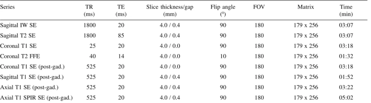
Series TR TE Slice thickness/gap Flip angle FOV Matrix Time
(ms) (ms) (mm) (0) (min)
Sagittal IW SE 1800 20 4.0 / 0.4 90 180 179 x 256 03:07
Sagittal T2 SE 1800 85 4.0 / 0.4 90 180 179 x 256 03:07
Coronal T1 SE 25 20 4.0 / 0.0 90 180 179 x 256 03:18
Coronal T2 FFE 40 14 4.0 / 0.0 10 180 179 x 256 01:32
Coronal T1 SE (post-gad.) 525 20 4.0 / 0.0 90 180 179 x 256 03:18
Sagittal T1 SE (post-gad.) 525 20 4.0 / 0.4 90 180 179 x 256 01:52
Axial T1 SE (post-gad.) 525 20 4.0 / 0.4 90 180 179 x 256 03:22
Axial T1 SPIR SE (post-gad.) 525 20 4.0 / 0.4 90 180 179 x 256 05:02
'
or >3.5 years), (2) clinical activity ofdisease, (3) previous use of corticoster-oids, (4) duration of corticotherapy, (5) dose of systemic corticosteroids, and (6) type of osteochondral changes were analyzed using the Fisher's exact test (2-tail)10, with a p value < 0.05
indi-cating statistical significance.
RESULTS
In Group I patient's joints (n1=number of joints) with disease du-ration longer than 3.5 years (n1 = 23), MRI depicted osteochondral changes both with (n1=14) and without (n1=3) associated synovial proliferation in 17 joints (Fig. 1 and Table 2). Four of these patients had clinically active dis-ease. In Group II children with longstanding disease (n1 = 21), 17 pa-tients demonstrated cartilaginous ab-normalities both with (n1=15) and without (n1=2) associated synovial changes (Fig. 2 and Table 2). Eight of these children had clinically active dis-ease.
Although the results between these two groups are comparable, significant differences in MRI findings with re-gard to osteochondral lesions and syn-ovial enhancement in patients with long and short disease duration were demonstrated only in Group I (p<0.001). In Group II, we did not find any significant relationship between disease duration (ie, £3.5 years or >3.5
years) and the 4 parameters analyzed by MRI (p=0.076), nor between the presence of osteochondral changes and clinical disease activity in any of the groups. However, we did find a signifi-cant difference between MRI findings of osteochondral abnormalities accord-ing to disease duration (p<0.001) (Table 2) in Group I patients. In this group (n1 = 36), among the 18 patients in whom MRI demonstrated osteo-chondral lesions, changes were more often associated with synovial
prolif-eration (Fig. 1A) than seen as isolated findings (Fig. 3A). Fifteen of these children demonstrated such prolifera-tion (p<0.001) (Table 2).
When we analyzed results accord-ing to disease duration for children with >3.5 years of disease in Group I patients (n1 = 23), presence of osteo-chondral lesions associated with syn-ovial proliferation was the most
com-mon finding, decom-monstrated in 14 (60.9%) knees. In contrast, in patients with at least 3.5 years of the disease (n1 = 13) this finding was demon-strated in only 1 joint (Table 2).
Regarding the effects of the dura-tion of corticosteroid treatment, we found a significant increase in osteo-chondral changes and synovial hyper-trophy in children receiving
corticos-Table 2 - Correlations between MRI findings and disease duration in joints of Group I and II patients. Disease Duration
MRI Findings (Group I) £ 3.5 years > 3.5 years Total
Osteochondral lesions 0 (0%) 3 (100%) 3
Synovial enhancement 6 (54.55%) 5 (45.45%) 11
Synovial enhancement and osteochondral lesions 1 (6.67%) 14 (93.33%) 15 No osteochondral/synovial pathologic findings 6 (85.71%) 1 (14.29%) 7
Total (number of joints) 13 23 36
Fisher's exact test (2-tail) p<0.001
Disease duration
MRI findings (Group II) £ 3.5 years > 3.5 years Total
Osteochondral lesions 1 (33.33%) 2 (66.67%) 3
Synovial enhancement 4 (66.67%) 2 (33.33%) 6
Synovial enhancement and osteochondral lesions 5 (25%) 15 (75%) 20 No osteochondral/synovial pathologic findings 5 (71.43%) 2 (28.57%) 7
Total (number of joints) 15 21 36
Fisher's exact test (2-tail) p=0.076
teroids for more than 1 year, as com-pared with children receiving corticos-teroids for less than 1 year. In the
group of patients using corticosteroids for >1 year (n1 = 23), osteochondral changes were seen in 13 children, and
in all 13 cases, synovial hypertrophy was also present. In the patients using corticotherapy for at least 1 year (n1 = 13), osteochondral changes were seen in 5 joints, in 3 as the only finding, and in 2 associated with synovial prolifera-tion, (p=0.015) (Table 3).
No statistically significant differ-ences were noted between MRI find-ings according to the total dose of sys-temic corticotherapy previously used in any of the Group I patients analyzed (n = 33) (p=0.205) (Table 4). Three pa-tients, each having 1 joint analyzed, were not included in this analysis be-cause of inconclusive data from clini-cal history and follow-up.
Table 5 outlines relationships be-tween types of osteochondral abnor-malities and MRI findings. A strong relationship was obtained in Group I (p<0.0001) but not in Group II. The majority of cartilaginous lesions were seen along with synovial proliferation in both Group I (n1 = 16) and Group II (n1 = 20). The most common osteo-chondral findings were the association of bone erosions and subchondral cysts in Group I (n1 = 9) (Fig. 3B) and iso-lated bone erosions in Group II (n1 = 15).
DISCUSSION
Because of its excellent soft tissue contrast, MRI allows detection of early articular changes5,6,11,12 and provides
in-formation for both diagnostic3,7,13 and
therapeutic purposes14-19. Before the
advent of MRI, radiography was the only noninvasive method available for direct visualization of bone changes in joints and was the “gold standard” mo-dality for identification of progression in rheumatoid arthritis20-25. Since the
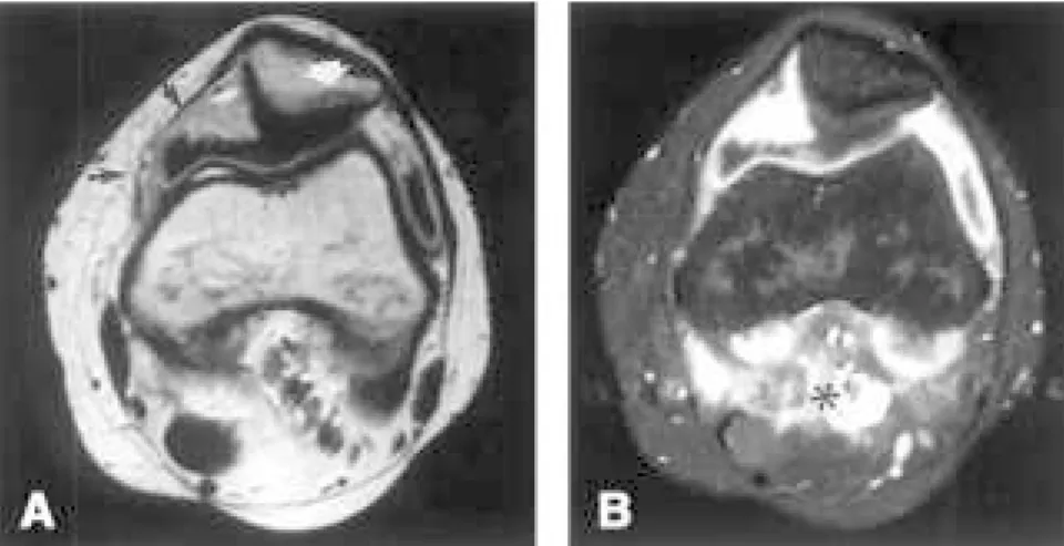
1950s, much concern has been ex-pressed about the effects of medication used in this disease which has led to large multicenter trials, such as those comparing the effects of oral steroids Figure 2 - Example of longstanding arthritis (12-year duration) in an 18-year-old girl with polyarticular
JRA with no previous use of systemic corticotherapy, represented by post-gadolinium MRI images of (A) an axial T1-weighted SE and (B) an axial fat-suppressed, T1-weighted SE sequence. Note the corresponding appearance of enhancing synovium (black arrows) and subchondral cyst on patella (white arrow) in panel A. In the axial fat-suppressed, T1-weighted SE image, the full extension of the hypertrophied synovium is masked by surrounding enhanced vessels (asterisk). This example shows that the natural progression of disease, independently of concomitant systemic therapy, may lead to formation of subchondral cysts. This evidence suggests that other factors than corticotherapy use – including natural progression of disease – may be responsible for osteochondral damage.
In this study, we found significant relationships between the MRI find-ings, including presence of osteochon-dral abnormalities (subchonosteochon-dral cysts and/or bone erosions), and disease du-ration in Group I (children with previ-ous history of corticotherapy); how-ever, no such relationship was found for Group II (children with no previ-ous history of corticotherapy). Pro-longed use of systemic corticosteroids (>1 year) also seemed to be related to a higher degree of deleterious effects on the cartilage, although no significant relationship to the total dose of corti-costeroids used during treatment was determined. Moreover, we found no evidence of a relationship between os-teochondral changes depicted on MRI and disease activity at the time of MRI performance either for Group I or for Group II. The use of systemic corticotherapy in the group of patients with more aggressive disease suggested the possibility of an interaction
be-Table 5 - Correlation between types of osteochondral changes and MRI abnormalities in the joints of Group I and II patients.
MR findings (Group I) SC Er + SC Er Absence Total
Osteochondral lesions 0 (0%) 1 (50%) 1 (50%) 0 (0%) 2
Synovial enhancement 0 (0%) 0 (0%) 0 (0%) 11 (100%) 11
Synovial enhancement and osteochondral lesions 2 (12.5%) 8 (50%) 5 (31.25%) 1 (6.25%) 16
No osteochondral/synovial pathologic findings 0 (0%) 0 (0%) 0 (0%) 7 (100%) 7
Total (number of joints) 2 9 6 19 36
Fisher's exact test (2-tail) p<0.0001
MR findings (Group II) SC Er + SC Er Absence Total
Osteochondral lesions 0 (0%) 0 (0%) 3 (100%) 0 (0%) 3
Synovial enhancement 0 (0%) 0 (0%) 0 (0%) 6 (100%) 6
Synovial enhancement and osteochondral lesions 0 (0%) 8 (40%) 12 (60%) 0 (0%) 20
No osteochondral/synovial pathologic findings 0 (0%) 0 (0%) 0 (0%) 7 (100%) 7
Total (number of joints) 0 8 15 13 36
Fisher's exact test (2-tail) p=0.526
SC= subchondral cysts; Er + SC = bone erosions associated to subchondral cysts; Er = bone erosions; Absence = no lesions Table 4 - Correlations between MRI findings and total dose of systemic corticotherapy used during
treatment of JRA in the joints of Group I patients.
Corticotherapy Group Cummulative dose of systemic corticosteroids (mg)
MR findings £ 5000 > 5000 Total
Osteochondral lesions 3 (100%) 0 (0%) 3
Synovial enhancement 6 (54.55%) 5 (45.45%) 11
Synovial enhancement and osteochondral lesions 4 (30.77%) 9 (69.23%) 13 No osteochondral/synovial pathologic findings 3 (50%) 3 (50%) 6
Total (number of joints) 16 17 33
Fisher's exact test (2-tail) p=0.205
Frequency missing = 3
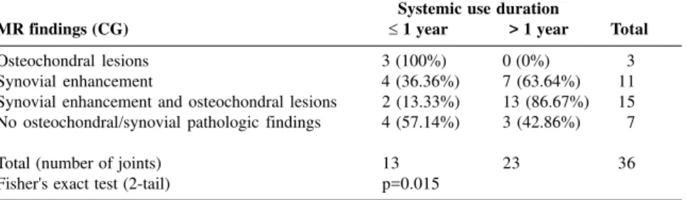
Table 3 - Correlations between MRI findings and duration of systemic corticotherapy in the joints of Group I patients.
Systemic use duration
MR findings (CG) £ 1 year > 1 year Total
Osteochondral lesions 3 (100%) 0 (0%) 3
Synovial enhancement 4 (36.36%) 7 (63.64%) 11
Synovial enhancement and osteochondral lesions 2 (13.33%) 13 (86.67%) 15 No osteochondral/synovial pathologic findings 4 (57.14%) 3 (42.86%) 7
Total (number of joints) 13 23 36
Fisher's exact test (2-tail) p=0.015
to effects of aspirin26-31. As an example
of such effects, adults treated with sys-temic or intra-articular corticosteroids have demonstrated disturbances of en-dogenous cortisol synthesis, including Cushing’s syndrome32.
There are recent studies about the local effects of intra-articular injection of corticosteroids14-17, 33, but few are
about the intra-articular results follow-ing prolonged systemic corticotherapy.
In 1990, Wilske and Healey19 proposed
an aggressive treatment plan for cases of destructive rheumatoid arthritis. Their study included the use of mul-tiple medications, including oral ste-roids, and challenged the conventional treatment that employed a slow, stepwise addition of medications34-37. A
few years earlier, Iannuzzi et al.38
cyclophospha-tween the severity of disease and osteo-chondral abnormalities rather than be-tween abnormalities and therapeutic medication. However, it was not pos-sible to establish statistically significant relationships between presence or ab-sence of lesions and previous use or lack of use of systemic corticotherapy, regardless of disease duration. Never-theless, our results corroborate previ-ous radiological studies39-41, providing
the possibility that over the course of the disease more severe deformities in articular joints could be produced fol-lowing treatment with corticosteroids. Because MRI allows identification of subtle cartilaginous abnormalities very early, we were able to show the abnor-malities in an earlier period of disease than previous radiological studies have shown. A limitation of this and previ-ous studies using radiographies is the difficulty to know whether patients re-ceiving steroids were already at higher risk of developing osteochondral ab-normalities, since they would belong to a more aggressive spectrum of JRA, which is bound to bias the results.
The goal of therapy for JRA is to retard clinical and radiographic joint degeneration, but the adverse systemic and intra-articular effects of long-term treatment should not outweigh the pur-pose of systemic therapy. Another point of concern is the disparity be-tween the clinical improvement in pa-tients following systemic medications and the worsening of radiographically
diagnosed damage. Our study evalu-ated children with JRA who received short- and long-term systemic corticos-teroids, comparing them with a noncorticotherapy group to determine the osteochondral abnormalities in the knees according to duration of immu-nosuppressive therapy. We did not con-sider other concurrent or confounding variables, such as combined therapy, that could influence the progression of articular damage in either Group I or Group II.
Numerous questions raised by this study reflect the problem of distin-guishing between effects of disease and effects of corticosteroids. Does sys-temic corticotherapy produce addi-tional and different changes than those caused by the disease? Were our results a consequence of the fact that the pa-tients given corticotherapy already had more destructive disease? Do these os-teochondral abnormalities also develop in non-rheumatoid experimental models given long-term systemic corticotherapy for other conditions?
It is possible that intra-articular cor-tisone injections that result in long-last-ing relief of symptoms with minor or few side effects17,42,43 could decrease the
need for systemic corticosteroids in the management of JRA.
Rheumatologists treating children with JRA must balance two opposing realities. First, prolonged cortico-therapy will invariably cause adverse systemic and articular effects in a
spec-trum of disease that does not carry the threat of mortality as, for example, ma-lignancies do. Second, a patient not given aggressive treatment after unag-gressive management is unsuccessful may suffer a lifetime of disability and functional impairment. Currently, labo-ratory data can identify those patients at greatest risk for destructive disease who should be treated more aggres-sively. However, this study suggests that after remission of the disease, noncorticotherapy would be preferred as a maintainance therapeutic strategy. Our statistical correlations raise the possibility that systemic corticotherapy may be a contributing factor in the in-tra-articular progression of the disease. However, differences in disease sever-ity between the corticosteroid-treated and not-treated groups should be con-sidered in the interpretation of our re-sults.
ACKNOWLEDGMENTS
We are grateful to Júlia Fukushima and Creusa dal Bo for statistical analy-sis and Angela C. Lorio, ELS for edi-torial assistance.
Study supported in part by FAPESP (Fundação de Amparo à Pesquisa do Estado de São Paulo) grant – 95 / 1890-0 and presented as a poster at the SPR Scientific Assembly on May 14, 1999.
RESUMO RHCFAP/3045
DORIA AS e col. - Correlação entre as alterações osteocondrais evidencia-das à ressonância magnética e a progressão da doença. Rev. Hosp. Clín. Fac. Med. S. Paulo 56(4): 107-114, 2001.
Objetivo: Determinar as conse-qüências do uso crônico de corticos-teróides sistêmicos em crianças com artrite reumatóide juvenil através da avaliação dos efeitos osteocondrais à ressonância magnética.
cortico-!
terapia sistêmica (22 pacientes do sexofeminino; 12 pacientes do sexo mascu-lino; idade média: 11.3 anos; duração média da doença: 5.9 anos; duração média da corticoterapia: 2.9 anos; dose média cumulativa de corticosteróides: 5000 mg); 35 (50.7%) pacientes não haviam feito uso prévio de cortico-terapia sistêmica (27 pacientes do sexo feminino; 8 pacientes do sexo masculi-no; idade média: 11.7 anos; duração média da doença: 5.3 anos).
Resultados: No grupo que recebeu corticoterapia sistêmica prévia (Grupo
I) a presença de alterações osteo-condrais à ressonância magnética rela-cionou-se de uma forma estatistica-mente significativa com longo tempo de duração da doença (>3.5 years; P<0.001). Tal relação não foi estabe-lecida no grupo de pacientes sem uso prévio de corticosteróides sistêmicos (Group II). Da mesma forma, não hou-ve relação entre a dose média de corticosteróides e os achados à resso-nância magnética.
Conclusão:É importante avaliar-se
os efeitos intra-articulares a longo pra-zo do uso crônico de corticoterapia sistêmica em artrite reumatóide juvenil à ressonância magnética, uma vez que os efeitos colaterais de uma terapia agressiva não devem ser potencialmen-te mais deletérios às articulações que os efeitos do curso natural da doença.
DESCRITORES: Corticoterapia sistêmica. Artrite reumatóide juvenil. Crianças. Joelho. Ressonância magnética.
REFERENCES
1. SCHALLER JG - Juvenile rheumatoid arthritis. Pediatr Rev 1980; 2:163-174.
2. JACOBS JC - Juvenile rheumatoid arthritis. Pediatric Rheumatology for the Practitioner. New York, Springer, 1993. p. 231-359.
3. YULISH BS, LIEBERMAN JM, NEWMAN AJ et al. - Juvenile rheumatoid arthritis: assessment with MR imaging. Radiology 1987; 165:149-152.
4. HERVÉ-SOMMA CMP, SEBAG GH, PRIEUR AM et al. - Juvenile rheumatoid arthritis of the knee: MR evaluation with Gd-DOTA. Radiology 1992; 182:93-98.
5. GRAHAM TB, BLEBEA JS, GYLYS-MORIN V et al. - Magnetic resonance imaging in juvenile rheumatoid arthritis. Semin Arthritis Rheum 1997; 27:161-168.
6. LAMER S & SEBAG GH - MRI and ultrasound in children with juvenile rheumatoid arthritis. Eur J Radiol 2000; 33:85-93. 7. UHL M, KRAUSS M, HERGET G et al. - The knee joint in early
juvenile idiopathic arthritis. Acta Radiol 2001; 42:6-9. 8. MELO-GOMES JA - Problems related to systemic glucocorticoid
therapy in children. J Rheumatol 1993; 20 (suppl 37):35-39. 9. BREWER EJ, BASS J, BAUM J et al. - Current proposed revision of
JRA criteria. Arthritis Rheum 1977; 20 (suppl):195-199. 10. SAS Institute Inc., SAS/STATâUser’s Guide, version 6, 4th edition,
Cary, NC: SAS Institute Inc.,1989. v.1.
11. REED MH & WILMOT DM - The radiology of juvenile rheumatoid arthritis. A review of the English language literature. J Rheumatol 1991; 18 (suppl 31):2-22.
12. GILKESON G, POLISSON R, SINCLAIR H et al. - Early detection of carpal erosions in patients with rheumatoid arthritis: a pilot study of magnetic resonance imaging. J Rheumatol 1988; 15 (9):1361-1366.
13. SENAC MO JR., DEUTSCH D, BERNSTEIN BH et al. - MR imaging in juvenile rheumatoid arthritis. Am J Rheumatol 1988; 150 :873-878.
14. OSTERGAARD M, STOLTENBERG M, HENRIKSEN O et al. -Quantitative assessment of synovial inflammation by dynamic gadolinium-enhanced magnetic resonance imaging. A study of the effect of intra-articular methylprednisolone on the rate of early synovial enhancement. Br J Rheumatol 1996; 35:50-59. 15. OSTERGAARD M, STOLTENBERG M, GIDEON P et al. - Changes
in synovial membrane and joint effusion volumes after intraarticular methylprednisolone. Quantitative assessment of inflammatory and destructive changes in arthritis by MRI. J Rheumatol 1996; 23:1151-1161.
16. EICH GF, HALLÉ F, HODLER J et al. - Juvenile chronic arthritis: imaging of the knees and hips before and after intraarticular steroid injection. Pediatr Radiol 1994; 24:558-563.
"
18. WALLACE CA & LEVONSON JE - Juvenile rheumatoid arthritis: outcome and treatment for the 1990s. Rheum Dis Clin North Am 1991; 17(4):891-905.
19. WILSKE KR & HEALEY LA - Challeging the therapeutic pyramid: a new look at treatment strategies for rheumatoid arthritis. J Rheumatol 1990; 17: 4-7.
20. JULKUNEN H, ROKKANEN P & LAINE H - Chloroquine treatment and bone changes in rheumatoid arthritis. Scand J Rheumatol 1976; 5:36-38.
21. WRIGHT V & AMOS R - Do drugs change the course of rheumatoid arthritis? Br Med J 1980; 280:964-966.
22. BROOK A, FLEMING A & CORBETT M - Relationship of radiological change to clinical outcome in rheumatoid arthritis. Ann Rheum Dis 1977; 36:274-275.
23. DE CARVALHO A & GRAUDAL H - Relationship between radiologic and clinical findings in rheumatoid arthritis. Acta Radiol [Diagn] (Stockholm) 1980; 21:797-802.
24. SHARP JT, LIDSKY MD, COLLINS LC et al. - Methods of scoring the progression of radiologic changes in rheumatoid arthritis: correlation of radiologic, clinical and laboratory abnormalities. Arthritis Rheum 1971; 14:706-720.
25. COHEN PA, JOB-DESLANDRE CH, LALANDE G et al. – Overview of the radiology of juvenile rheumatoid arthritis (JIA). Eur J Radiol 2000; 33:94-101
26. EMPIRE Rheumatism Council - Multi-centre controlled trial comparing cortisone acetate and acetyl salicylic acid in the long-term treatment of rheumatoid arthritis: results up to one year. Ann Rheum Dis 1955; 14:353-370.
27. EMPIRE Rheumatism Council - Multi-centre controlled trial comparing cortisone acetate and acetyl salicylic acid in the long-term treatment of rheumatoid arthritis: results of three years’ treatment. Ann Rheum Dis 1957; 16:277-289.
28. JOINT COMMITTEE of the Medical Research Council and Nuffield Foundation on Clinical Trials of Cortisone, A C.T.H., and other Therapeutic Measures in Chronic Rheumatic Diseases - A comparison of cortisone and aspirin in the treatment of early cases of rheumatoid arthritis. Br Med J 1954; 1:1223-1227.
29. EMPIRE Rheumatism Council - A comparison of cortisone and aspirin in the treatment of early cases of rheumatoid arthritis. Br Med J 1955; 2:695-700.
30. JOINT COMMITTEE of the Medical Research Council and Nuffield Foundation on Clinical Trials of Cortisone, A C.T.H., and other Therapeutic Measures in Chronic Rheumatic Diseases - Long-term results in early cases of rheumatoid arthritis treated with either cortisone or aspirin. Br Med J 1957; 1:847-850.
31. JOINT COMMITTEE of the Medical Research Council and Nuffield Foundation - A comparison of prednisolone with aspirin or other analgesics in the treatment of rheumatoid arthritis. Ann Rheum Dis 1959; 18:173-188.
32. O’SULLIVAN MM, RUMFELD WR, JONES MK et al. - Cushing’s syndrome with suppression of the hypothalamic-pituitary-adrenal axis after intra-articular steroid injections. Ann Rheum Dis 1985; 44:561-563.
33. SPARLING M, MALLESON P, WOOD B et al. - Radiographic follow-up of joints injected with triamcinolone hexacetonide for the management of childhood arthritis. Arthritis Rheum 1990; 33(6):821-826.
34. RUDDY S - The management of rheumatoid arthritis. In: KELLEY WN, HARRIS ED Jr., RUDDY S et al. eds. Textbook of Rheumatology. Philadelphia, Saunders, 1981. p. 1000-1014. 35. LIGHTFOOT RW Jr. - Treatment of rheumatoid arthritis. Arthritis
and allied conditions: A textbook of rheumatology. Philadelphia, Lea, 1979. p. 513-518.
36. BUNCH TW & O’DUFFY TD - Disease-modifying drugs for progressive rheumatoid arthritis. Mayo Clin Proc 1980; 55 :161-179.
37. ELLMAN P, LAWRENCE JS & THOROLD GP - Gold therapy in rheumatoid arthritis. Br Med J 1940; 2:314-316.
38. IANNUZZI L, DAWSON N, ZEIN N et al. - Does drug therapy slow radiographic deterioration in rheumatoid arthritis? N Engl J Med 1983; 309(17):1023-1028.
39. RASKER JJ & COSH JA - Radiological study of cervical spine and hand in patients with rheumatoid arthritis of 15 years’ duration: an assessment of the effects of corticosteroid treatment. Ann Rheum Dis 1978; 37:529-535.
40. MEIKLE JAK & WILKINSON M- Rheumatoid involvement of the cervical spine. Ann Rheum Dis 1971; 30:154-161.
41. SMITH P, BENN RT & SHARP J - Natural history of rheumatoid cervical luxations. Ann Rheum Dis 1972; 31:431-439.
42. ALLEN RC, GROSS KR, LAXER RM et al. - Intraarticular triamcinolone hexacetonide in the management of chronic arthritis in children. Arthritis Rheum 1986; 29:997-1001.
43. EARLEY A, CUTTICA RJ, MCCULLOUGH C et al. - Triamcinolone into the knee in juvenile chronic arthritis. Clin Exp Rheumatol 1988; 6:153-155.