REV. HOSP. CLÍN. FAC. MED. S. PAULO 56(4):115-118, 2001 JULY-AUGUST
From the Division of Gynecology, Hospital das Clínicas, Faculty of Medicine, University of São Paulo.
REVIEW
MICROLAPAROSCOPY IN GYNECOLOGY: ANALYSIS
OF 16 CASES AND REVIEW OF LITERATURE
Fabio Ikeda, Maurício Simões Abrão, Sérgio Podgaec, Alexandre Pupo Nogueira, Rosa Maria Neme and José Aristodemo Pinotti
RHCFAP/3046 IKEDA F et al. - Microlaparoscopy in gynecology: analysis of 16 cases and review of literature. Rev. Hosp. Clín. Fac. Med.
S. Paulo 56(4):115-118, 2001.
Microlaparoscopy represents the development of endoscopic surgery towards a minimally invasive surgical procedure. The advantages include fewer surgical complications, faster return to daily activities, more comfortable postoperative recovery, and satisfactory aesthetic results. The possibility of performing surgery under sedation may result in shorter hospitalization, lower hospital costs, and easier anesthetic procedures.
The authors report their preliminary experience with the use of microlaparoscopy, using optics and 2mm instruments, as well as a review of the literature since the introduction of this new technique. The report of these 16 cases demonstrates that microlaparoscopy is a feasible technique with satisfactory results. On the other hand, this new technique requires precise indications and a training period for the development of the skills necessary for performing these surgeries.
DESCRIPTORS: Laparoscopy. Gynecology. Microsurgery. Surgical Procedures. Laparoscopic instruments.
Laparoscopy has been used since the beginning of the 20th century and became known as a diagnostic and therapeutic procedure in gynecology in the 1970s. In the 1980s with the com-ing of video-laparoscopy, it was much more widespread, offering more com-fort during the surgery and more ad-vanced procedures17. In the beginning
of the 1990s, with the concept of mini-mally invasive surgery, a new tech-nique, microlaparoscopy, started being developed using optics and instruments smaller than 5mm in diameter18.
Dorsey & Tabb8 and Risquez et al.22
were the first to mention this endo-scopic method8,22. However, these cases
did not have the expected impact be-cause of the poor quality of the image. Risquez et al.21, performed 30
microlaparoscopies obtaining an im-provement of the image and presenting more indications for the use of this method21.
At that time, technology had greatly improved, and the development of endoscopes with better fiber optics permitted clearer images with a scope of a smaller diameter. Therefore, it be-came possible to perform diagnostic laparoscopy and small treatments, such as cauterization of endometriosis foci, adhesiolysis, tubal sterilization, appen-dectomy, and assisted reproduction procedures, such as ZIFT (zygote intrafallopian transfer) and GIFT (ga-mete intrafallopian transfer)4,7,9,11,23,24.
Later, a microlaparoscopic hysterec-tomy was performed, demonstrating that this technique is feasible even for more complex procedures25.
The main advantages obtained with the reduction in the caliber of the in-struments are decrease in the surgical trauma and the patients’ costs due to the shorter period of recovery1.
The aim of this study is to report 16 cases in which microlaparoscopy with diagnostic and therapeutic purpose was performed. These cases had satisfactory results and open promising perspectives for many different indications.
The first cases using sedation and local anesthesia were also analyzed, demonstrating benefits over general an-esthesia.
ANALYSIS OF 16 CASES
REV. HOSP. CLÍN. FAC. MED. S. PAULO 56(4):115-118, 2001 JULY-AUGUST
sociated intercurrent factor for any of the patients.
Twelve female patients underwent general anesthesia with fentanyl (5µg/kg), propofol (2mg/kg) and atracurium (0.4 to 0.5mg/kg) following inhalation of isoflurane and subsequent orotracheal intubation. Four female pa-tients underwent conscious sedation and local anesthesia with satisfactory results. These patients showed a nor-mal body mass index with no suspicion of large pelvic adhesions. The sedation was administered using midazolam (5 to 10 mg) and fentanyl chloridrate (2µg/kg), and using an additional 1µg/kg as necessary.
In all the cases, local anesthesia was administered with infiltration of 5mL 0.5% bupivacaine into the surgi-cal sites and instillation of 40mL 0.5% lidocaine over pelvic structures.
The surgery was performed with a 2-mm microfiberoptic microlapa-roscope (Auto-Suture, United States Surgical Corporation, USA), which was introduced into the abdominal cav-ity with a 2mm trocar, acting simulta-neously as a Veress needle. Thus, be-fore placing the pneumoperitoneum, the microlaparoscope may be intro-duced in the cavity to check correct
position of the cannula in order to avoid the injection of carbonic gas er-roneously into the pre-peritoneum.
The accessory punctures were per-formed with trocars similar to those de-scribed above, 2-mm or 5-mm in diam-eter when there was need to remove larger samples from the abdominal cav-ity. The 2-mm forceps used were made of titanium, offering adequate precision and satisfactory resistance. The pneu-moperitoneum was maintained with intraabdominal pressure of 9mm Hg.
A uterine manipulator was used in most of the cases, which was funda-mental to providing an adequate expo-sition of the pelvic organs.
In 14 surgeries, the postoperative di-agnostic was minimal or mild en-dometriosis, exposing a disease appropri-ately approached by microlaparoscopy. In 1 case, endometriosis was character-ized as moderate (according to standards from the American Society for Repro-ductive Medicine, 1996)5. In all the cases,
biopsy, resection, and cauterization of en-dometriosis foci were done. Salpingos-tomy for an ectopic pregnancy, myomec-tomy, and adhesiolysis were other per-formed surgeries.
Table 1 describes reported cases with more details.
DISCUSSION
Nowadays, after lengthy studies on microlaparoscopy, many authors have been using this new surgical approach, which has become a feasible new di-agnostic and therapeutic option in se-lected cases.
In several studies, the diagnostic accuracy between the 2-mm and the 10-mm telescope was the same10,12,16.
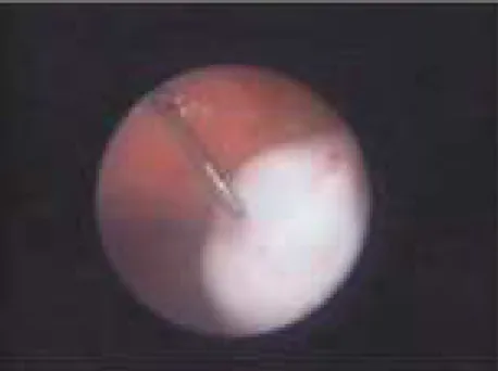
The patients described in this article were operated on with adequate im-ages, and the performance of micro-laparoscopy was a feasible technique (Fig.1). The endometriosis lesions that were the indications for most of the surgeries were easily identified, leav-ing no doubt about their presence.
The possibility of simplification of anesthesia in some cases of microlapa-roscopy may bring interesting benefits, such as reduction in the hospitalization and postoperative pain. Bauer et al.6
studied the administration of microlaparoscopy procedures with se-dation and local infiltration, listing the anesthetic methods.
The achieved results in our cases were satisfactory, emphasizing that the incidence of nausea was insignificant, and no orotracheal discomfort with
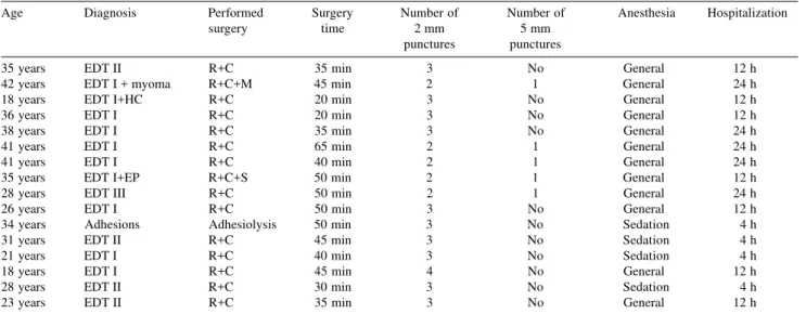
se-Table 1 – Analysis of 16 cases of microlaparoscopy.
Age Diagnosis Performed Surgery Number of Number of Anesthesia Hospitalization
surgery time 2 mm 5 mm
punctures punctures
35 years EDT II R+C 35 min 3 No General 12 h
42 years EDT I + myoma R+C+M 45 min 2 1 General 24 h
18 years EDT I+HC R+C 20 min 3 No General 12 h
36 years EDT I R+C 20 min 3 No General 12 h
38 years EDT I R+C 35 min 3 No General 24 h
41 years EDT I R+C 65 min 2 1 General 24 h
41 years EDT I R+C 40 min 2 1 General 24 h
35 years EDT I+EP R+C+S 50 min 2 1 General 12 h
28 years EDT III R+C 50 min 2 1 General 24 h
26 years EDT I R+C 50 min 3 No General 12 h
34 years Adhesions Adhesiolysis 50 min 3 No Sedation 4 h
31 years EDT II R+C 45 min 3 No Sedation 4 h
21 years EDT I R+C 40 min 3 No Sedation 4 h
18 years EDT I R+C 45 min 4 No General 12 h
28 years EDT II R+C 30 min 3 No Sedation 4 h
23 years EDT II R+C 35 min 3 No General 12 h
REV. HOSP. CLÍN. FAC. MED. S. PAULO 56(4):115-118, 2001 JULY-AUGUST
dation was observed. The adequate se-lection of the patients is fundamental. The surgery should be rapid and accu-rate because the patient is awake.
The reduction in the amount of car-bonic gas used for the pneumoperito-neum was essential, because when the abdominal pressure was higher than 15-mm Hg or the carbonic gas volume was higher than 2,5 liters, patients ex-perienced discomfort23.
The mean time of postoperative hospitalization was shorter for the pa-tients that had received sedation (mean time 4 hours) compared with those who had received general anesthesia (mean time 17 hours).
The utilization of the uterine ma-nipulator was essential to a satisfactory pelvic structure exposure. An auxiliary puncture was needed in a virgin pa-tient, since this use was not possible.
In some cases, a diode laser was used, which was portable, easily man-aged, and adaptable to the 2-mm tro-car. This innovation has brought more accuracy to the procedure, with desired benefits to these cases2.
The postoperative recovery was shorter with reduction of pain and ab-dominal discomfort than conventional laparoscopy.
The aesthetic results were adequate, with no need for skin suture. The incision was closed with adhesive tape. There was no report of postoperative infection.
The simplification of the anesthesia allowed the intervention to be done in an ambulatory surgery center. However, there are still doubts whether it can be
performed in a gynecologist office. Palter & Olive in 1996 and Almeida & Val-Gallas in 1998 per-formed office microlaparoscopies3,19.
These authors recommend this tech-nique to patients with chronic pelvic pain, and it may be performed with lo-cal anesthesia and light conscious se-dation. However, some authors do not agree with office microlaparoscopy and suggest an ambulatory surgical room for these procedures11.
Analyzing the postoperative pain, a minor incidence of abdominal and scapular pain has been reported, as well as the necessity for analgesics in microlaparoscopy14. Abdominal
instil-lation and local infiltration of anesthet-ics have benefited the patients13,20. This
procedure was performed in all the cases reported in this paper.
Risquez et al.23 published a
multicen-tric report of 408 cases, the biggest study of this technique. The patients were di-vided into 3 groups according to the type of anesthesia. The first group received only local anesthesia and oral diazepam; the second, sedation and local anesthe-sia; and the third, general anesthesia without orotracheal intubation. From this work, we conclude that microlapa-roscopy is effective as a diagnostic tech-nique and for some therapeutic proce-dures, such as adhesiolysis, endometrio-sis cauterization, embryo transfer, and salpingostomy. The most satisfactory an-esthetic technique depends on the char-acteristics of each patient.
Another possible innovation with this method is conscious pain mapping, where the patient helps the surgeon lo-calize the painful areas, making it pos-sible to perform appendectomy by mapping4. In patients without pelvic
diseases, some areas are painful when touched, extended, or compressed. Thus, the stretching of the fallopian tube or of the utero-ovarian ligament caused a lot of discomfort, while in the manipulation of omentum, bowels, and ovary, the pain was not notable26.
An additional use of the
micro-laparoscopy consists of the preliminary visualization of the umbilical region in patients with extensive previous abdomi-nal surgeries. In these cases, a microlaparoscope could be introduced in the left hypochondrium (Palmer point) to verify the presence of adhesions in the umbilical area. If possible, the lysis could be accomplished by introducing the tra-ditional forceps into the iliac fossas15.
With microlaparoscopy, there has been a reduction in surgical complica-tions due to the possibility of visualiz-ing the abdominal cavity before the pneumoperitoneum is installed, avoid-ing the insufflation of erroneous places such as the subcutaneous layer or retroperitoneum9. Incisional
her-niations are rare since the cannula is smaller11. With the reduction of the
pneumoperitoneum pressure, a smaller amount of carbonic gas is utilized, which prevents the pain in the shoul-der that is usually experienced in con-ventional laparoscopies. In the patients included in this study, there were no surgical complications, and none of the patients complained about pain in the scapula in the postoperative period.
As a disadvantage of this method, microlaparoscopy requires new train-ing even for the experienced lapa-roscopic surgeons because there is a 30 to 40% reduction of the skill using the 2-mm instruments18. When compared
with traditional laparoscopy using a 10-mm telescope, microlaparoscopy offers a 40% smaller visual field, mak-ing it more difficult to centralize and focalize the image12,23. Additionally, the
instruments are more delicate and re-quire adequate skill to operate.
Probably in the near future, micro-laparoscopy will be a routinely used technique for endoscopic procedures. Nowadays, this method represents an interesting alternative to some easier interventions, and the simplification of anesthesia may still bring more ben-efits, drastically reducing the socioeco-nomic and psychologic costs of surgi-cal procedures.
REV. HOSP. CLÍN. FAC. MED. S. PAULO 56(4):115-118, 2001 JULY-AUGUST
RESUMO RHCFAP/3046
IKEDA F e col. – Microlaparoscopia em ginecologia: análise de 16 casos e revisão da literatura. Rev. Hosp. Clín. Fac. Med. S. Paulo 56(4):
115-118, 2001.
A microlaparoscopia representa a evolução da cirurgia endoscópica, vi-sando um procedimento cirúrgico mi-nimamente invasivo. As suas vantagens incluem a redução das complicações cirúrgicas, retorno às atividades mais
rápido, período pós-operatório mais confortável e resultado estético satis-fatório. A possibilidade da realização sob sedação pode resultar em inter-nação hospitalar menor, diminuição dos custos hospitalares, além da sim-plificação do procedimento anestésico. Os autores relatam sua experiência inicial com a microlaparoscopia, utili-zando óptica e instrumentais de 2mm, assim como uma revisão dos trabalhos
publicados desde a introdução dessa nova técnica. O relato destes 16 casos mostrou ser a microlaparoscopia exe-qüível, apresentando resultados satisfa-tórios. Por outro lado, o seu uso requer indicações precisas e treinamento para manipular o instrumental.
DESCRITORES: Laparoscopia. Ginecologia. Microcirurgia. Procedimentos Cirúrgicos.
REFERENCES
1. ABRÃO MS & IKEDA F - O impacto da laparoscopia no diagnóstico da endometriose. In: ABRÃO MS, editor. Endometriose: uma visão contemporânea. São Paulo, Revinter, 2000. p.111-125. 2. ABRÃO MS, IKEDA F, PODGAEC S et al. - Microlaparoscopy for
an intact ectopic pregnancy and endometriosis with the use of a diode laser. Hum Reprod 2000; 15(6):1369-1371.
3. ALMEIDA OD Jr & VAL-GALLAS JM - Office microlaparoscopy under local anaesthesia in the diagnosis and treatment of chronic pelvic pain. J Am Assoc Gynecol Laparosc 1998; 5 (4): 407-410. 4. ALMEIDA OD Jr, VAL-GALLAS JM & RISK B - Appendectomy under local anaesthesia following conscious pain mapping with microlaparoscopy. Hum Reprod 1998; 13(3):588-590.
5. AMERICAN society for reproductive medicine - Revised American Society for Reproductive Medicine classification of endometriosis: 1996. Fertil Steril 1997; 67(5):817-821.
6. BAUER O, DEVROEY P, WISANTO A et al. - Small diameter laparoscopy using a microlaparoscope. Hum Reprod 1995; 10(6):1461-1464.
7. CASTELLOTTI DS, MOTTA ELA, ALEGRETTI JR et al. - Successful birth after intrafallopian transfer of microhatched embryos. Fertil Steril 1997; 68(2):367-369.
8. DORSEY JM & TABB CR - Mini-laparoscopy and fiber optic lasers. Obstet Gynecol Clin North Am 1991; 18:613-617.
9. DOWNING BG & WOOD C - Initial experience with a new microlaparoscope 2 mm in external diameter. Aust N Z Obstet Gynaecol 1995; 35(2):202-204.
10. FABER BM & CODDINGTON CC - Microlaparoscopy: a comparative study of diagnostic accuracy. Fertil Steril 1997; 67(5):952-954. 11. FULLER PN - Microendoscopic surgery: A comparison of four
microendoscopes and a review of the literature. Am J Obstet Gynecol 1996; 174:1757-1762.
12. HAEUSLER G, LEHNER R, HANZAL E et al. - Diagnostic accuracy of 2 mm microlaparoscopy. Acta Obstet Gynecol Scand 1996; 75:672-675.
13. HELVACIOGLU A & WEIS R - Operative laparoscopy and postoperative pain relief. Fertil Steril 1992; 57(3):548-552. 14. KOVACS GT, BAKER G, DILON M et al. - The microlaparoscope
should be used routinely for diagnostic laparoscopy. Fertil Steril 1998; 70(4):698-701.
15. LEE PI, CHI YS, CHANG YK et al. - Minilaparoscopy to reduce complications from cannula insertion in patients with previous pelvic or abdominal surgery. J Am Assoc Gynecol Laparosc 1999; 6(1):91-95.
16. MOLLOY D - The diagnostic accuracy of a microlaparoscope. J Am Assoc Gynecol Laparosc 1995; 2(2):203-206.
17. NEZHAT C, NEZHAT F & NEZHAT C - Laparoscopia Operatória (Cirurgia Minimamente Invasiva): Estado de Conhecimento. In: UENO J, editor. Cirurgia Video-endoscópica em Ginecologia. São Paulo, Roca, 1996. p. 247-70.
18. PALTER SF - Office microlaparoscopy under local anesthesia. Obstet Gynecol Clin North Am 1999; 26(1):109-120.
19. PALTER SF & OLIVE DL - Office microlaparoscopy under local anesthesia for chronic pelvic pain. J Am Assoc Gynecol Laparosc 1996; 3(3):359-364.
20. PELLICANO M, ZULLO F, DI CARLO C et al. - Postoperative pain control after microlaparoscopy in patients with infertility: a prospective randomized study. Fertil Steril 1998; 70(2):289-292. 21. RISQUEZ F, PENNEHOUAT G, FERNANDEZ R et al. - Microlapa-roscopy: a preliminary report. Hum Reprod 1993; 8(10):1701-1702. 22. RISQUEZ F, PENNEHOUAT G, FOULOT H et al. - Transcervical tubal cannulation and falloposcopy for the management of tubal pregnancy. Hum Reprod 1992; 7:375-376.
23. RISQUEZ F, PENNEHOUAT G, MC CORVEY R et al. - Diagnostic and operative microlaparoscopy: a preliminary multicentre report. Hum Reprod 1997; 12(8):1645-1648.
24. STEELE SJ - The potential for improved abdominal procedures and approaches for tubal occlusion. Int J Gynaecol Obstet 1995; 51(Suppl 1):S17-S22.
25. WATTIEZ A, GOLDCHMIT R, DURRUTY G et al. - Minilaparoscopy hysterectomy. J Am Assoc Gynecol Laparosc 1999; 5(4):97-100. 26. ZUPI E, SBRACIA M, MARCONI D et al. - Pain mapping during minilaparoscopy in infertile patients without pathology. J Am Assoc Gynecol Laparosc 1999; 6(1):51-4.