Cop
yright
© ABE&M t
odos os dir
eit
os r
eser
vados
.
1 Núcleo de Pesquisa e
Pós-Graduação, Hospital Santa Casa de Belo Horizonte, Belo Horizonte, MG, Brazil
Correspondence to:
José Augusto Nogueira Machado Rua Domingos Vieira, 590 30150-240 – Belo Horizonte, MG, Brazil
[email protected] Received on Dec/22/2011 Accepted on Aug/23/2012
Oxidative stress and interleukin-6
secretion during the progression
of type 1 diabetes
Estresse oxidativo e secreção de interleucina-6 durante a progressão do diabetes tipo 1
Janice Sepúlveda Reis1, Clara Araújo Veloso Amaral1, Caroline Maria Oliveira
Volpe1, Jamille Silveira Fernandes1, Erica Abreu Borges1, Camila Armond
Isoni1, Paula Martins Ferreira dos Anjos1, José Augusto Nogueira Machado1
ABSTRACT
Objective: To evaluate inlammatory, oxidizing, and reducing responses during the progression of type 1 diabetes mellitus (T1DM) in patients without chronic complications. Subjects and methods: Plasma antioxidant status, reactive oxygen species (ROS), and interleukin-6 (IL-6) were measured in 42 patients with T1DM and in 24 healthy subjects. Results: Signiicant incre-ases were detected in the median values of ROS and IL-6 in patients with T1DM compared with healthy subjects (ROS ~ 4,836 vs. 2,036 RLU/min, respectively; P < .05: IL-6 ~ 14.2 vs. 9.7 pg/mL, respectively; P = .002). No signiicant between-group differences (P > 0.05) were observed in oxidizing responses or in IL-6 concentrations when diabetic patients were grouped according to time after diagnosis (0 - 10, 10 - 20 and > 20 years). Plasma antioxidant responses were similar in patients with T1DM and in healthy subjects.Conclusions: Our results demonstrate that oxidizing and inlammatory responses are increased at the onset of T1DM, but remain un-changed during disease progression. These indings suggest that functional changes involved in diabetic complications may commence in the irst years after diagnosis. Arq Bras Endocrinol Metab. 2012;56(7):441-8
Keywords
Type 1 diabetes; disease progression; oxidative stress; plasma antioxidant status
RESUMO
Objetivo: Avaliar as respostas inlamatória, oxidativa e redutora na progressão do diabetes melito tipo 1 (DM1) em pacientes sem complicações crônicas. Sujeitos e métodos: Capacidade antioxidante do plasma, espécies reativas de oxigênio (ROS) e interleucina-6 (IL-6) foram avalia-das em 42 pacientes com DM1 e 24 indivíduos saudáveis. Resultados: Aumentos signiicativos foram detectados nas medianas de ROS e IL-6 em pacientes com DM1 comparados com indiví-duos saudáveis (ROS ~ 4.836 vs. 2.036 RLU/min, respectivamente, P < 0,05: IL-6 ~ 14,2 vs. 9,7 pg/ mL, respectivamente, P = 0,002). Diferenças não signiicativas (P > 0,05) foram observadas na resposta oxidante e IL-6 quando os diabéticos foram agrupados de acordo com o tempo após o diagnóstico (0-10, 10-20 e > 20 anos). A resposta antioxidante do plasma foi semelhante em pa-cientes com DM1 e em indivíduos saudáveis. Conclusões: Nossos resultados demonstram que as respostas oxidante e inlamatória estão aumentadas desde o início do DM1, mas mantêm-se inalteradas durante a progressão da doença, sugerindo que as mudanças funcionais envolvidas nas complicações diabéticas podem começar nos primeiros anos após o diagnóstico. Arq Bras Endocrinol Metab. 2012;56(7):441-8
Descritores
Cop
yright
© ABE&M t
odos os dir
eit
os r
eser
vados
.
INTRODUCTION
T
ype 1 diabetes mellitus (T1DM) is an inlammatory disease of the pancreatic islets in which the destruc tion of beta cells is mediated by cytotoxic T cells, self antibodies, and inlammatory mediators (1). High le vels of inlammatory biomarkers can be detected in pa tients that have been recently diagnosed with T1DM, and this indicates that the inlammatory response is ac tivated during very early stages of the disease (2,3).It has been suggested that a number of different hyperglycemiainduced biochemical mechanisms may be involved in the pathogenesis of complications that are characteristic of diabetes. In this context, it is known that hyperglycemia is linked with the activation of protein kinase C (PKC) and nuclear factor kappa B, enhanced polyol activity, increased formation of advanced glyca tion endproducts (AGEs), and elevated lux through the hexosamine pathway (4,5). A mechanism that is common to all the pathways implicated in these oxidati ve processes involves the generation of reactive oxygen species (ROS) (6), which lead to changes in blood low, vascular permeability, and angiogenesis. Increased levels of ROS stimulate PKC via diacylglycerol (DAG), and give rise to greater production of inlammatory cytoki nes, mainly interleukin6 (IL6), which is considered the primary mediator of acute inlammatory response. Ho wever, all inlammatory cytokines exhibit cytotoxic and cytostatic activities and have the ability to induce apop tosis in pancreatic islet cells (7). It has been hypothesized that alterations in oxidative metabolism without simulta neous increases in antioxidant response are characteristic of oxidative stress which, in endothelial cells, results in endothelial dysfunction and vascular damage (4,6).
Over the last few decades, studies on atherosclerosis and other inlammatory disorders have shown that le vels of inlammatory markers can be used to predict car diovascular risk, thus reinforcing the need to deine the impact of inlammatory activity in diabetes (8). While the main determinants of tissue injury in diabetes are believed to be the duration and severity of hyperglyce mia (9), there are very few reports on the changes that occur in inlammatory, oxidizing, and reducing respon ses during the progression of T1DM. Several authors have stated that, in patients with T1DM, no signiicant pathological consequences of diabetes can be detected earlier than 5 years from onset (10), even though sig niicant increases in ROS generation can be detected in patients at the onset of the disease (11). These indings suggest that the structural and functional changes in
volved in diabetic complications may commence at the onset of the hyperglycemic trigger, and it is possible that, in the presence of persistent acute peaks or chro nic hyperglycemia, inlammatory metabolic and immu nological pathways can become activated at all times after diagnosis (12).
In the light of the information presented above, the objective of the present study was to investigate inlam matory and antioxidant status in patients with T1DM but without chronic complications, during the irst years after diagnosis (010 years) and during further progres sion of the disease (1020 and > 20 years after diagnosis).
METHODS
Details of the study were submitted to, and approved by the Ethical Committee of the Santa Casa Hospital in Belo Horizonte, Minas Gerais, Brazil. The investiga tion was conducted according to the principles of the Declaration of Helsinki, and written informed consent was obtained from each participant before the study.
Subjects
The study population was made of 42 patients with T1DM and 24 healthy subjects. Diabetic patients exhi biting micro or macrovascular complications or ketoa cidosis in the previous year, and those who were taking statin and/or metformin and/or vitamins, or suffering from infection, dementia, inlammation, or malignant disease, or addicted to smoking or alcohol, or were pregnant, were excluded from the study. Patients with T1DM who were selected to be included in the study showed clinical onset of the disease 13.03 ± 9.42 years before, and presented history of diabetic ketoacidosis and/or were positive for glutamic acid dehydrogenase (GAD) antibodies and C peptide (< 0.5 ng/mL). The patients selected for the study, all of whom were recei ving intensive insulin therapy by means of multiple dai ly injections, were divided into three groups according to the time elapsed since diagnosis, namely, 010 years, 1020 years, and > 20 years. Clinical and biochemical evaluations were performed in all participants accor ding to standard procedures.
Preparation of plasma and granulocyte samples
Cop
yright
© ABE&M t
odos os dir
eit
os r
eser
vados
.
heparinized venous blood (10.0 mL), and was stored at −20°C until the moment of analysis. Granulocytes were puriied using a FicollHypaque gradient according to the method described by Bicalho and cols. (13).
Estimation of ROS production in granulocytes
Luminometric determination of the modulation of gra nulocyte ROS generation was carried out using a quan titative chemiluminescence assay. Granulocytes (1 x 10⁶
cells/mL; 200 μL) suspended in phosphate buffered saline (PBS; 300 μL) were transferred to an unsealed luminescence tube together with luminol (200 μL) dis solved in 0.4 M dimethyl sulfoxide. Chemiluminescen ce of the assay mixture was monitored for 30 min. After that, ROS production was stimulated by the addition of a 10μL aliquot of 104 M phorbol 12,13dibutyrate
(PDB), and chemiluminescence monitored for other 30 min (13). Results of the assay were expressed in re lative light units (RLU) per minute.
Evaluation of plasma antioxidant status
Plasma antioxidant status was determined from the direct reduction of 3(4,5dimethylthiazol2yl)2,5 diphenyltetrazolium bromide (MTT; Sigma Chemi cal Co.) according to the method of Medina and cols. (14). Briely, a 200μL aliquot of plasma was mixed with 25 μL of MTT solution (5.0 mg/mL in PBS), the inal volume was adjusted to 500 μL with PBS, and the whole mixture was incubated for 120 min at 37°C. The reaction was subsequently terminated by the addition of 1.0 mL of 0.04 M hydrochloric acid in isopropanol. The mixture was centrifuged (200 g, 10 min) and ab sorbance of the supernatant measured at 570 nm.
Determination of IL-6
Plasma concentrations of IL6 were determined by en zymelinked immunosorbent assay (ELISA) using As say Designs (Enzo Life Sciences) Human IL6 Enzyme Immunometric kits.
Statistical analyses
The ShapiroWilk test was applied in order to verify that data were normally distributed and, where applica ble, mean values were compared using Student’s ttests for paired samples. Betweengroup comparisons of me dian values of inlammatory biomarkers and plasma re ducing responses of diabetic patients were performed
using the nonparametric MannWhitney test. Within group correlations were determined using Spearman rank correlation tests. In all cases, statistical signiicance was accepted at P values < 0.05.
RESULTS
Baseline characteristics of the study populations
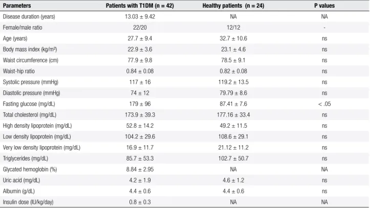
Clinical characteristics of patients with T1DM and heal thy subjects were very similar, as shown in Table 1. In relation to biochemical characteristics, fasting plasma glucose values were signiicantly increased in diabetic patients in comparison with healthy subjects (P < .05), while levels of glycated hemoglobin (HbA1c) in patients with T1DM (8.84%) were somewhat higher than the accepted range for healthy subjects (< 7%). When clini cal and biochemical characteristics of the population of diabetic patients were distributed according to the time elapsed since diagnosis (Table 2), signiicant between group differences were observed in some parameters, although these were not signiicant in clinical practice.
Inlammatory biomarkers and oxidative stress
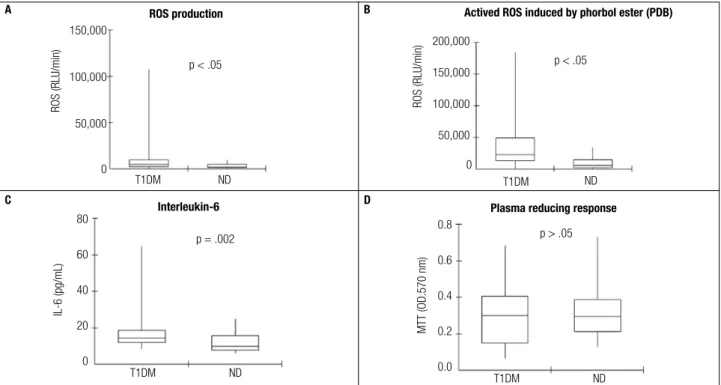
A signiicant difference was detected between patients with T1MD and healthy subjects in relation to the me dian values of ROS production in unstimulated granu locytes (4,836 versus 2,036 RLU/min, respectively; P < 0.05) (Figure 1A). The presence of phorbol ester stimulated granulocyte ROS production differently in diabetic patients and healthy subjects (22369 and 5780 RLU/min, respectively; P < 0.05) (Figure 1B). Addi tionally, IL6 plasma levels of patients with T1MD were signiicantly higher than those of healthy subjects (14.2 versus 9.7 pg/mL, respectively; P = 0.002) (Figure 1C). In contrast, no signiicant difference (P > 0.05) was ob served between diabetic patients and healthy subjects in relation to plasma reducing responses (as determined by direct reduction of MTT), although patients with T1DM showed a slightly lower response (median va lues 0.3 versus 0.29 OD
570) (Figure 1D).
Cop
yright
© ABE&M t
odos os dir
eit
os r
eser
vados
.
Table 2. Clinical and biochemical characteristics of patients with T1DM stratiied according to time elapsed after diagnosis
Parameters < 10 years (n = 18) 10 - 20 years (n = 13) > 20 years (n = 11)
Disease duration (years) 4.3 ± 2.2* 15.2 ± 2.8** 24.7 ± 7.2‡
Female/male ratio 9/9 8/5 5/6
Age (years) 21.4 ± 6.7* 26.5 ± 4.7† 39.6 ± 5.7‡
Body mass index (kg/m²) 21.8 ± 3.5 22.5 ± 2.5 25.1 ± 4.4 Waist circumference (cm) 74.9 ± 7.3 76.8 ± 5.4 84.5 ± 14.3 Waist-hip ratio 0.8 ± 0.1 0.8 ± 0.1† 0.9 ± 0.1‡
Systolic pressure (mmHg) 108.8 ± 9.6* 119.8 ± 15.5 128.2 ± 18.3‡
Diastolic pressure (mmHg) 69.8 ± 8.5* 79.5 ± 13.4 77.7 ± 13.7‡
Fasting glucose (mg/dL) 174.9 ± 101.5 182.7 ± 89.5 181.8 ± 104.9 Total cholesterol (mg/dL) 168.5 ± 37.2 186 ± 49.1 168.5 ± 28.8 High density lipoprotein (mg/dL) 53.4 ± 12.5 55.7 ± 15.5 48.5 ± 27.3 Low density lipoprotein (mg/dL) 101.6 ± 30 113.8 ± 30.7 97.1 ± 27.3 Very low density lipoprotein (mg/dL) 13.5 ± 6.5 16.5 ± 14.8 22.9 ± 12.8 Triglycerides (mg/dL) 67.3 ± 32.3 70.5 ± 38.1† 133.9 ± 68.2‡
Glycated hemoglobin (%) 9.7 ± 3.5 7.9 ± 2.7 8.31 ± 1.7 Uric acid (mg/dL) 3.3 ± 1.1 3.7 ± 1.4† 6.1 ± 2.2‡
Albumin (g/dL) 4.6 ± 0.4 4.2 ± 0.8 4.3 ± 0.6 Insulin dose (IU/kg/day) 1.0 ± 0.3* 0.7 ± 0.2 0.6 ± 0.2‡
Data are expressed as means ± standard deviation. Signiicant differences between the groups (* < 10 and 10 – 20 years; † 10 – 20 and > 20 years; ‡ < 10 and > 20 years) were determined
using Student’s t test (P < 0.05).
Table 1. Clinical and biochemical characteristics of the studied population
Parameters Patients with T1DM (n = 42) Healthy patients (n = 24) P values
Disease duration (years) 13.03 ± 9.42 NA NA Female/male ratio 22/20 12/12 -Age (years) 27.7 ± 9.4 32.7 ± 10.6 ns Body mass index (kg/m²) 22.9 ± 3.6 23.1 ± 4.6 ns Waist circumference (cm) 77.9 ± 9.8 78.5 ± 9.1 ns Waist-hip ratio 0.84 ± 0.08 0.82 ± 0.08 ns Systolic pressure (mmHg) 117 ± 16 119.2 ± 13.5 ns Diastolic pressure (mmHg) 74 ± 12 79.79 ± 8.6 ns Fasting glucose (mg/dL) 179 ± 96 87.41 ± 7.6 < .05 Total cholesterol (mg/dL) 173.9 ± 39.3 177.16 ± 33.4 ns High density lipoprotein (mg/dL) 52.8 ± 14.2 49.2 ± 11.5 ns Low density lipoprotein (mg/dL) 104.2 ± 29.6 108.6 ± 29.1 ns Very low density lipoprotein (mg/dL) 16.9 ± 11.7 21.12 ± 11.2 ns Triglycerides (mg/dL) 85.7 ± 53.3 102.7 ± 50.7 ns Glycated hemoglobin (%) 8.84 ± 2.95 NA NA Uric acid (mg/dL) 4.2 ± 1.9 4.6 ± 1.2 ns Albumin (g/dL) 4.4 ± 0.6 4.4 ± 0.6 ns Insulin dose (IU/kg/day) 0.8 ± 0.3 NA NA
NA: not applicable; ns: not signiicant.
Data are expressed as means ± standard deviation. Signiicant differences between the groups were determined using Student’s t test (P < 0.05).
ding levels measured in healthy subjects. In contrast, plasma antioxidant status did not differ signiicantly
Cop
yright
© ABE&M t
odos os dir
eit
os r
eser
vados
.
Figure 1. Inlammatory biomarkers and plasma reducing responses in patients with T1DM and in healthy subjects, A: production of reactive oxygen species (ROS) in the absence of phorbol 12,13-dibutyrate (PDB); B: production of ROS in the presence of 10-4 M PDB; C: plasma concentration of interleukin-6 (IL-6); D: reducing response evaluated by direct reduction of 3-(4,5-dimethylthiazol-2-yl)-2,5-diphenyltetrazolium bromide (MTT) dye, ND: Non diabetic control;
T1DM: Patients with type 1 diabetes mellitus; OD: optical density, Statistical analyses were performed using Mann-Whitney test at 5% signiicance level,
Figure 2. Inlammatory biomarkers and plasma reducing responses in patients with T1DM stratiied according to time elapsed after diagnosis and in healthy
subjects. A: production of reactive oxygen species (ROS) in the absence of phorbol 12,13-dibutyrate (PDB); B: production of ROS in the presence of 10-4 M
PDB; C: plasma concentration of interleukin-6 (IL-6); D: reducing response evaluated by direct reduction of 3-(4,5-dimethylthiazol-2-yl)-2,5-diphenyltetrazolium
bromide (MTT) dye. ND: non diabetic control; T1DM: patients with type 1 diabetes mellitus; OD: optical density. Statistical analyses were performed using
Mann-Whitney test at 5% signiicance level; * P < .05 compared with healthy individuals. # P > .05 compared with healthy individuals.
ROS production
150,000
100,000
50,000
0
ROS (RLU/min)
ND
Time after diagnosis (years) 0-10 10-20
* * *
> 20
Interleukin-6
80
60
40
20
0
IL-6 (pg/mL)
ND
Time after diagnosis (years) 0-10 10-20
* * *
> 20
Plasma reducing response
0,8
0,6
0,4
0,2
0,0
MTT (OD,570 nm)
ND
Time after diagnosis (years) 0-10 10-20
# # #
> 20
Actived ROS induced by phorbol ester (PDB)
150,000 200,000
100,000 50,000 0
ROS (RLU/min)
ND
Time after diagnosis (years) 0-10 10-20
* * *
> 20
ROS production
150,000
100,000
50,000
0
ROS (RLU/min)
T1DM ND p < .05
Actived ROS induced by phorbol ester (PDB)
150,000 200,000
100,000 50,000 0
ROS (RLU/min)
T1DM ND p < .05
Interleukin-6
80
60
40
20
0
IL-6 (pg/mL)
T1DM ND p = .002
B
Plasma reducing response
0.8
0.6
0.4
0.2
0.0
MTT (OD.570 nm)
T1DM ND p > .05
D A
C
D
A B
Cop
yright
© ABE&M t
odos os dir
eit
os r
eser
vados
.
Correlations of parameters within the groups of diabetic patients
There were no signiicant correlations between the levels of inlammatory biomarkers in the diabetic pa tients, either considered as a single group or when divi ded according to time since diagnosis, or between the se markers and the clinical and biochemical parameters evaluated. Similar results were observed in relation to plasma antioxidant status. Additionally, there were no correlations between fasting glucose, HbA1c and in lammatory biomarkers or plasma antioxidant status.
DISCUSSION
Results obtained in the present study suggest the pre sence of systemic inlammation in diabetic patients at all moments after T1DM diagnosis. Such condition results from an increased oxidizing metabolic response in the absence of compensating increase in plasma antioxidant status, and is indicative of oxidative stress at disease on set and in longestablished T1DM.
A considerable body of evidence has been accumula ted over the last few decades supporting the hypothesis that ROS generation, and the resultant oxidative stress, plays a central role in the pathogenesis of diabetic com plications. In this context, it is welldocumented that pathways stimulated by hyperglycemia (i.e., glucose oxidation, enhanced polyol pathway activity, and for mation of complexes between AGEs and their recep tors) bring about cell injury in response to oxidative stress (15). In addition, ROS triggers an inlammatory cascade by means of the production of proinlamma tory cytokines, such as IL6, which are involved in the pathogenesis of micro and macrovascular complica tions. Indeed, these signaling cytokines are often em ployed as markers of the inlammatory response (16).
Studies focusing on the extent of free radical forma tion in diabetic patients are scarce because of the inhe rent dificulties involved in measuring low concentra tions of such highly reactivity and shortlived species in body luids. Instead, surrogate markers are commonly employed in order to evaluate oxidative stress. In the present study, ROS production by granulocytes from patients with T1DM was signiicantly higher than in healthy subjects. This result is in accordance with that of Reis and cols. (11) who reported levels of ROS that were signiicantly elevated in 16 young patients with T1DM (age, 15.25 ± 3.83 years; duration of disease, 2.62 ± 2.24 years; HbA1c, 10.18 ± 2.5%) in compari
son with their healthy counterparts. Ceriello and cols. (17) were able to demonstrate a signiicant increase in the production of superoxide anions (SOA) in 10 patients with T1DM, although the study was limited to adult subjects. On the other hand, Hsu and cols. (18) analyzed a group of 47 diabetic children (age, 12.7 ± 4.8 years; duration of disease, 4.94 ± 3.28 years; HbA1c, 9.22 ± 2.63%), and reported signiicantly hi gher levels of SOA compared with healthy individuals.
Increased levels of diacylglycerol (DAG) and the activation of PKC induced by hyperglycemia have also been associated with the initiation of diabetic complica tions. Phorbol esters are analogues of DAG and mimic the in vitro hyperglycemia effect of the glyceride by me ans of direct stimulation of PKC. In the present study, ROS generation in the presence of phorbol ester was increased both in patients with T1DM and in healthy subjects, but the enhancement was much more intense in diabetic patients (Figure 1). This suggests that hyper glycemia activates the expression of membrane PKC.
A signiicant increase in IL6 levels between diabetic patients and healthy subjects was detected in the pre sent study. This inding is consistent with other reports of signiicant increases in the concentration of IL6 in patients with T1DM, but without microor macrovas cular complications, in comparison with healthy indivi duals (19,20). Similarly, a recent SEARCH study (21) focusing on the association of between inlammation and obesity, dyslipidemia, and hyperglycemia in 553 young patients with T1DM, and 215 healthy indivi duals, found signiicantly higher levels of IL6 in the diabetic group irrespective of weight or HbA1c con centration. In contrast, Erbagci and cols. (22) and Alexandraki and cols. (3) reported that there were no differences in IL6 levels between children and adults with T1DM and their healthy counterparts.
Cop
yright
© ABE&M t
odos os dir
eit
os r
eser
vados
.
previously reported ones. However, Hsu and cols. (18) detected no signiicant differences between groups of children with T1DM when concentrations of SAO were compared in relation to disease duration (< 5 years versus > 5 years), even though levels of the biomarker were signiicantly higher in the diabetic children (grouped together) compared with healthy children. Erbagci and cols. (22) observed no differences in the levels of IL6 between healthy children and those with T1DM, but when the diabetic children were divided according to time after diagnosis, IL6 levels were higher in recen tly diagnosed (< 1 year) patients compared with long standing (> 1 year) patients. In contrast, the SEARCH study found no differences in IL6 concentrations be tween young patients with T1DM when grouped ac cording to disease duration (< 1 year versus > 1 year after diagnosis) (21).
In the present study, no signiicant differences in plasma antioxidant status were observed between he althy subjects and patients with T1DM (considered together or grouped according to time elapsed since diagnosis). However, results revealed a slight, but not signiicant, increase in antioxidant status in diabetic patients who had been diagnosed for > 20 years. This increase was not attributable to the use of drugs, since none of the patients studied was receiving any medica tion other than insulin. To the best of our knowledge, only one published study is available reporting antioxi dant concentrations (superoxide dismutase, glutathio ne peroxidase, glutathione reductase, and vitamins A, C, and E) in patients with T1DM grouped according to disease progression (< 5 years versus > 5 years after diagnosis), and it showed no betweengroup differen ces in the parameters (18).
The present study revealed that inlammatory bio markers were not correlated with glycemic control (i.e., fasting glucose and HbA1c) or with any of the clinical and biochemical parameters studied. The possible cor relation between inlammatory biomarkers and glyce mic control is somewhat controversial. Acute hyper glycemia and poor glycemic control during the early stages of diabetes has been associated with increased inlammation in children with T1DM (3), but it is un clear whether chronic hyperglycemia, caused by long established diabetes, is associated with inlammation. Thus, according to a study of 22 children with T1DM, Rosa and cols. (23) reported that acute hyperglycemia was associated with increased levels of IL6, IL4, and IL1, and these elevated levels persisted for at least 2
h after glucose control. On the other hand, a number of studies have found no such associations (16,24,25). The lack of correlation between inlammatory bioma rkers and levels of fasting glucose or HbA1c may sug gest that, once triggered, inlammatory response can be modulated positively or negatively by parameters other than those studied, including, for example, glucose excursions (12). This hypothesis simply reinforces the complexity and multifactorial nature of T1DM.
The results obtained in the present study suggest that, during the irst years after diagnosis of the disease, when the majority of patients do not present chronic complications, T1DM is characterized by a proinlam matory proile and absence of compensatory antioxidant response. However, this type of proile does not differ from that exhibited by patients with longestablished T1DM in whom micro and macrovascular complica tions are more prevalent. In other words, typical oxi dative stress exists since early stages of the disease, and structural and functional changes involved in diabetic complications may start in the irst years after diagnosis.
Funding: This study was supported by the Conselho Nacional de De senvolvimento Cientíico e Tecnológico (CNPq), the Coordenação de Aperfeiçoamento de Pessoal de Nível Superior (Capes), and Fun dação de Amparo à Pesquisa do Estado de Minas Gerais (Fapemig). Disclosure: no potential conlict of interest relevant to this article was reported.
REFERENCES
1. Donath MY, Størling J, Maedler K, et al. Inlammatory mediators and islet beta-cell failure: a link between type 1 and type 2 diabe-tes. J Mol Med. 2003;81:455-70.
2. Dogan Y, Akarsu S, Ustundag B, et al. Serum IL-1beta, IL-2, and IL-6 in insulin-dependent diabetic children. Mediators Inlamm. 2006;2006(1):59206.
3. Alexandraki KI, Piperi C, Ziakas P, et al. Cytokine secretion in long-standing diabetes mellitus type 1 and 2: associations with low-grade systemic inlammation. J Clin Immunol. 2008;28:314-21. 4. Brownlee M. Biochemistry and molecular cell biology of diabetic
complications. Nature. 2001;414:813-20.
5. Reis JS, Veloso CA, Mattos RT, et al. Oxidative stress: a review on metabolic signaling in type 1 diabetes. Arq Bras Endocrinol Metabol. 2008;52:1096-105.
6. Eizirik DL, Sandler S, Welsh N, et al. Cytokines suppress human islet function irrespective of their effects on nitric oxide genera-tion. J Clin Invest. 1994;93:1968-74.
7. Brownle M. The pathobiology of diabetic complications – A unify-ing mechanism. Diabetes. 2005;56:1615-25.
8. Maritim AC, Sanders RA, Watkins JB. Diabetes, oxidative stress, and antioxidants: a review. J Biochem Mol Toxicol. 2003;17:24-38. 9. The Diabetes Control and Complications Trial Research Group
de-Cop
yright
© ABE&M t
odos os dir
eit
os r
eser
vados
.
velopment and progression of long-term complications in insu-lin-dependent diabetes mellitus. N Engl J Med. 1993;329:977-86. 10. Danne T, Weber B, Hartmann R, et al. Long-term glycemic
con-trol has a nonlinear association to the frequency of background retinopathy in adolescents with diabetes. Follow-up of the Berlin Retinopathy Study. Diabetes Care. 1994;17:1390-6.
11. Reis JS, Bosco AA, Veloso CA, et al. Oxidizing and reducing re-sponses in type 1 diabetic patients determined up to 5 years after the clinical onset of the disease. Acta Diabetol. 2008;45:221-4. 12. Wentholt IM, Kulik W, Michels RP, et al. Glucose luctuations and
activation of oxidative stress in patients with type 1 diabetes. Dia-betologia. 2008;51:183-90.
13. Bicalho HM, Gontijo CM, Nogueira-Machado JA. A simple tech-nique for simultaneous human leukocytes separation. J Immunol Methods. 1981;40:115-6.
14. Medina LO, Veloso CA, Borges EA, et al. Determination of the an-tioxidant status of plasma from type 2 diabetic patients. Diab Res Clin Prac. 2007;77:193-7.
15. Giugliano D, Ceriello A, Paolisso G. Oxidative stress and diabetic vascular complications. Diabetes Care. 1996;19:257-67.
16. Davì G, Chiarelli F, Santilli F, et al. Enhanced lipid peroxidation and platelet activation in the early phase of type 1 diabetes mellitus: role of interleukin-6 and disease duration. Circulation. 2003;107:3199-203. 17. Ceriello A, Giugliano D, Quatraro A, et al. Metabolic control may
inluence the increased superoxide generation in diabetic serum. Diabet Med. 1991;8:540-2.
18. Hsu WT, Tsai LY, Lin SK, et al. Effects of diabetes duration and gly-cemic control on free radicals in children with type 1 diabetes mellitus. Ann Clin Lab Sci. 2006;36:174-8.
19. Myrup B, de Maat M, Rossing P, Gram J, et al. Elevated ibrinogen and the relation to acute phase response in diabetic nephropathy. Thromb Res. 1996;81:485-90.
20. Targher G, Zenari L, Bertolini L, et al. Elevated levels of interleu-kin-6 in young adults with type 1 diabetes without clinical evi-dence of microvascular and macrovascular complications. Diabe-tes Care. 2000;23:577-9.
21. Snell-Bergeon JK, West NA, Mayer-Davis EJ, et al. Inlam-matory markers are increased in youth with type 1 diabetes: the SEARCH case-control study. J Clin Endocrinol Metab. 2010;95:2868-76.
22. Erbagci AB, Tarakcioglu M, Coskun Y, et al. Mediators of inlam-mation in children with type I diabetes mellitus: cytokines in type I diabetic children. Clin Biochem. 2001;34:645-50.
23. Rosa JS, Flores RL, Oliver SR, et al. Sustained IL-1, IL-4, and IL-6 elevations following correction of hyperglycemia in children with type 1 diabetes mellitus. Pediatr Diabetes. 2008;9:9-16.
24. Güzel S, Seven A, Satman I, et al. Comparison of oxidative stress indicators in plasma of recent-onset and long-term type 1 dia-betic patients. J Toxicol Environ Health. 2000;14:59:7-14. 25. Dominguez C, Ruiz E, Gussinye M, et al. Oxidative stress at onset