O
RIGINALA
RTICLE Revista Brasileira de FisioterapiaNegative effects of chronic kidney failure on
lung function and functional capacity
Efeitos negativos da insuficiência renal crônica sobre a função pulmonar e a
capacidade funcional
Juliana L. Cury1, Antonio F. Brunetto2†, Ricardo D. Aydos3
Abstract
Objective: To evaluate lung function and functional capacity in patients with chronic kidney failure (CKF) undergoing dialysis and in patients after kidney transplant. Methods: Seventy-two participants were evaluated: 32 patients with CKF on dialysis (DG) for at least six months, ten patients who had kidney transplants (TG) at least six months earlier, and 30 healthy subjects as a control group (CG). All groups were evaluated using spirometry, with maximum inspiratory pressure (MIP) and maximum expiratory pressure (MEP), and using the six-minute walking test (6MWT). The SPSS 12.0 software was used for statistical analysis, with a minimum significance level of α<0.05. Results: There was a decreased lung function in the DG for FVC, FEV1, MVV, VC, MIP and MEP, and decreased FEV1 and MVV in the TG compared to the CG (one-way ANOVA/Fisher’s post-hoc; p<0.01). There was also an association (chi-square) between decreased MIP and belonging to the DG (ג=0.5, p<0.001), between lower performance in the 6MWT for the DG and TG (p<0.01) compared to the CG (one-way ANOVA/Fisher’s post-hoc), and between MIP and MEP (Pearson’s coefficient; r=0.752; p<0.01).
Conclusions: Patients with CKF undergoing dialysis showed impaired functional capacity and lung function that were not completely
reverted in the kidney transplant patients.
Key words: dialysis; kidney transplant; spirometry; respiratory muscles; functional capacity.
Resumo
Objetivo: Avaliar a função pulmonar e a capacidade funcional em pacientes com insuficiência renal crônica (IRC) em hemodiálise e
em pacientes após transplante renal. Métodos: Foram avaliados 72 indivíduos, sendo 32 pacientes com IRC em hemodiálise (GD) há mais de 6 meses, 10 pacientes transplantados renais (GT) há, pelo menos, 6 meses e 30 sujeitos saudáveis para grupo controle (GC). Todos os grupos foram avaliados utilizando espirometria, pressões inspiratória (PImax) e expiratória (PEmax) máximas e teste da caminhada em seis minutos (TC6min). Para análise estatística, foi utilizado o programa SPSS 12.0, com nível mínimo de significância
α<0,05. Resultados: Foram encontrados resultados estatisticamente significativos (p<0,01) para: diminuição da função pulmonar no GD para Capacidade vital forçada (CVF), Volume expirado forçado (VEF1), Ventilação voluntária máxima (VVM), Capacidade vital (CV), PImax, PEmax e, para o GT, diminuição do VEF1 e VVM, quando comparados ao GC (ANOVA uma via/post hoc Fischer); associação (qui-quadrado) entre diminuição da PImax e pertencer ao GD (ג=0,5, p<0,001); menor desempenho no TC6min no GD e GT (p<0,01) quando comparados ao GC (ANOVA uma via/post hoc Fischer). Encontrou-se correlação significativa (coeficiente de Pearson) entre PImax e PEmax (r=0,752, P<0,01). Conclusões: Pode-se concluir que existem alterações na capacidade funcional e na função pulmonar do paciente com IRC em hemodiálise, as quais são indicativas de prejuízos funcionais que não se apresentam completamente revertidos no paciente transplantado renal.
Palavras-chave: hemodiálise; transplante renal; espirometria; músculos respiratórios; capacidade funcional.
Received: 13/03/08 – Revised: 31/03/09 – Accepted: 18/08/09
1 Department of Physical Therapy, Centro Universitário da Grande Dourados (UNIGRAN), Dourados (MS), Brazil 2 Department of Physical Therapy, Universidade Estadual de Londrina (UEL), Londrina ( PR), Brazil
3 Faculty of Medicine, Universidade Federal do Mato Grosso do Sul (UFMS), Campo Grande (MS), Brazil † In Memorian
Correspondence to: Juliana Loprete Cury, Rua Antônio de Carvalho, 2.535, COHAFABA 3º Plano, CEP 79826-250, Dourados (MS), Brazil, email: [email protected]
Introduction
Chronic kidney failure (CKF) is an irreversible pathologi-cal condition characterized by loss of the kidneys’ ability to maintain homeostasis. he kidneys regulate the body’s vital functions such as water, acid-base and electrolyte balance, and participate in hormonal functions and blood pressure regulation. Patients with CKF require dialysis in the form of hemodialysis or peritoneal dialysis for survival, because these can partially replace the impaired kidney function while the patient awaits a deinitive solution through kidney transplant, if possible1.
he number of patients with CKF has been growing over recent years. In 1994, Brazil had 24,000 patients maintained through dialysis programs2. In 2004, data from around the
world showed that the United States, Japan and Brazil were the top three in numbers of patients with CKF, and Brazil had more than 58,000 cases. Worldwide, it is expected that the igure of 1,371,000 patients undergoing dialysis in 2004 will have jumped to more than 2,000,0000 patients in 2010, thus showing an in-crease in the prevalence of this disease2,3.
Patients with CKF undergoing dialysis can develop dysfunc-tion in multiple systems such as the musculoskeletal, cardiovas-cular, metabolic and respiratory systems. he musculoskeletal system is seriously afected, and there are several interrelated causal factors in the development of muscle problems in pa-tients with CKF. Among them are decreased protein-calorie intake, muscle atrophy through disuse and muscle protein imbalance, which mostly afect type II muscle ibers; reduction of the vascular and capillary bed; presence of intravascular cal-ciication and decreased local blood low. hese results are part of the pathogenesis of uremic myopathy and are commonly de-scribed in the literature in relation to skeletal muscles such as the deltoid, quadriceps and abdominal muscles4-9.
he muscles responsible for respiratory function, such as the diaphragm and intercostals, among others, are classiied as skeletal muscles and may show decreases in muscle strength and endurance properties resulting from uremic myopathy. Some authors10 who have studied the involvement of uremia
in the diaphragm have concluded that loss of strength occurs through severe uremia. he ventilatory deicit due to this im-pairment in respiratory muscles, combined with other lung tis-sue impairments, compromises the functioning of this system, thereby contributing towards decreased lung capacity11,12.
Other complications in lung tissue are found in patients with CKF, such as pulmonary edema, pleural efusion (mainly in terminal patients with CKF), pulmonary and pleural ibrosis and calciication, pulmonary hypertension, decreased pulmo-nary capillary blood low and hypoxemia13,14. here are also
deicits in oxygen supply to the muscles as a result of decreased
peripheral microcirculation, decreased muscle ATP synthesis due to deiciencies in the use of carbohydrates, signs of insulin resistance and changes to glycolytic enzymes, and decreased oxidation of fatty acids15-17 .
Some changes found in patients with CKF undergoing dialysis are also observed in transplant patients, even after restoration of kidney function. hese changes can be partially attributed to immunosuppressive therapy, which commonly uses corticosteroids. his medication is associated with de-creased synthesis and inde-creased protein catabolism, which could hamper full return of the functions of kidney transplant patients18-20.
From the above, it can be seen that the changes to the mus-culoskeletal, metabolic, circulatory and respiratory systems may be directly involved in the decreased lung function and functional capacity of patients with CKF and would appear not to be fully reversed after kidney transplantation. It is unknown which factor most afects the functional capacity of these patients.
he aim of this study was to evaluate pulmonary function and functional capacity among patients with CKF undergoing dialysis and among kidney transplant patients. Hence, the hy-potheses for the present study were that the muscle complica-tions due to CKF signiicantly afect the respiratory muscles, thereby impairing the lung function and functional capacity of patients undergoing dialysis, and that such lung and func-tional capacity changes are present in patients even after kidney transplantation. Methods for evaluating lung function using spirometry and maximum respiratory pressure and for evaluating functional capacity using the 6MWT enable precise analysis and easy clinical assessment of the parenchymal, air-way, respiratory muscle pump components and the circulatory and metabolic functional performance of these patients.
Methods
his study was approved by the Ethics Committee for Human Research (CEP/UNIGRAN) of the University Center of Grande Dourados, under the number 010/2006. A cross-sectional observational study was conducted between July and November 2006, in which all the 72 subjects between 24 and 71 years of age who underwent dialysis and those on the list of kidney transplant patients during this period, in a town in the interior of Mato Grosso do Sul (MS), were evaluated. All the sub-jects participated voluntarily in the study and signed a consent form that was prepared in accordance with resolution 196/96 of the National Health Council of the Ministry of Health.
uncontrolled hypertension, recent ischemic heart disease (no more than three months ago), unstable angina, severe cardiac arrhythmias, skeletal disease limiting physical activity and re-spiratory and neurological diseases were not included.
he subjects were divided into three groups: dialysis group (DG), transplant group (TG) and control group (CG). For the DG, all 32 patients with CKF were included (27 men and ive women). hese individuals had been undergoing dialysis regu-larly for at least six months; they were clinically stable, without anemia, and were under clinical follow-up. his group included two former smokers who had quit more than 10 years earlier. he TG was composed of 10 individuals (9 men and 1 woman) who had undergone kidney transplant at least six months ear-lier. hese patients were stable from a clinical and surgical point of view and were also under regularly clinical follow-up. To form the CG, 30 healthy subjects chosen for convenience were evaluated: these were of the same age and gender as the other two groups and fulilled the same criteria for non-inclusion.
All the subjects underwent functional evaluation for the following parameters: pulmonary function (spirometry and respiratory muscle strength) and functional capacity. All tests were performed by a trained evaluator. For DG, the evaluations were conducted on the second and third day of dialysis in the week (Wednesday and Friday or hursday and Saturday)21.
he pulmonary function evaluation was performed us-ing spirometry, and followed the criteria established by the American horacic Society22, with reference values as reported
by Knudson et al.23. he interpretation of the tests followed
the guidelines for pulmonary function tests published by the Brazilian Society of Pulmonology and Phthisiology24. he Pony
MicroQuark spirometer (Cosmed; Pavona di Albano, Rome, Italy) was used in the tests, and the following parameters were obtained: forced vital capacity (FVC), forced expiratory volume in one second (FEV1), Tüfenau index (FEV1/%FVC), forced expiratory low 25%-75% (FEF25%-75%), peak expiratory low (PEF), maximal voluntary ventilation (MVV) held directly, vital capacity (VC), tidal volume (VT) and minute volume (MV). Only reproducible evidence with variation of less than 5% was taken into consideration, and the largest value was selected for the study.
Respiratory muscle strength was evaluated through the maximal respiratory pressure test, following the protocol of Black and Hyatt25. he subjects’ MIP and MEP were evaluated,
using an analog manovacuometer (Commercial Médica M120; São Paulo, SP, Brazil). he measurements were performed three times or until the value became reproducible, and the largest value obtained was used for this study. he reference values for normal populations followed those described by Neder et al.26
for the Brazilian population. MIP values were classiied accord-ing to the risk values for postoperative complications proposed
by Bellinetti and homson27, as less than or equal to 75% of
pre-dicted and greater than 75% of prepre-dicted.
To evaluate functional capacity, the 6MWT was performed, as validated by Guyatt et al.28 for patients with heart failure,
with reference values as described by Troosters et al.29. he test
was performed in a wide ventilated corridor of 30 meters in length, and the patients were encouraged with standardized phrases every minute. Along with the test, measurements of vital signs were made to monitor the patients’ performance: heart rate (HR), systolic blood pressure (SBP), diastolic blood pressure (DBP), respiratory rate (RR) and the dyspnea value reported by the individual through viewing the Borg scale at the beginning (b) and at the end (e) of the test30.
he results were shown as means (and standard deviations) with the signiicance level established at α < 0.05. To compare the groups in relation to parameters with normal distribution, one-way ANOVA with post-hoc Fisher’s LSD (least signiicant diference) was used. For parameters without normal distribu-tion, Kruskal-Wallis with post-hoc Mann-Whitney (only for the weight parameter) was used. For correlations, Pearson’s corre-lation coeicient was used, since the correlated variables were normally distributed. he chi-square test was used for associa-tions of groups and variables. For statistical analysis, the SPSS 12.0 software was used.
Results
It can be seen in Table 1 that the groups were homoge-neous, showing a diference only in the weight parameter, in which the DG was lower than the other groups.
From the spirometry results (Table 2), it can be seen that there were diferences between the groups in relation to the parameters evaluated. he DG had lower values for FVC, FEV1, MVV, MIP and MEP, while the TG had lower values for FEV1 and MVV, in comparison with CG. Interestingly, the FVC and MVV parameters showed values within the normal range (mean > 80% of predicted) in all three groups.
In classifying the spirometry results from the subjects in the three groups, into normal ventilatory function or obstruc-tive, restrictive and mixed disorders, there was only one case of
Table 1. Characteristics of the study subjects. Dialysis
(n=32)
Transplantation (n=10)
Control
(n=30) p-value
Age (years) 43.91 (2.32) 50.4 (2.79) 48.4 (2.6) 0.26
Weight (kg) 65.93 (2.2) 75.27 (5.18) 71.78 (2.23) 0.05 *
Height (m) 1.67 (0.01) 1.71 (0.02) 1.67 (0.01) 0.4
Bmi (kg/m2) 23.67 (0.69) 25.87 (1.61) 25.68 (0.78) 0.15
Time (years) 2.77 (0.32) 4.0 (0.58)
* Kruskal-Wallis p<0.05 between groups: χ2=6.215, gl=2, p=0.045. CG>DG (p=0.031;
Mann-Whitney).
mixed disorder in the DG (a former smoker who quit 11 years earlier), seven cases of restrictive disorder in the DG and one case of restrictive disorder in the TG. he remaining subjects had normal ventilatory function.
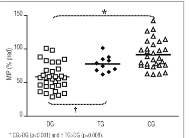
In the evaluation of respiratory muscle strength (Table 2 and Figures 1 and 2), lower values for MIP and MEP were ob-served in the DG group than in the CG. he DG also showed a lower value for MIP than seen in the TG, but the latter group showed a tendency for the MIP to be lower than the MIP of the CG. he MEP had lower value only in the DG, compared with the CG, but with a tendency for the TG also to be lower than the CG.
he classiication of inspiratory muscle strength in rela-tion to the percentage of predicted values showed that 78.1% of DG subjects, 50% of TG subjects and 20% of CG subject showed values less than or equal to 75% of predicted values. his result showed statistical signiicance in the chi-square test (X2=20.93, gl=2, p<0.001) and strength of association of
50% (ג=0.5, p<0.001), when analyzing the MIP as a dependent variable. herefore, individuals in the DG had a higher chance of showing inspiratory muscle strength lower than the general population.
Functional capacity was lower for both DG and TG, com-pared with CG (Table 3). In analyzing the values of total trav-eled distance in each group, it was observed that in the DG, only three individuals (15.63%) walked more than 500 meters, whereas 56.67% of the CG walked more than 500 meters (which is the expected minimum normal value, according to authors who mention this test)31. hese results did not show signiicant
associations in the chi-square test, but there was a tendency for individuals with CKF to walk shorter distances than would be expected for the general population. his can be shown best through comparing the result from the 6MWT with the refer-ence values30. ANOVA showed signiicant diferences between
Figure 1. MIP variation between three groups.
DG TG CG
0 50 100 150
MIP (% pred)
*
†
* CG>DG (p<0.001) and † TG>DG (p=0.008).
Figure 2. MEP variation between three groups.
DG TG CG
0 30 60 90 120 150 180
MEP
(%
p
re
d)
*
*CG>DG (p<0.001).
Table 2. Pulmonary function of the study subjects. Dialysis
(n=32)
Transplantation (n=10)
Control
(n=30) p-value
FVC (%pred) 91.17 (2.88) 94.81 (4.33) 104.33 (2.17) <0.01 *
FEV1 (%pred) 91.13 (3.17) 97.98 (4.54) 110.03 (2.69) <0.01 *
FEV1/FVC% (%pred) 100.06 (1.65) 101.57 (2.34) 104.43 (2.2) 0.26
FEF25-75% (%pred) 95.43 (5.5) 104.19 (7.02) 109.86 (6.23) 0.2
Peak Flow (L/s) 7.47 (0.32) 8.25 (0.44) 6.69 (0.44) 0.09
MVV (%pred) 82.05 (3.52) 93.17 (5.46) 118.37 (4.39) <0.01 *
MIP (cmH2O) 67.19 (4.1) 87.0 (5.1) 94.13 (3.5) <0.01 *
MEP (cmH2O) 76.25 (5.06) 89.2 (6.1) 107.6 (4.59) <0.01 *
* ANOVA p<0.01 between groups; post-hoc Fisher’s LSD (least significant difference); FVC (F2, 69=6.777, p=0.002; LSD CG>DG, p=0.001); FEV1 (F2, 69=10.592, p<0.001; LSD CG>DG,
p<0.001; CG>TG, p=0.046); MVV (F2, 69=22.613, p<0.001; LSD CG>DG, p<0.001; CG>TG, p=0.002); MIP (cmH2O): (F2, 69=13.527, p<0.001; LSD TG>DG, p=0.010; CG>DG, p<0.001);
MEP (cmH2O): (F2, 69=11.182, p<0.001; LSD CG>DG, p<0.001).
Table 3. Six-minute walking distance test on the study subjects.
Dialysis (n=32)
Transplantation (n=10)
Control
(n=30) p-value
6MWT (m) 434.69 (13.25) 456.9 (18.06) 502.53 (8.01) <0.01 **
HRi (ppm) 81.88 (2.21) 76.9 (4.54) 74.77 (2.25) 0.09
HRe (ppm) 99.0 (3.67) 88.9 (5.1) 95.1 (2.24) 0.25
RFi (bpm) 18.44 (0.49) 17.30 (0.79) 16.4 (0.59) 0.03 *
RFe (bpm) 22.5 (0.71) 24.1 (0.82) 21.43 (0.73) 0.16
SAPi (mmHg) 144.38 (3.2) 132.5 (5.54) 116.5 (2.29) <0.01 **
SAPe (mmHg) 152.66 (4.15) 145.0 (5.22) 131.83 (2.94) <0.01 **
DAPi (mmHg) 92.81 (2.74) 84.5 (2.83) 75.0 (1.9) <0.01 **
DAPe (mmHg) 92.5 (2.91) 82.0 (3.27) 77.5 (1.71) <0.01 **
Borgi 1.06 (0.04) 1.1 (0.1) 1.0 (0.0) 0.3
Borge 1.88 (0.26) 2.2 (0.36) 1.33 (0.15) 0.09
*ANOVA p<0.05; ** ANOVA p<0.01 between groups; post-hoc Fisher’s LSD (least significant difference): 6MWT: (F2,69=9.612, p<0.001; LSD CG>DG, p<0.001; CG>TG,
p=0.045); HRi: (2.492, p=0.09); HRe: (1.402, p=0.253); RFi: (F2,69=3.738, p=0.029; LSD DG>CG, p=0.008); RFe: (1.892, p=0.158); SAPi: (F2,69=23.753, p<0.001; LSD DG>TG,
p=0.043; DG>CG, p<0.001); SAPe: (F2,69=8.620, p<0.001; LSD DG>CG, p<0.001); DAPi: (F2,69=14.976, p<0.001; LSD DG>CG, p<0.001; TG>CG, p=0.046); DAPe: (F2,69=10.365,
the groups (p<0.0001): CG>DG (LSD p<0.001); CG>TG (LSD p<0.05); mean 6MWT in the DG, 56.9% of predicted; in the TG, 62.3% of predicted; and in the CG, 70.2% of predicted.
here were positive correlations between FVC and MIP (r=0.310; p<0.05), FVC and MEP (r=0.332; p<0.05), MVV and MIP (r=0.463; p<0.001), MVV and MEP (r=0.430; p<0.001) and MIP and MEP (r=0.752; p<0.001) (Figure 3).
here were correlations between FVC and 6MWT (r=0.355, p=0.046) and between FVC and MVV (r=0.469, p=0.007) only in the DG.
Discussion
In this study, it was seen that the DG showed the worst results for lung function (FVC, FEV1, MVV, MIP and MEP) and functional capacity (6MWT), in comparison with the CG. It is noteworthy that the worst result found for the DG was the signiicant decrease in inspiratory muscle strength and its correlation with proportional loss of expiratory muscle strength. he TG also showed lower results for lung function (MVV and MIP) and functional capacity (6MWT), compared with the CG. Attention is drawn to the positive correlation results between respiratory muscle strength (MIP and MEP) and the volumetric parameters (FVC) and overall functioning of the respiratory system (MVV) in the study groups, thus suggesting that the muscle strength parameter was the main component with the greatest inluence on impairment of lung function in patients undergoing dialysis and in kidney transplant patients.
he mechanisms proposed to explain the worse results from the volumetric component found in the present study, as deined by decreased FVC but FEV1/FVC% index within the normal range24, with a tendency for subjects to show restrictive
disorders, are not entirely clear in the literature. However, the present authors propose that the main disorders associated with these results are the following: chronic and often subclini-cal pulmonary edema; decreased serum albumin with conse-quent water and protein imbalance in the microcirculation; interstitial ibrosis and calciication of the lung parenchyma and bronchial tree; recurrent infections; and alveolitis and ibrosis due to corticosteroid therapy in immunosuppressed patients. Studies that evaluate lung function in patients with CKF undergoing dialysis and after kidney transplantation have described results similar to those found in the present study and help to explain the lesion mechanisms14,31-33.
One of the irst studies to demonstrate the behavior of lung function in patients with CKF at diferent stages of the disease was carried out by Bush and Gabriel34. hese authors studied 80
patients: 20 patients with CKF under medical treatment only (pre-dialysis), 20 patients undergoing continuous ambulatory
peritoneal dialysis (CAPD), 20 patients undergoing dialysis and 20 kidney transplantation patients. hey evaluated the FVC, FEV1, FEV1/FVC%, PEF, TLC (total lung capacity) and RV (residual volume) parameters as spirometry parameters, along with CO (carbon monoxide) difusion. hey found values within normal range for the pre-dialysis group; a small reduction in spirometric parameters and large reduction in CO difusion in patients undergoing CAPD; a small reduction in spirometric parameters but increased RV in the dialysis group; and normal spirometric values for the post-transplantation group, but with decreased TLC and CO difusion and the lowest RV value. hey did not ind any correlation between the lung function parameters and the biochemical tests and duration and severity of CKF.
Another component that was lower in the DG and TG, in the spirometric evaluation, was the MVV. he three groups were within the normal range (>80% of predicted), but with lower values than in the CG, thus showing that patients with CKF undergoing dialysis and kidney transplant patients have limitations to their ventilatory capacity. here has only been one report of this parameter in evaluating lung function35 in
patients with CKF. However, that author did not compare the value obtained with normal values. A correlation between MVV and FVC was also found in the DG, thus suggesting that the reduction in MVV appears in individuals who have lower FVC. his may be another factor indicative of the negative efect of decreased lung volume, even if still within normal limits, which may cause a functional impairment for individuals with CKF.
he decrease in both inspiratory and expiratory muscle strength found in the DG and TG groups demonstrates that CKF signiicantly afects both the inspiratory and the expira-tory respiraexpira-tory muscles. his can be interpreted through the positive correlation found between MIP and MEP, thereby showing that respiratory muscle strength is decreased overall, Figure 3. Correlation between MIP and MEP (r=0.752; p<0.01).
□ – DG; ♦ - TG; ∆ - CG
and that patients have a linear decrease in the two components (inspiratory and expiratory). Even after kidney transplant, pa-tients do not seem to fully recover respiratory muscle strength, thus showing that factors other than uremia maintain the muscle deicit in this population.
he causal factors relating to the decrease in the respira-tory muscle strength component have been described in the literature as due to the causal mechanisms of uremic myopa-thy. hey include decreases in muscle mass (cross-sectional area, mainly of type II ibers) found in studies on mice and humans, decreases in oxidative metabolism, decreases in muscle protein synthesis and decreases in calcium plasmatic concentration 6-10.
For transplantation patients, it is believed that the use of corticosteroid immunosuppressant therapy impedes the re-covery of muscle ibers after kidney transplantation by caus-ing a decrease in muscle protein synthesis and impairment of oxidative metabolism18-20. Other factors such as the age of the
population, sedentary lifestyle and lack of systematic rehabili-tation programs for kidney transplanrehabili-tation patients in Brazil, may lead these individuals to maintain deicits that may have negative inluences on their functional outcome.
Some authors have reported on evaluations of respiratory muscles among patients with CKF. Gómez-Fernández et al.36
were among the irst authors to report on evaluations of maxi-mal respiratory pressures in this population. hey evaluated CKF patients who underwent CAPD and found decreased MIP in patients with CKF (59.6% of predicted values), compared with controls (82.7% of predicted values). Other authors14,33,35
have also found results similar to those found in the present study. here is a consensus that respiratory muscle strength is decreased, and the pathogenesis of this condition is similar to what is observed in peripheral muscles.
he results from the 6MWT in this study demonstrated that individuals in the DG and TG had worse results than did those in the CG. Oh-Park et al.37 evaluated the 6MWT and reported
that the CKF patients walked distances that were shorter than what is considered to be normal, with a mean of 405 meters for dialysis patients, i.e. a value slightly lower than what was found in the present study. Becker-Cohen et al.38 evaluated the
6MWT in children and young adults with CKF and with kid-ney transplants who were still undergoing dialysis. hey found values within normality and, although there were no speciic predictive values for children, they found that on average, the distance that they were able to walk was only 100 meters less than what the adults who were evaluated could achieve. hose authors therefore considered this result to be normal.
Decreased functional capacity is caused by multiple factors, including cardiovascular, respiratory and muscle problems, in which the capability to capture, transport and use O2 might
be harmed. In this study, it was found that the component that showed further injury and was thus a negative inluence on functional capacity was lung function, with a positive cor-relation between FVC and functional capacity in the DG. his suggests that even a small reduction in FVC may inluence these individuals’ performance in the functional capacity test, although the FVC values were within normal ranges.
Another factor not evaluated in this study but of func-tional interest is that patients with CKF may show decreased O2 consumption (VO2)4, as reported by Sietsema et al.39. hey
demonstrated in their study that maximum O2 consumption values greater than 17.5 ml/min/kg are strong and important predictors of survival among patients with CKF, thereby indi-cating that functional capacity evaluation is essential within the follow-up for patients with CKF.
he 6MWT provides important measurements for following up patients’ evolution during the disease and also for evaluating the beneits of rehabilitation programs developed among these individuals. Although this test is still infrequently used for eval-uating patients with CKF, and this disease does not appear as an indication for 6MWT, as described by the American horacic Society30, the results from the 6MWT can be of practical use for
physical therapists working in dialysis units and care centers for kidney transplantation patients. his idea is reinforced in the study by Reboredo et al.40, who evaluated functional capacity by
means of the 6MWT and correlated the results with cardiopul-monary tests. hey concluded that the 6MWT can be used as a means of evaluating patients with CKF.
Regarding the anthropometric characteristics of the study population, it was observed that the sample was homogeneous in relation to the parameters of age, height and BMI. here was a signiicant diference regarding weight. Although this study did not include the aim of evaluating nutritional status, it was found that the means of the groups were within the normal range for BMI. his result is positive, since low weight is a fac-tor of negative prognosis for chronic diseases, and overweight is a risk factor for cardiovascular disease41.
Some diiculties were found in carrying out the tests, since the subjects with CKF (DG) showed limitations and complica-tions after dialysis sessions, which hampered their performance in all the functional tests. It was impossible to match the num-ber of subjects in the TG with the numnum-bers in the other groups, since many of these individuals did not join in the project, thus impairing the homogeneity among the groups.
Conclusions
1. Parmar MS. Chronic renal disease: early identification and active management of patients with renal impairment in primary care can improve outcomes. BMJ. 2002; 325(7355):85-90.
2. Romão Jr JE. Doença renal crônica: definição, epidemiologia e classificação. J Bras Nefrol. 2004;26(3 Supl 1):S1-3.
3. Grassmann A, Gioberge S, Moeller S, Brown G. ESRD patients in 2004: global overview of patient numbers, treatment modalities and associated trends. Nephrol Dial Transplant. 2005;20(12):2587-93.
4. Violan MA, Pomes T, Maldonado S, Roura G, De la Fuente I, Verdaguer T, et al. Exercise capacity in hemodialysis and renal transplant patients. Transplant Proc. 2002;34(1):417-8.
5. McIntyre CW, Selby NM, Sigrist M, Pearce LE, Mercer TH, Nais PF. Patients receiving maintenance dialysis have more severe functionally significant skeletal muscle wasting than patients with dialysis-independent chronic kidney disease. Nephrol Dial Transplant. 2006;21(8):2210-6.
6. Quintanilla AP, Sahgal V. Uremic myophaty. Inter J Artif Org. 1984;7(5):239-42.
7. Fahal IH, Bell GM, Bone JM, Edwards RH. Physiological abnormalities of skeletal muscle in dialysis patients. Nephrol Dial Transplant. 1997;12(1):119-27.
8. Adey D, Kumar R, McCarthy JT, Nair KS. Reduced synthesis of muscle proteins in chronic renal failure. Am J Physiol Endocrinol Metab. 2000;278(2):E219-25.
9. Cupisti A, Licitra R, Chisari C, Stampacchia G, D’Alessandro C, Galetta F, et al. Skeletal muscle and nutritional assessment in chronic renal failure patients on a protein-restricted diet. J Inter Med. 2004;255(1):115-24.
10. Tarasuik A, Heimer D, Bark H. Effect of chronic renal failure on skeletal and diaphragmatic muscle contraction. Am Rev Respir Dis. 1992;146(6):1383-8.
11. Kemp GJ, Crowe AV, Anijeet HK, Gong QY, Bimson WE, Frostick SP, et al. Abnormal mitochondrial function and muscle wasting, but normal contractile efficiency, in haemodialysed patients studied non-invasively in vivo. Nephrol Dial Transplant. 2004;19(6):1520-7.
12. Sakkas GK, Sargean AJ, Mercer TH, Baal D, Koufaki P, Karatzaferi C, et al. Changes in muscle morphology in dialysis patients after 6 months of aerobic exercise training. Nephrol Dial Transplant. 2003;18(9):1854-61.
13. Marrades RM, Roca J, Campistol JM, Diaz O, Barberá JÁ, Torregosa JV, et al. Effects of erythropoietin on muscle O2 transport during exercise in patients with chronic renal failure. J Clin Invest. 1996;97(9):2092-100.
14. Karacan O, Tutal E, Colak T, Sezer S, Eyüboglu FO, Haberal M. Pulmonary function in renal transplant recipients and end-stage renal disease
in patients with CKF undergoing dialysis and in kidney trans-plantation patients show lower values than those of the general population, and that patients undergoing dialysis have greater impairment of muscle and lung function than do kidney
transplant patients. Based on the correlations found between respiratory muscle strength and FVC and MVV parameters, it appears that muscle strength is the respiratory component that is most afected in individuals with CKF.
patients undergoing maintenance dialysis. Transplant Proc. 2006;38(2): 396-400.
15. Moreira PR, Barros E. Atualização em fisiologia e fisiopatologia renal: bases fisiopatológicas da miopatia na insuficiência renal crônica. J Bras Nefrol. 2000;22(1):34-8.
16. Sarnak MJ, Levey AS, Schoolwerth AC, Coresh J, Culleton B, Hamm LL, et al. Kidney disease as a risk factor for development of cardiovascular disease: a statement from the american heart association councils on kidney in cardiovascular disease, high blood pressure research, clinical cardiology, and epidemiology and prevention. Hypertension. 2003;42(5):1050-65.
17. Bardin T. Musculoskeletal manifestations of chronic renal failure. Curr Opin Rheumatol. 2003;15(1):48-54.
18. Van Balkom RH, Zhan WZ, Prakash YS, Dekhuijzen PN, Sieck GC. Corticosteroid effects on isotonic contractile properties of rat diaphragm muscle. J Appl Physiol. 1997;83(4):1062-7.
19. Koerts-de Lang E, Schols AM, Rooyackers OE, Gayan-Ramirez G, Decramer M, Wouters EF. Different effects of corticosteroid-induced muscle wasting compared with undernutrition on rat diaphragm energy metabolism. Eur J Appl Physiol. 2000;85(5-6):493-8.
20. Mitsui T, Azuma H, Nagasawa M, Iuchi T, Akaike M, Odomi M, et al. Chronic corticosteroid administration causes mitochondrial dysfunction in skeletal muscle. J Neurol. 2002;249(8):1004-9.
21. Kovelis D, Pitta FO, Probst VS, PeresCPA, Delfino VDA, Mocelin AJ, et al.
Função pulmonar e força muscular respiratória em pacientes com doença renal crônica submetidos à hemodiálise. J Bras Pneumol. 2008;34(11): 907-12.
22. Standardization of spirometry, 1994 Update. American Thoracic Society. Am J Respir Crit Care Med. 1995;152(3):1107-36.
23. Knudson RJ, Lebowitz MD, Holberg CJ, Burrows B. Changes in the normal maximal expiratory flow-volume curve with growth and aging. Am Rev Respir Dis. 1983;127(6):725-34.
24. Sociedade brasileira de pneumologia e tisiologia. Diretrizes para testes de função pulmonar. J Bras Pneumol. 2002;28(Supl 3):S1-238.
25. Black LF, Hyatt RE. Maximal respiratory pressures: normal values and relationship to age and sex. Am Rev Respir Dis. 1969;99(5):969-74.
26. Neder JA, Andreoni S, Lerario MC, Nery LE. Reference values for lung function tests. II. Maximal respiratory pressures and voluntary ventilation. Braz J Med Biol Res. 1999;32(6):719-27.
27. Bellinetti LM, Thomson JC. Avaliação muscular respiratória nas toracotomias e laparotomias superiores eletivas. J Bras Pneumol. 2006;32(2):99-105.
28. Guyatt GH, Sullivan MJ, Thompson PJ, Fallen EL, Pugsley SO, Taylor DW, et al. The 6-minute walk: a new measure of exercise capacity in patients with chronic heart failure. Can Med Assoc J. 1985;132(8):919-23.
29. Troosters T, Gosselink R, Decramer M. Six minute walking in healthy elderly subjects. Eur Respir J. 1999;14(2):270-4.
30. ATS Committee standards for clinical pulmonary function laboratories. ATS Statement: guidelines for the six-minute walk test. Am J Respir Crit Care Med. 2002;166(1):111-7.
31. Kalender B, Erk M, Pekpak MA, Apaydin S, Ataman R, Serdengeçti K, et al. The effect of renal transplantation on pulmonary function. Nephron. 2002;90(1):72-7.
32. Guleria S, Agarwal RK, Guleria R, Bhowmik D, Agarwal SK, Tiwari SC. The effect of renal transplantation on pulmonary function and respiratory muscle strength in patients with end-stage renal disease. Transplant Proc. 2005;37(2):664-5.
33. Karacan O, Tutal E, Uyar M, Eyüboglu FO, Sezar S, Ozdemir FN. Pulmonary function in uremic patients on long-term hemodialysis. Ren Fail. 2004;26(3):273-8.
34. Bush A, Gabriel R. Pulmonary function in chronic renal failure: effects of dialysus and transplantation. Thorax. 1991;46(6):424-8.
35. Ulubay G, Akman B, Sezer S, Calik K, Eyuboglu Oner F, Ozdemir N, et al. Factors affecting exercise capacity in renal transplantation candidates
on continuous ambulatory peritoneal dialysis therapy. Transplant Proc. 2006;38(2):401-5.
36. Gómez-Férnandez P, Sánchez Agudo L, Calatrava JM, Escuin F, Selgas R, Martínez ME, et al. Respiratory muscle weakness in uremic patients under continuous ambulatory peritoneal dialysis. Nephron. 1984;36(4):219-23.
37. Oh-Park M, Fast A, Gopal S, Lynn R, Frei G, Drenth R, et al. Exercise for the dialyzed: aerobic and strength training during hemodialysis. Am J Phys Med Rehabil. 2002;81(11):814-21.
38. Becker-Cohen R, Nir A, Rinat C, Feinstein S, Algur N, Farber B, et al. Risk factors for cardiovascular disease in children and young adults after renal transplantation. Clin J Am Soc Nephrol. 2006;1(6):1284-92.
39. Sietsema KE, Amato A, Adler SG, Brass EE. Exercise capacity as a predictor of survival among ambulatory patients with end-stage renal disease. Kidney Int. 2004;65(2):719-24.
40. Reboredo MM, Henrique DMN, Faria RS, Bergamini BC, Bastos MG, Paula RB. Correlação entre a distância obtida no teste de caminhada de seis minutos e o pico de consumo de oxigênio em pacientes portadores de doença renal crônica em hemodiálise. J Bras Nefrol. 2007;29(2): 85-9.