Results of a control quality strategy
in cervical cytology
Resultados de uma estratégia de controle de qualidade em colpocitologia
Caio Eduardo Gullo1, Anna Luiza Tannús Dami1, Amanda Pires Barbosa1, Aline Maria de Vita Marques1,2,
Marianna Angelo Palmejani1, Luiz Guilherme Cernaglia Aureliano de Lima3, Jane Lopes Bonilha3
Study carried out at Hospital de Base de São José do Rio Preto - São Jose do Rio Preto (SP), Brazil.
1 Undergraduate Course of Medicine (2011), Faculdade de Medicina de São José do Rio Preto – FAMERP, São Jose do Rio Preto (SP), Brazil. 2 Graduate Program (Master´s degree) in Microbiology, Faculdade de Medicina de São José do Rio Preto – FAMERP, São Jose do Rio Preto (SP), Brazil. 3 Department of Pathology and Forensic Medicine, Faculdade de Medicina de São José do Rio Preto – FAMERP, São Jose do Rio Preto (SP), Brazil.
Corresponding author: Jane Lopes Bonilha – Faculdade de Medicina de São José do Rio Preto (FAMERP) – Departamento de Patologia e Medicina Legal – Av. Brigadeiro Faria Lima, 5416 – Vila São Pedro – Zip code 15090-000 – São José do Rio Preto – (SP), Brazil – Phone: (17) 3210-5700 – E-mail: [email protected]
Received on: Apr 5, 2011– Accepted on: Dec 12, 2011
Conflict of interest: None
aBstRact
objective: To determine the efficacy of a quality control strategy in cervical cytology in the detection of high-grade squamous intraepithelial lesions. methods: Forty-two patients were selected who underwent a Pap smear and cervical uterine biopsy between April 2008 and December 2009, with evidence of a high-grade squamous intraepithelial lesion in one or both tests. The statistical parameters of the smear test were calculated before and after systematic meetings for review of the archived test results (6 years), in which the following was done: interobserver diagnostic consensus; cytohistological correlation, with the latter as gold standard; and evaluation of the therapeutic status of each patient. Results: Once these controls were applied, it was noted that sensitivity and positive likelihood ratio of the test for high-grade squamous intraepithelial lesion increased 9.5% (34.5 to 44%) and 0.45% (1.64 to 2.09%), respectively, while specificity remained at 79%. Reduction in interference of false-negative results associated with errors in the analytical phase of the cytological productive process gave an estimate of failures in collection of the specimens (pre-analytical phase). conclusion: In addition to improving the performance of the cytological diagnosis of the high-grade squamous intraepithelial lesion, the proposed quality control strategy allows a reflection on the causes of incorrect or conflicting scrutiny.
Keywords: Efficacy; Quality control; Intraepithelial cervical neoplasia; Cervix uteri/pathology
ResUmo
objetivo: Determinar a eficácia de uma estratégia de controle de qualidade em colpocitologia na detecção da lesão intraepitelial escamosa de alto grau. métodos: Foram selecionadas 42 pacientes
que realizaram Papanicolaou e biópsia cervicouterina entre abril de 2008 e dezembro de 2009, com evidência de lesão intraepitelial escamosa de alto grau em um ou em ambos os exames. Os parâmetros estatísticos do esfregaço foram calculados antes e após reuniões sistematizadas de revisão dos exames arquivados (6 anos), nas quais se procedeu a: consensualização diagnóstica interobservadores; correlação cito-histológica, sendo a última padrão-ouro; e avaliação do status terapêutico de cada paciente. Resultados: Aplicados tais controles, observou-se que a sensibilidade e a likelihood ratio
positiva do teste para lesão intraepitelial escamosa de alto grau aumentaram 9,5% (34,5 para 44%) e 0,45% (1,64 para 2,09%), respectivamente, enquanto sua especificidade se manteve em 79%. A redução da interferência dos falso-negativos associados a erros na fase analítica do processo produtivo citológico traz estimativa das falhas de coleta do material (fase pré-analítica). conclusão: Além de melhorar o desempenho do diagnóstico colpocitológico de lesão intraepitelial escamosa de alto grau, a estratégia de controle de qualidade proposta permite refletir sobre as causas de escrutínio incorreto ou discordante.
descritores: Eficácia; Controle de qualidade; Neoplasia intra-epitelial cervical; Colo do útero/patologia
intRodUction
programs for disease control, there has been an accentuated increase in the number of cases, with an estimated 19000 new cases/year for 2008 and 2009(2).
Colpocytology, or the Papanicolaou test, is the most widely used for population tracking of asymptomatic and potentially curable lesions of the lower female genital tract, as it is simple, safe, and low-cost. Currently, the impact of oncotic cytology on the natural progression of uterine cervix cancer, specifically in secondary prevention, has been questioned. This is because even in cities with adequate gynecological assistance coverage, women who develop the invasive form of the carcinoma displayed a similar percentage of prior cytology tests relative to healthy individuals(3).
There is a considerable variation (5 to 70%) in false-negative (FN) rates for the test(4). Limitations
in its sensitivity are attributed, in general, to care in patient preparation and techniques for collection of specimens(5). Additionally, the existence of numerable
cases with cytomorphological alterations with imprecise diagnostic resolution(6)and interobserver variability(7)
are factors that interfere in the analytical phase of the test. As an example, variations of the usual cellular pattern may go by undetected and are associated with atypical squamous cells of underdetermined significance (ASC-US) and low-grade squamous intraepithelial lesions (LGSIL). In the presence of these conditions, precise diagnosis has little influence on the subsequent follow-up, since they evolve with a high percentage of spontaneous regression.
On the other hand, interpretation by different observers of borderline deviations between minor and major gravity may cause discrepancy due to the subjectivity of its qualitative and quantitative aspects(5).
When there is a high-grade squamous intraepithelial lesion (HGSIL), specific detection is of vital importance to impede its progression to invasive carcinoma(8).
Thus, the adoption of quality control programs is necessary for any laboratory that performs cytology analyses. Numerous studies(6,9,10) demonstrated that
critical review of the smear tests is an indispensable practice for improving accuracy of the test. Also emphasized is the importance of cytohistological correlation in discordant cases as a method of error detection and evaluation of the performance of the service(8,11,12).
oBJectiVe
To determine and evaluate the results of a quality control strategy of the analytical phase of cervical cytology in diagnosis of HGSIL.
metHods
This is a study with a quantitative, descriptive, and cross-section approach carried out at the Pathology Service (SAP) of the Hospital de Base de São José do Rio Preto (SP).
Patients who participated in the study were those who were followed-up at the Gynecology Outpatient Clinic of the hospital during the period between April 2008 and December 2009. Inclusion criteria were results during this period from colpocytology and histopathology tests of the uterine cervix positive for at least one of the following: HGSIL, cervical intraepithelial neoplasia (CIN) II or III, or carcinoma in situ. The diagnosis of invasive carcinoma of the uterine cervix was an exclusion criterion.
Procedures
Since the identity of selected patients was preserved and their data were obtained from clinical records and tests, this study did not require an Informed Consent Form. The study was approved by the Research Ethics
Committee of the Faculdade de Medicina de São José
do Rio Preto (FAMERP), in accordance with official
statement number 001-004414/2009, dated July 29, 2009. Initially, a review of the selected tests was performed in order to exclude possible false-positive (FP) diagnoses of the cervical condition under study. Next, we developed a quality control strategy in cervical cytology structured into two phases: critical and systematic review of the prior cervical vaginal smear tests; and cytohistological correlation and evaluation of the treatment adopted, case-by-case.
In the first part of the study, we analyzed prior cervical cytology tests (April 2003 to March 2008) of each patient in weekly meetings with the cytopathology team of the SAP, composed of a cytopathologist with more than 30 years of experience; nine medical residents in Pathology, equally distributed in their rotations of the first (R1), second (R2), and third (R3) years of the Medical Residency Program; a medical resident active in the area of Cytopathology (R4); and a cytology technician. We used a light microscope connected to an image projection multimedia device and another multi-head type apparatus, which enabled the collective investigation proposed and colpocytological diagnosis by consensus.
The cytological classification of the uterine cervix lesions followed the Bethesda System 2001
of nomenclature for cervical cytology(13). We also
biopsies or surgical products were classified as per the World Health Organization (WHO)(14).
In the second phase of the study, all the clinical charts were reviewed to obtain information regarding the course of treatment followed, i.e., whether an expectant approach or active therapy, such as the indication for outpatient electrocauterization (ECT) and/or ectocervical or endocervical high frequency electrocautery (HFEC). The choice of hysterectomy was also mentioned and determined the exclusion of tests posterior to this procedure. Lastly, the cytohistological correlation and the impact of interventions undergone by the patients were weighed in order to establish the significance of each colpocytological diagnosis, according to the possibilities identified in figure 1. In case of disagreement as to the results among cytology and biopsy, even after review of the tests, the histology technique was considered the gold standard for the cervical pathology under study.
The data were tabulated with the Microsoft Office Excel 2003 for Windows program, grouped into tables, and analyzed as per the objective of the study, presented as frequencies and percentages.
statistical analysis
Sensitivity and specificity were evaluated by calculation of the positive predictive values (PPV) and negative predictive values (NPV) of the method under scrutiny
(control) and after review of the slides (Chart 1). In both situations, a positive likelihood ratio was calculated.
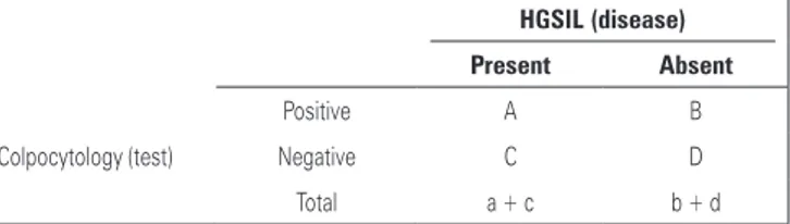
chart 1. Possible results of cytological technique (diagnostic test) in detecting high-grade squamous intraepithelial lesion
Hgsil (disease)
Present absent
Colpocytology (test)
Positive A B
Negative C D
Total a + c b + d
HSIL: high-grade squamous intraepithelial lesion; a: true positive; b: false positive; c: false negative; d: true negative.
C: colpocytology method; H: histopathology test; HGSIL: high-grade squamous intraepithelial lesion.
Figure 1. Characterization of diagnoses in cervical-vaginal smears
Applicable formulas(15):
- S = a / (a + c); - E = d / (b + d); - PPV = a / (a + b); - NPV = d / (c + d).
Likelihood ratio positive when S / (100-E).
ResUlts
with satisfactory suitability of the samples, collected as of April 2003 in this study population; colposcopy-directed biopsies and HFEC products of the ectocervix and endocervix totaled 98 tests.
Initially, a critical and systematic review was made of all slides obtained in meetings with physicians from the SAP for definition of consensus as to pathological classification of the microscopic alterations found. At these meetings, eight cytology tests (3%) had their diagnoses modified for HGSIL and four of them (50%) had initially been documented as LGSIL in routine scrutiny. As to histological material, four (4%) biopsies also underwent correction of their results and were all consistent with LGSIL; in two of them, the presence of HGSIL had been first considered. No prior record of HGSIL, NIC II, or III, was reclassified into other parts.
Later, we verified the cytohistological correlation as to the diagnosis of HGSIL and, when necessary, the interventions suffered by patients in order to establish the validity of the colpocytology. Agreement at least in some points between both techniques was noted in 26 patients throughout the follow-up performed. Of the remaining 16, seven presented with cytology positive for HGSIL with no corresponding findings in biopsies or product of HFEC; in the other nine, the
inverse situation was observed, that is, the detection of moderate or severe dysplasia of the uterine cervix was possible only with the histological method. Still, 28 patients were submitted to therapeutic procedures, HFEC and/or ECT of the uterine cervix, and in the absence both of a detectable alteration upon colposcopy and a biopsy with evidence of posterior residual lesion, were considered free of disease.
Therefore, according to criteria established in table 1, we found the following values of true-positive (TP) and true-negative (TN) results, as well as of FP and FN, for cervical cytology before (Chart 2) and after (Chart 3) review of the slides.
Finally, we calculated the statistical parameters of the test in both situations (Table 1).
discUssion
Quality control in cervical cytology has the objective of improving the performance of the test, in order to eliminate FN results. These are more worrisome in a routine examination than the FP, since non-diagnosed women may lose follow-up and continue at risk of developing severe lesions. Nevertheless, failures in the opposite situation are not harmless, either, since they lead to unnecessary surgical procedures that can alter the reproductive and sexual life of women(11), in
addition to the evident psychological impacts.
In order to establish the goals of intervention, it is necessary to assess all steps in which failures might occur, from the collection of samples to the routine screening and interpretation of microscopic findings. Problems in scrutiny happen when neoplasic cells are not recognized, albeit present, due to attention deficit, insufficient time, and lack of experience of the examiner. On the other hand, problems related to diagnostic interpretation are attributed to the judgment of these cells as benign, in the case of an inexperienced professional and inadequate clinical information(16).
Due to the non-feasibility of review in all cases, sampling strategies are adopted with several designs. In the present study, we investigated the effect of review of prior tests of patients with HGSIL as per cytology and/or histology according to different observers, associated with the correlation of both methods and evaluations of the therapeutic status. This is because there is a significant reduction in accuracy of the cytology technique in women who are known to have the lesion(8,17).
We verified that regarding the diagnosis of HGSIL, the sensitivity of the test went from 34.5 to 44%, accompanying the 0.45 increase of the positive likelihood ratio, while specificity remained at 79%.
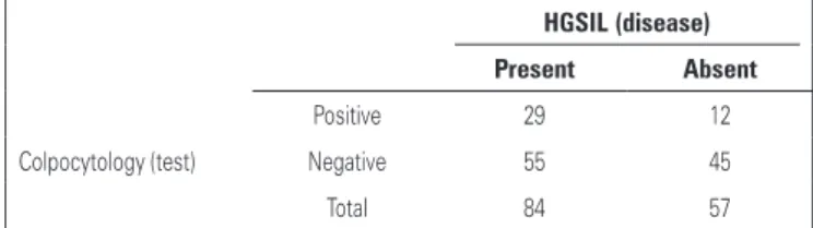
chart 2. Results of cytological technique (diagnostic test) in detecting high-grade squamous intraepithelial lesion in routine screening
Hgsil (disease)
Present absent
Colpocytology (test)
Positive 29 12
Negative 55 45
Total 84 57
HSIL: high-grade squamous intraepithelial lesion.
chart 3. Results of cytological technique (diagnostic test) in detecting high-grade squamous intraepithelial lesion (HSIL) after slide review
Hgsil (disease)
Present absent
Colpocytology (test)
Positive 37 12
Negative 47 45
Total 84 57
HSIL: high-grade squamous intraepithelial lesion.
table 1. Statistical parameters of cytological technique in detecting high-grade squamous intraepithelial lesion (HSIL) before and after slide review
s (%) e (%) PPV (%) nPV (%) PlR
Screening 34.5 79 71 45 1.64
Review 44 79 76 49 2.09
The positive likelihood ratio, or positive odds ratio, relates sensitivity to the complement of specificity, that is, the probability of the test being positive in the individual that has the disease against the probability of the test being negative in the individual that has the disease. The greater the positive likelihood, the better the diagnostic test will be, so that our results allow establishing a better performance of review proposed as to screening.
Nevertheless, little can be done in the laboratory in terms of reducing the FN results when the uterine cervix is not adequately represented(5). In this sense, we
are limited by the fact that the samples were collected by medical students and Gynecology residents with different degrees of experience. This allows us to infer that the errors in sampling have a significant repercussion on the detection rates of HGSIL at our institution.
Pinho and Mattos(17) found high sensitivity (96%) for
the test; however, they considered as positive results the presence of any alterations of the cervical epithelium. Specificity, however, was 51.5%; therefore, many women with a suspect diagnosis would have continued investigation unnecessarily, generating excessive costs, besides the potential iatrogenesis that this action might cause in many of them. It is worth pointing out that the cutoff point established to delimit the positive results from the negative has a great influence on the values of sensitivity and specificity(15).
In the study by Santos et al.(18), sensitivity and specificity
found for HGSIL were 79% and 84%, respectively. It was also evident that the review of the slides by a second observer from the same laboratory did not increase the performance of cytology, a fact that, although juxtaposed to our results, is justified by the lesser influence that a poor sampling technique exerted on the quality of the smears. The researchers concluded that the repetition of the test and its evaluation with the knowledge of clinical data is sufficient for improving diagnosis of HGSIL with prior results of altered cytology.
Indeed, the review of negative cases in the screening selected by clinical risk criteria, such as postmenopausal genital hemorrhage, sexually transmitted diseases, macroscopic alterations upon the speculum examination, and prior alterations on cytological testing could detect a greater number of FN in this population(16).
Considering this information and the correlated therapeutic history, systematic meetings to review slides guarantee that a greater number of observers will reflect on the marked cytology fields and on the possible causes of incorrect or discordant diagnoses, and do not generate additional costs.
Finally, we suggest that cytopathology laboratories periodically issue a consolidated report of the test of
each professional or institution, describing aspects related to suitability of the samples collected. Quality indicators should be established in order to guide and indicate which collection technique needs revision.
conclUsions
The strategy of HGSIL detection with multiple cases and examiners associated with cytohistological and clinical correlation enables an evaluation of the performance of cervical cytology services and of the team relative to borderline findings. Standardization of procedures, with improvements in techniques and diagnostic interpretation, is an important tool that raises the quality of the entire cytological productive process.
acKnoWledgments
We thank the National Council of Scientific and Technological Development (CNPq) for the financial aid (Process # 119.407/2009-4).
ReFeRences
1. Uchimura NS, Nakano K, Nakano LC, Uchimura TT. [Quality and performance of pap smears in the cervical cancer screening program in a city of southern Brazil]. Rev Assoc Med Bras. 2009;55(5):569-74. Portuguese.
2. Brasil. Ministério da Saúde. Secretaria de Atenção à Saúde. Instituto Nacional de Câncer. Coordenação de Prevenção e Vigilância de Câncer. Estimativas 2008: incidência de câncer no Brasil. Rio de Janeiro: INCA, 2007; 2008 [citado 2010 Ago 18]. Disponível em: http://www.inca.gov.br/ estimativa/2008/versaofinal.pdf
3. Murillo R, Cendales R, Wiesner C, Piñeros M, Tovar S. [Effectiveness of cytology-based cervical cancer screening in the Colombian health system]. Biomedica. 2009;29(3):354-61. Spanish.
4. Rama CH. Idade e prevalência da infecção genital por papilomavírus humano de alto risco em mulheres submetidas a rastreamento para o câncer cervical [tese]. São Paulo: Universidade de São Paulo; 2006.
5. Franco R, Amaral RG, Montemor EB , Montis DM, Morais SS, Zeferino LC. Fatores associados a resultados falso-negativos de exames citopatológicos do colo uterino. Rev Bras Ginecol Obst. 2006;28(8):479-85.
6. Maeda MY, di Loreto C, Barreto E, Cavaliere MJ, Utagawa ML, Sakai YI, et al. Estudo preliminar do SISCOLO-Qualidade na rede de saúde pública de São Paulo. J Bras Patol Med Lab. 2004;40(6):425-9.
7. Amaral RG, Souza NL, Tavares SB, Manrique EJ, Assem DZ, Azevedo LL, et al. Controle externo da qualidade dos diagnósticos citológicos no rastreamento do câncer cervical: estudo piloto. Rev Bras Anal Clin. 2006;38(2):79-81. 8. Tuon FF, Bittencourt MS, Panichi MA, Pinto AP. Avaliação da sensibilidade
e especificidade dos exames citopatológico e colposcópico em relação ao exame histológico na identificação de lesões intra-epiteliais cervicais. Rev Assoc Med Bras. 2002;48:140-4.
9. Jones B. Rescreening in gynecologic cytology: rescreening of 3762 previous cases for current high-grade squamous intraepithelial lesions and carcinoma: a College of American Pathologists Q-Probes study of 312 institutions. Arch Pathol Lab Med. 1995;119(12):1097-103.
10. Collaço LM. Monitoramento externo da qualidade em citopatologia cérvico-vaginal [tese]. Curitiba: Universidade Federal do Paraná; 2002.
cytologic-histologic correlation data on cervical cancer screening performance. Arch Pathol Lab Med. 2008;132(1):16-22.
13. Solomon D, Nayar R. Sistema Bethesda para citopatologia cervicovaginal: definições, critérios e notas explicativas. Rio de Janeiro: Revinter; 2005. 14. Tavassoli FA, Devilee P. Pathology and genetics of tumours of the breast and
female genital organs: World Health Organization; 2003.
15. Medronho RA, Perez MA. Testes diagnósticos. In: Medronho RA, Carvalho DM, Block KV, Luiz RR, Werneck GL, organizadores. Epidemiologia. São Paulo: Atheneu; 2003. p. 259-70.
16. Tavares SB, Amaral RG, Manrique EJ, Sousa NL, Albuquerque ZB, Zeferino
LC. Controle de qualidade em citopatologia cervical: revisão de literatura. Rev Bras Cancerol. 2007;53(3):355-64.
17. Pinho AA, Mattos MC. Validade da citologia cervicovaginal na detecção de lesões pré-neoplásicas e neoplásicas de colo de útero; Validity of cervicovaginal cytology for detection of cancerous and precancerous lesions of the cervix. J Bras Patol Med Lab. 2002;38(3):225-31.