EVALUATION AND CLASSIFICATION OF VAGINAL STENOSIS AFTER
BRACHYTHERAPY
Luciana Martins da Rosa1, Karina Silveira de Almeida Hammerschmidt2, Vera Radünz3, Patrícia Ilha4, Andrelise Viana Rosa Tomasi5, Rafaela Vivian Valcarenghi6
1 Ph.D. in Nursing. Professor, Departamento de Enfermagem,Programa de Pós-Graduação Gestão do Cuidado em Enfermagem, Universidade Federal de Santa Catarina (UFSC). Florianópolis, Santa Catarina, Brazil. E-mail: [email protected]
2 Ph.D. in Nursing. Professor, Departamento de Enfermagem, UFSC. Florianópolis, Santa Catarina, Brazil. E-mail: [email protected] 3 Ph.D. in Nursing. Professor, Departamento de Enfermagem,Programa de Pós-Graduação em Enfermagem (PEN), UFSC. Florianópolis,
Santa Catarina, Brazil. E-mail: [email protected]
4 Doctoral student, PEN/UFSC. Florianópolis, Santa Catarina, Brazil. Email: [email protected]
5 Doctoral student, PEN/UFSC. Professor, Centro Universitário Estácio de Sá, Santa Catarina. Florianópolis, Santa Catarina, Brazil. E-mail: [email protected]
6 Doctoral student, PEN/UFSC. Florianópolis, Santa Catarina, Brazil. E-mail: [email protected]
ABSTRACT: This narrative review identiied, in the scientiic production, the methods used for evaluating and classifying vaginal stenosis in women who have undergone brachytherapy. Data collection was undertaken in July 2013 in the publications of SciELO, MEDLINE and PubMed, without time limits, and in studies cited by two scientiic reviews which addressed the issue investigated here. The search protocol included the description of the method for evaluating and classifying vaginal stenosis. Comparative analysis between the indings showed there to be diversity among the methods used by different researchers. In the light of this inding, this study proposes elements for making an evaluative instrument to be applied by nurses. The standardization of the technique will help in the early detection of vaginal stenosis and in the care for women subsequent to vaginal brachytherapy.
DESCRIPTORS: Uterine cervical neoplasm. Brachytherapy. Constriction, pathologic. Nursing.
AVALIAÇÃO E CLASSIFICAÇÃO DA ESTENOSE VAGINAL
PÓS-BRAQUITERAPIA
RESUMO: Revisão narrativa que identiicou, na produção cientíica, os métodos utilizados para avaliação e classiicação da estenose vaginal em mulheres pós-braquiterapia. A coleta de dados foi realizada em julho 2013 nas publicações da SciELO, MEDLINE e PubMed, sem limite de tempo, e em estudos citados por duas revisões cientíicas que abordam a temática aqui investigada. O protocolo de busca incluiu a descrição do método para avaliação e classiicação da estenose vaginal. Análise comparativa entre os achados mostrou que há diversidade entre os métodos utilizados por diferentes pesquisadores. Diante deste achado, este estudo propõe constituintes para composição de instrumento avaliativo a ser aplicado por enfermeiros. A padronização da técnica auxiliará na detecção precoce da estenose vaginal e nos cuidados à mulher pós-braquiterapia vaginal.
DESCRITORES: Neoplasias do colo do útero. Braquiterapia. Constrição patológica. Enfermagem.
EVALUACIÓN Y CLASIFICACIÓN DE ESTENOSIS DESPUÉS DE LA
BRANQUITERAPIA VAGINAL
RESUMEN: Revisión narrativa que identiicó en los métodos de producción cientíicos utilizados para la evaluación y clasiicación de las estenosis vaginal en mujeres después de braquiterapia vaginal. La recolección de datos se llevó a cabo en julio de 2013 en las publicaciones de la SciELO, Medline y PubMed, sin límite de tiempo, y estudios citados dos revisiones cientíicas que abordan el tema investigado aquí. El
protocolo de búsqueda incluyó la descripción del método para la evaluación y clasiicación de la estenosis vaginal. Análisis comparativo de los resultados mostró que existen diferencias entre los métodos utilizados por diferentes investigadores. Teniendo en cuenta este resultado, este estudio propone constituyentes para hacer un instrumento de evaluación para ser utilizado por las enfermeras. La estandarización de la técnica le ayudará en la detección precoz de la estenosis vaginal y atención a la mujer después de la braquiterapia vaginal.
DESCRIPTORES: Neoplasias del cuello uterino. Braquiterapia. Constricción patológica. Enfermería.
INTRODUCTION
Among the gynecological cancers, cervical can-cer is an important public health problem worldwide. The most recent international incidences indicate that it is the type of cancer which is fourth-most common
among women, with 527,000 new cases; and was
responsible for the deaths of 265,000 women in 2012,
with 87% of the deaths taking place in developing countries. Approximate survival at ive years is 70%, and prevalence for ive years was deined at 1,547,161
cases.1 The International Agency of Research on Can-cer published that in Brazil, in 2012, there were 18,503
cases of the disease and 8,414 deaths; and it estimated 60,498 prevalent cases for the next ive years.1
Brazil’s National Cancer Institute (INCA)
es-timated that for 2014, one can expect 15,590 cases of
cervical cancer, with an estimated risk of 15.33 cases per 100,000 women. Among the regions of Brazil,
the South region has the ifth highest rate for cases of the disease, and 2,320 new cases are expected in this region, with an estimated risk of 15.87 cases per
100,000 women. In the State of Santa Catarina, 480
new cases are expected, with an estimated risk of 14.97 cases per 100,000 women.1
The pap smear, the Papanicolaou test, was the principal strategy used in tracking programs for control of cervical cancer. For the appropriate treatment of the precursor lesions, the Brazilian guidelines recommend – after colposcopic or
his-tological conirmation – the excisional treatment
of the high grade squamous intraepithelial lesions,
through exeresis of the transformation zone through
electrosurgery. In the case of unsatisfactory
colpos-copy or when the lesion extends beyond the irst
centimeter of the canal, the treatment indicated is conization, which is ideally undertaken using the technique of electrosurgery.2
Among the most common treatments for cervi-cal cancer are surgery and radiotherapy. The type of treatment depends on the staging of the disease, the size of the tumor, and personal factors, such as age and the wish to preserve fertility.3
Radiotherapy is the localized treatment which uses ionizing radiation to destroy or inhibit the growth of the neoplastic or ill cells. This therapy can be used in isolation, combined with surgery, combined with chemotherapy, or with both, as it is an effective mode of cure for many neoplasias. It
can also be used exclusively, adjuvantly, curatively
and palliatively.3
In radiotherapy, there are two distinct modes
of treatment; tele-therapy and brachytherapy.
Tele-therapy is radiotherapy which uses ionizing
radiation via external ray, which is placed at a
distance from the skin varying from 1 cm to 1 m. Brachytherapy is radiotherapy treatment which uses sources of radiation applied a few centimeters from the tumor or within the tumor to be irradiated.
It can be used as an exclusive therapy or in thera -peutic association, depending on the volume, type and localization of the tumor. The most common therapeutic associations of brachytherapy involve
external radiotherapy, surgery, chemotherapy and
hormone therapy.3
Currently, in brachytherapy, the type of remote unit indicated most for treatment of cervi-cal cancer is high dose rate brachytherapy, which, in only a few minutes, liberates high doses. This
allows a reduction in exposure to the personnel
involved, as well as the possibility of attending the public on an outpatient basis. In Brazil, high dose rate brachytherapy (HDRB) was incorporated into
clinical practice in January 1991, when the irst
HDRB equipment in Latin America was installed, located in the Radiotherapy Service of the Teaching Hospital of the Faculty of Medicine of the University of São Paulo.4-5
In the State of Santa Catarina, HDRB was in-corporated into clinical practice in September 2006, in the outpatient unit of a State oncology institute which attends all of the women of Santa Catarina with gynecological tumors requiring brachytherapy for control of their diseases.
HDRB proposes to distribute the therapeutic dose prioritizing the points of greatest neoplastic
activity, aiming, through this, for greater eficacy
with a lower probability of complications. However, due to the localization of the tumor, and the conse-quent directing of the irradiation in the treatment, some pelvic organs affected by the irradiation may
become deicient, leading to an inadequate sexual response and, consequently, to sexual dysfunction.6
The toxicity affecting the irradiated tissues
depends on the radiosensitivity or tolerance of the tissue itself, as well as on the therapeutic scheme
employed. Other factors which can inluence the appearance of toxicities are the tumor volume and
the dose administered, besides the facts inherent to the clinical condition of the person being treated.3 However, the occurrence of some complications resulting from this treatment may be inevitable. These include: vesical irritability, diarrhea,
cutane-ous changes, intestinal istulas, vesical istulas, and vaginal ibrosis.7 These changes can cause various
repercussions on the sexual health of the women
and of their partners. The principal impact in these cases is attributed to vaginal stenosis, which can
be associated directly with sexual dysfunction and
dyspareunia.7-8
Vaginal stenosis results from the involve-ment of the vaginal mucosa, the connective tissues, and the small blood vessels, leading to epithelial denudation and reduction in the blood supply,
with subsequent hypoxia and the development of
teleangectasia. The tissue atrophy following the treatment with gynecological radiotherapy leads to a reduction in the thickness of the vaginal mu-cosa, and absence of lubrication and the formation
of adhesions and ibroses, resulting in the loss of
vaginal elasticity.7,9
These changes are intensiied by the absence
or reduction of ovarian function induced by the
radiotherapy, which provokes estrogen deiciency.
The combination of these effects, in the long term,
besides leading to sexual dysfunction, can hinder routine clinical gynecological examinations, which
are indispensable in the clinical follow-up of these women.7,9
As a result, it is extremely important for
these women’s quality-of-life to address possible
sexual dysfunctions in women who have undergone
brachytherapy, due to cervical cancer and other gynecological cancers.6,10
In this context, nurses working in the only
Brachytherapy Outpatient Center of the State of
Santa Catarina reported the dificulty for evaluat -ing and classify-ing post-brachytherapy vaginal stenosis, due to the absence of standardization for undertaking this.
This report adds to what was presented by two review studies, which assert the shortage and
diversity of standardization for veriication and classiication of vaginal stenosis.7,9
One literature review which classiied vaginal
stenosis in patients who had undergone radiother-apy concluded that further studies are necessary in order to standardize the method of evaluation for vaginal stenosis for patients who have undergone radiotherapy. It asserted the necessity of developing a method which is capable of objectively measuring the vaginal area, which would assist in the more precise diagnosis of vaginal stenosis, leading to
bet-ter therapeutic proposals; and that such a deinition would allow the veriication of the actual impact and
incidence of stenosis according to its variables, in a standardized form. Furthermore, it asserted that various methods are described for assessing vaginal
stenosis but that there is an absence of standardiza-tion and that there is inconsistency in relastandardiza-tion to the variability of methodological rigor. As a result, these factors make the analysis of the incidence of this condition questionable, and hinder the undertaking
of a detailed patient history, diagnostic conirma -tion and the preventive work undertaken by health professional.7
A survey study undertaken in 21 Oncology Centers in Australia investigated whether common
practices existed in preventing vaginal stenosis.9
In that study, the authors indicated that few
re-searchers deine vaginal stenosis resulting from the toxicity of the brachytherapy; and that those who do so deine it – and the method for assessing
it – inconsistently.9
Considering these aspects, this study was based on the following guiding question: What are
the methods used for the veriication of vaginal
stenosis in women who have undergone vaginal
brachytherapy? The study objective was deined
as: to identify, in the Brazilian and international
scientiic production, methods used for evaluating
or classifying vaginal stenosis in women who had undergone vaginal brachytherapy.
This study is being proposed such that it may be possible to adopt a method for evaluating and classifying vaginal stenosis by nurses working in the Radiotherapy Outpatient Center of the State of Santa Catarina.
METHOD
This is a narrative review, which collected data in the publications in the following databases: Medical Literature Analysis and Retrieval System Online (MEDLINE) via the Virtual Health Library (Biblioteca Virtual da Saúde), the Scientiic Electronic Library Online (SciELO) and in the PubMed data-base of the US National Library of Medicine, with
the following criteria: no time limit, text available
for complete access online, in English, Portuguese or
Spanish, with all indexes; and in the studies which make up two scientiic reviews which address the
issue investigated.7,9 These review studies were
included due to their scientiic quality and because they included research which is signiicant for this
investigation.
Data collection was undertaken in July 2013. The search protocol included: the reference of the article selected, the concept of vaginal stenosis, the
For the search in the databases, the following descriptors were used: Brachytherapy and vaginal
stenosis; Radiotherapy and vaginal stenosis; Uterine
Cervical Neoplasms and vaginal stenosis.
The following results were obtained in the initial selection on MEDLINE: Brachytherapy and
vaginal stenosis, ten articles; Radiotherapy and vagi
-nal stenosis, 13 articles; Uterine Cervical Neoplasms
and vaginal stenosis, 12 articles. In the initial selec-tion on SciELO, the following results were obtained:
Brachytherapy and vaginal stenosis, one article; Radiotherapy and vaginal stenosis, two articles;
Uterine Cervical Neoplasms and vaginal stenosis, one article. In the initial selection on PubMed, the following results were obtained: Brachytherapy
and vaginal stenosis, one article; Radiotherapy
and vaginal stenosis, two articles; Uterine Cervical Neoplasms and vaginal stenosis, three articles. In the two review studies, 13 articles were found ad-dressing the issue investigated here.
After checking the titles, authors and abstracts,
with the objective of ascertaining the repeated publications, availability for complete access, and the issue investigated (articles which presented the description of the technique of evaluation and
classiication of vaginal stenosis), 31 articles were excluded, and 12 articles were included.
It is emphasized that the evaluation of the articles included in this analysis was undertaken by
peers who are experts in the area of investigation.
After the inclusion of the articles, the follow-ing was undertaken: the analytical readfollow-ing of each article, the recording of the concept and method used for evaluating and classifying of the vaginal
stenosis, comparative analysis between the indings,
and theoretical discussion.
RESULTS AND DISCUSSION
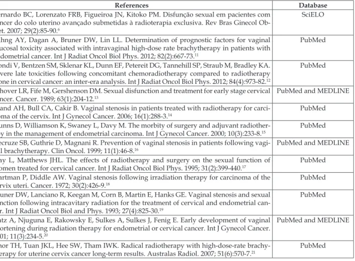
The articles included in this study (12 articles), which present the method of evaluation and
classi-ication of vaginal stenosis are presented in Table 1.
Table 1 – Publications which present the method of evaluation and classiication of vaginal stenosis
by database. Florianópolis, Santa Catarina, Brazil, 2013
References Database
Bernardo BC, Lorenzato FRB, Figueiroa JN, Kitoko PM. Disfunção sexual em pacientes com câncer do colo uterino avançado submetidas à radioterapia exclusiva. Rev Bras Ginecol Ob -stet. 2007; 29(2):85-90.6
SciELO
Bahng AY, Dagan A, Bruner DW, Lin LL. Determination of prognostic factors for vaginal mucosal toxicity associated with intravaginal high-dose rate brachytherapy in patients with endometrial cancer. Int J Radiat Oncol Biol Phys. 2012; 82(2):667-73.11
PubMed
Gondi V, Bentzen SM, Sklenar KL, Dunn EF, Petereit DG, Tannehill SP, Straub M, Bradley KA. Severe late toxicities following concomitant chemoradiotherapy compared to radiotherapy alone in cervical cancer: an inter-era analysis. Int J Radiat Oncol Biol Phys. 2012; 84(4):973-82.12
PubMed
Schover LR, Fife M, Gershenson DM. Sexual disfunction and treatment for early stage cervical cancer. Cancer. 1989; 63(1):204-12.13
PubMed and MEDLINE
Brand AH, Bull CA, Cakir B. Vaginal stenosis in patients treated with radiotherapy for carci-noma of the cervix. Int J Gynecol Cancer. 2006; 16(1):288-3.14
PubMed
Nunns D, Williamson K, Swaney L, Davy M. The morbity of surgery and adjuvant radiother-apy in the management of endometrial carcinoma. Int J Gynecol Cancer. 2000; 10(3):233-8.15
PubMed
Decruze SB, Guthrie D, Magnani R. Prevention of vaginal stenosis in patients following vagi -nal brachytherapy. Clin Oncol. 1999; 11(1):46-8.16
PubMed and MEDLINE
Flay L, Matthews JHL. The effects of radiotherapy and surgery on the sexual function of
women treated for cervical cancer. Int J Radiat Oncol Biol Phys. 1995; 31(2):399-440.17 PubMed
Hartman P, Diddle AW. Vaginal stenosis following irradiation therapy for carcinoma of the cervix uteri. Cancer. 1972; 30(2):426-9.18
PubMed
Bruner DW, Lanciano R, Keegan M, Corn B, Martin E, Hanks GE. Vaginal stenosis and sexual function following intracavitary radiation for the treatment of cervical and endometrial can-cer. Int J Radiat Oncol Biol and Phys. 1993; 27(4):825-30.19
PubMed
Katz A, Njuguna E, Rakowsky E, Sulkes A, Sulkes J, Fenig E. Early development of vaginal shortening during radiation therapy for endometrial or cervical cancer. Int J Gynecol Cancer. 2001; 11(3):234-5.20
PubMed and MEDLINE
Khor TH, Tuan JKL, Hee SW, Tham IWK. Radical radiotherapy with high-dose-rate brachy-therapy for uterine cervix cancer long-term results. Australas Radiol. 2007; 51(6):570-7.21
Evaluative method used, by study investigated
The study ‘Determination of prognostic factors
for vaginal mucosal toxicity associated with intrava -ginal high-dose rate brachytherapy in patients with endometrial cancer’11 considered vaginal stenosis to be the narrowing and/or shortening of the vagina, interfering with the use of internal sanitary
protec-tion, sexual activity or physical examinations. Ma -nifesting as vaginal dryness or dyspareunia, this has severe discomfort as a result, which was reported by the women. The method used for evaluating vaginal stenosis in this study was based on the Common Terminology Criteria for Adverse Events (CTCAE) Version4.0 - 2009, Version4.03 - 2010, published by
the National Cancer Institute, which classiies vagi -nal stenosis as:Grade 1: asymptomatic, mild short
-ening or narrowing of the vagina; Grade 2: vaginal
narrowing and/or shortening, not interfering with
the undertaking of the physical examination; Grade
3: vaginal narrowing and/or shortening interfering
with the use of tampons, sexual activity, or physical examination; Grade 4: unspeciied; Grade 5: death.22
The study ‘Severe late toxicities following
concomitant chemoradiotherapy compared to radiotherapy alone in cervical cancer: an inter-era analysis’,12 considered vaginal stenosis in the same way as the study cited above, and also used the criteria established in the Common Terminology Criteria for Adverse Events (CTCAE) Version 4.0 -
2009, Version4.03 – 2010,in order to establish the evaluative criteria for vaginal stenosis used in the investigation.22
The study ‘Disfunção sexual em mulheres com câncer do colo uterino avançado submetidas à radiote-rapia exclusiva’ (Sexual dysfunction in women with advanced cervical cancer who have undergone
exclusive radiotherapy),6 deined vaginal stenosis
as narrowing of the vaginal canal to the extent
that it was impossible to completely introduce the number 1 gynecological speculum through the
lower extremity of the vagina for the gynecological examination (the number 1 gynecological speculum, or size ‘small’, is approximately 110 mm on the longitudinal axis of its articulated elements, 27 mm maximum distal width, and 24 mm greatest proxi -mal width).6The methods indicated for evaluation
of vaginal stenosis found in these irst three studies
are described clearly, and in a way that is easy to understand and to undertake. However, there is no detailed description of the systematic stages for undertaking it.
In the study ‘Sexual disfunction and treatment
for early stage cervical cancer,13 the classiication
used was limited to the division of the stenosis into
‘mild’ or ‘severe’; in addition to this, a questionnaire
was used to be applied during the gynecological
examination, in which the mucosa and the size of the vagina were classiied as ‘normal’, ‘partially
changed’ or ‘severely changed’, and the pain dur-ing palpation was considered as ‘mild discomfort’,
‘severe’, or ‘absent’. It was observed in this classiica -tion that the descrip-tion of the technique used does not mention technical or objective detail allowing
the classiication of the characteristics evaluated.
The study ‘Vaginal stenosis in patients treated
with radiotherapy for carcinoma of the cervix’,14
classiied the compromising of the vaginal canal in
various grades: absence of stenosis - grade 1, partial
stenosis - grade 2 and total obliteration - grade 3;
in addition to this, the authors also considered the presence of severe complications associated with the tissue changes caused by the radiotherapy, such as ulcers and necrosis - grade 4, and vesical
and intestinal istulas - grade 5. It was observed that in this method there are no criteria for deining
stenosis of grades 1 and 2, but that grades 3 and 4
are easily identiied.
In the study ‘The morbity of surgery and ad-juvant radiotherapy in the management of endome-trial carcinoma’,15 the presence of vaginal stenosis
in the clinical examination was characterized by the inability to introduce two ingers into the vaginal canal, this test being undertaken by two examiners.
The technical description in this study is easy to understand and undertake. It is emphasized that the literature indicates that the vagina is highly distensible and generally allows the introduction
of two ingers for the internal examination, thus conirming that described by the authors.23
In the study ‘Prevention of vaginal stenosis in patients following vaginal brachytherapy’,16 the
pre-sence or abpre-sence of vaginal stenosis was identiied through the gynecological examination which com -pared the vaginal dimension before and one year after the conclusion of the intracavitary treatment. It is emphasized that small anatomical changes can
occur in women, principally with advancing age;
however, these manifestations may not compromise
the woman’s sex life and sexual health, that is to say,
small changes may not represent vaginal stenosis. The study ‘The effects of radiotherapy and
surgery on the sexual function of women treated
the vaginal stenosis – they merely relate the signs reported by the women to the results found in the
gynecological examination. The results in this stu -dy show that one third of the women included in
the study (ive women) experienced a sensation of
shortening and narrowing of the vagina and that
these women, in the physical examination, had a
vaginal length inferior to eight centimeters. It is emphasized that anatomical studies describe that the mean calculated for the length of the vagina is
from 7 to 9 centimeters and the width, from three
to four cm.23 This being the case, the length of 8 cm may be considered normal length, or within the
mean expected.
In the study ‘Vaginal stenosis following
irra-diation therapy for carcinoma of the cervix uteri’,18
the classiication of the vaginal stenosis was also established in grades; however, the authors limited
themselves to the possible obliteration of the vaginal canal in its length, with grade 1 being absence of
ste-nosis; grade 2 being the stenosis in the irst proximal
third of the vagina, and grade 3, the stenosis beyond
the irst third of the vagina, up to total obliteration. Thus, this method does not objectively deine how
to evaluate the degrees of the stenosis.
The study ‘Vaginal stenosis and sexual func -tion following intracavitary radia-tion for the treat-ment of cervical and endometrial cancer’19 classiied vaginal stenosis as vaginal length inferior to the
normal 8 – 9 cm. This technique has already been
presented above. The study reasserts that the length
of the vagina can oscillate between 7 to 9 cm.23 In the study ‘Early development of vaginal shortening during radiation therapy for endometrial or cervical cancer’,20 vaginal stenosis was evaluated through comparison and measuring of the diffe-rence between the distance from the superior edge
of the pubis and the apex of a cylinder introduced
into the vagina. For this evaluation, a radiological
examination was used, which was undertaken prior
to the treatment and after the second application of brachytherapy. However, vaginal stenosis, in gene-ral, is manifested at a late stage11 and the requesting
of the radiological examination may limit the action
of the nurse, as the prescription of the same is linked to the practice of medicine.
The study Radical radiotherapy with
high-dose-rate brachytherapy for uterine cervix cancer
- long-term results21 classiied vaginal stenosis as
severe (grade 1) or moderate (grade 2); however,
the way this evaluation was undertaken, and the criteria used, were not described.
Comparing the indings, it was observed that
there is diversity in evaluative and classiicatory
methods for vaginal stenosis, as well as a lack of standardization for the undertaking of the same.
This reafirms what was evidenced by the review
studies: Métodos avaliativos para estenose vaginal pós-radioterapia (Evaluative methods for post-radio-therapy vaginal stenosis)7 and ‘Preventing vaginal stenosis after brachytherapy for gynaecologial cancer: an overview of Australian practices’,9 cited in the introduction of this article.
One review study published in 2012 also as-serts that studies which evaluate
post-brachyther-apy vaginal toxicity and its determining clinical
factors are rare.11
In the light of these results, general constitut-ing elements were organized for the elaboration of
an instrument for the evaluation and classiication
of vaginal stenosis, to be applied by nurses
work-ing in the context of care for the woman who has
undergone vaginal brachytherapy, based on the
professional experience of this study’s authors, and
of the studies included in this research.
The deining of the evaluative and classiica -tory methods of vaginal stenosis could minimize risks and increase the safety and quality of life of the women, and also meet the professional needs.
General constituting elements for the
composition of an instrument for evaluating
and classifying vaginal stenosis
The data collection indicated for the nurse’s in-terview must investigate the patient’s questions and
the discomfort that she experiences; the frequency of sexual relations; the frequency of undertaking vagi
-nal physiotherapy; the presence of dificulties for
undertaking vaginal physiotherapy for maintaining
sexual relations; the presence of pain in the vagina and its intensity and characteristics; the undertaking of the gynecological examination after terminating brachytherapy; the presence of dificulties for un
-dertaking the gynecological examination; the need
for the use of vaginal lubricant and the presence of vaginal bleeding and/or secretion.
The data collection indicated for the
gyne-cological examination must follow the technical recommendations for this purpose;24 whether the
exam was undertaken, the size of the speculum
used, and characteristics of the vaginal canal and of
the cervix; and reasons which could have impeded the undertaking of the gynecological examination must be recorded; if indicated, the pap smear must
undertake the gynecological examination, that is to say, if the presence of ulcers, necrosis, or istulas in the vaginal canal should be identiied, the patient
must be referred for medical evaluation. Finally, the nurse must record the decision-making, that is, the nursing interventions undertaken.
For classification of the vaginal stenosis,
the following classiication is proposed: Grade 0: asymptomatic woman; Grade 1: the woman who
mentions some vaginal change or discomfort, but which does not impede the use of internal sanitary
protection, sexual activity or the gynecological ex
-amination; Grade 2: a woman who presents vaginal
shortening that partially interferes in the use of
tam-pons, sexual activity and in the undertaking of the gynecological examination; Grade 3: a woman who presents total constriction of the vagina, identiied
in the visual inspection during the undertaking of
the gynecological examination, and that makes it impossible to undertake the gynecological examina
-tion or sexual activity; Grade 4: the woman presents ulcers or necrosis in the vaginal canal; Grade 5: the woman presents vesical and/or intestinal istulas.
Vaginal stenosis induced by radiation can be prevented by the undertaking of vaginal
physiother-apy or by maintaining sexual relations as advised
during the nursing consultation, before and during the undertaking of HDRB.
Women who were only advised regarding
the importance of sexual relations present a greater
incidence of vaginal stenosis when compared with women who undertook vaginal physiotherapy,9 thus evidencing the importance of this resource for the preventive intervention.
However, the choices of each woman and the responses of the organism can be diverse. It falls, therefore, to the nurses and doctors to evaluate the presence or absence of vaginal stenosis to maintain the preventive care, but also the interventions in the light of the complications which may be manifested.
For this to happen, the deinition of the evaluative
method guides the professional to identifying the possible physiological changes and also serves as
guidance for deining the necessary interventions.
CONCLUSION
Considering the magnitude of cervical cancer, the importance of women’s health and the need for evaluation of the nursing care given to women during HDRB, one can assert the relevance of this research and the construction of the knowledge produced.
In the identiication of the methods used for the veriication of vaginal stenosis in women fol
-lowing vaginal brachytherapy, the indings were diverse and divergent. This inding is similar to
that of other studies which assert the inconsistency
of the criteria for veriication of vaginal stenosis.
The differences found support the elaboration of a guidance instrument to be applied by nurses.
The establishing of a speciic instrument for nurses standardizes the technique for veriication
of vaginal stenosis, contributes to the evaluation of the nursing care provided, and assists in the care to be instituted for self-care with or without the pres-ence of vaginal stenosis in women post-HDRB. The instrument will be validated by a subsequent study
by judges who are experts in the area in question.
REFERENCES
1. Instituto Nacional de Câncer José Alencar Gomes da Silva. Coordenação de Prevenção e Vigilância Estimativa 2014: Incidência de Câncer no Brasil. Rio de Janeiro (RJ): INCA; 2014.
2. Instituto Nacional de Câncer José Alencar Gomes da
Silva. Programa Nacional de Controle do Câncer do colo de Útero. Rio de Janeiro (RJ): INCA; 2012. 3. Salvajoli JV, Souhami L, Faria SL. Radioterapia em
oncologia. São Paulo (SP): Atheneu; 2013.
4. Eifel, PJ. High-dose-rate brachytherapy for carcinoma of the cervix: high tech or high risk? Int J Radiat Oncol Biol Phys [internet]. 1992 [cited 2014 Out 27]; 24:383-6. Available from: http://ac.els-cdn. com/036030169290696F/1-s2.0-036030169290696F-main.pdf?_tid=07f4546c-c26d-11e4-9a32-00000aab0 f02&acdnat=1425473606_4288606cbe05dee2553205e e6a8f4217
5. Esteve SCB, Oliveira OCZ, Feijó LFA. Braquiterapia de alta taxa e dose no Brasil. Radiol Bras [internet]. 2004 [cited 2014 Out 27]; 37(5):337-41 Available from: http://www.scielo.br/pdf/rb/v37n5/22113.pdf 6. Bernardo BC, Lorenzato FRB, Figueiroa JN, Kitoko
PM. Disfunção sexual em pacientes com câncer do colo uterino avançado submetidas à radioterapia exclusiva. Rev Bras Ginecol Obstet [internet]. 2007 [cited 2014 Out 27]; 29(2):85-90. Available from: http://www. scielo.br/pdf/rbgo/v29n2/05.pdf
7. Silva MPPS, Gannuny CS, Aiello NA, Higinio MAR, Ferreira NO, Oliveira MMF. Métodos avaliativos para estenose vaginal pós-radioterapia. Rev Bras Cancerol [internet]. 2010 [cited 2014 Out 27]; 56(1):71-83. Available from: http://www.inca.gov. br/rbc/n_56/v01/pdf/10_revisao_de_literatura_ metodos_avaliativos_estenose_vaginal.pdf
9. Lancaster L. Preventing vaginal stenosis after brachytherapy for gynecological cancer: an overview of Australian practices. Eur J Oncol Nurs [internet]. 2004 [cited 2014 Out 27]; 8(1):30-9. Available from; http://www.sciencedirect.com/science/article/pii/ S1462388903000590
10. Muniz RM, Zago MMF, Schwartz E. As teias da sobrevivência oncológica: com a vida de novo. Texto Contexto Enferm [internet]. 2009 [cited 2014 Out 27]; 18(1):25-32. Available from: http://www. scielo.br/scielo.php?script=sci_arttext&pid=S0104-07072009000100003&lng=pt
11. Bahng AY, Dagan A, Bruner DW, Lin LL. Determination of prognostic factors for vaginal mucosal toxicity associated with intravaginal high-dose rate brachytherapy in patients with endometrial cancer. Int J Radiat Oncol Biol Phys [internet]. 2012 [cited 2014 Out 27]; 82(2):667-73. Available from: http://www.sciencedirect.com/science/article/pii/ S0360301610036515
12. Gondi V, Bentzen SM, Sklenar KL, Dunn EF, Petereit
DG, Tannehill SP, Straub M, Bradley KA. Severe late toxicities following concomitant chemoradiotherapy compared to radiotherapy alone in cervical cancer: an inter-era analysis. Int J Radiat Oncol Biol Phys [internet]. 2012 [cited 2014 Out 2014]; 84(4):973-82. Available from: http://www.sciencedirect.com/ science/article/pii/S036030161200137X
13. Schover LR, Fife M, Gershenson DM. Sexual
disfunction and treatment for early stage cervical cancer. Cancer. 1989; 63(1):204-12.
14. Brand AH, Bull CA, Cakir B. Vaginal stenosis in patients treated with radiotherapy for carcinoma of the cervix. Int J Gynecol Cancer. 2006; 16(1):288-3. 15. Nunns D, Williamson K, Swaney L, Davy M. The
morbity of surgery and adjuvant radiotherapy in the management of endometrial carcinoma. Int J Gynecol Cancer. 2000; 10(3):233-8.
16. Decruze SB, Guthrie D, Magnani R. Prevention
of vaginal stenosis in pacients following vaginal brachytherapy. Clin Oncol. 1999; 11(1):46-8.
17. Flay L, Matthews JHL. The effects of raditherapy and
surgery on the sexual function of women treated for cervical câncer. Int J Radiat Oncol Biol Phys. 1995; 31(2):399-440.
18. Hartman P, Diddle AW. Vaginal stenosis following irradiation therapy for carcinoma of the cervix uteri. Cancer. 1972; 30(2):426-9.
19. Bruner DW, Lanciano R, Keegan M, Corn B, Martin
E, Hanks GE. Vaginal stenosis and sexual function following intracavitary radiation for the treatment of cervical and endometrial cancer. Int J Radiat Oncol Biol and Phys. 1993; 27(4):825-30.
20. Katz A, Njuguna E, Rakowsky E, Sulkes A, Sulkes J, Fenig E. Early development of vaginal shortening during radiation therapy for endometrial or cervical câncer. Int J Gynecol Cancer. 2001; 11(3):234-5. 21. Khor TH, Tuan JKL, Hee SW, Tham IWK. Radical
radiotherapy with high-dose-rate brachytherapy for uterine cervix cancer long-term results. Australas Radiol [internet]. 2007 [cited 2014 Out 27]; 51(6):570-7. Available from: http://onlinelibrary.wiley.com/ doi/10.1111/j.1440-1673.2007.01895.x/pdf
22. National Cancer Institute. National Institutes of
Health. U.S. Department of Health and Human Services. Common Terminology Criteria for Adverse Events (CTCAE) Version 4.0: 2009 May 28, v4.03: 2010 June 14 [internet]. U.S.A: National Cancer Institute; 2010 [cited 2014 Out 27]. Available from: http://evs. nci.nih.gov/ftp1/CTCAE/CTCAE_4.03_2010-06-14_ QuickReference_8.5x11.pdf
23. Moore KL, Dally AF, Agur AMR. Anatomia orientada para a clínica. Rio de Janeiro (RJ): Guanabara Koogan; 2012.
24. Ministério da Saúde (BR). Secretaria de Atenção à
Saúde. Departamento de Atenção Básica. Controle dos cânceres do colo do útero e da mama. 2ª ed. Brasília (DF): Editora do Ministério da Saúde, 2013.
Correspondence: Patrícia Ilha Rua Europa, 228, ap. 942
88036-135 – Trindade, Florianópolis, SC, Brazil Email: [email protected]