Fisioter. Mov., Curitiba, v. 30, n. 3, p. 443-451, Jul./Sep. 2017 Licenciado sob uma Licença Creative Commons DOI: http://dx.doi.org/10.1590/1980-5918.030.003.AO02
Therapeutic ultrasound associated with copaiba oil reduces pain
and improves range of motion in patients with knee osteoarthritis
Ultrassom terapêutico associado ao óleo de copaíba
reduz a dor e melhora amplitude de movimento
de pacientes com osteoartrite de joelho
Luis Fernando Sousa Filho, Jessica Batista de Souza, Grace Kelly Melo de Almeida, Itamar Couto Guedes de Jesus, Evaleide Diniz de Oliveira*
* Universidade Federal de Sergipe (UFS), Centro de Ciências Biológicas e da Saúde, São Cristovão, SE, Brazil
[R]
Abstract
Introduction: Osteoarthritis is a disease that affects millions of Brazilians.Therapeutic ultrasound has been
used in its treatment, either alone or associated with drugs. Objective: The aim of this study was to evaluate the effects of ultrasound (US) associated with Copaiba oil (CO) on knee osteoarthritis. Methods: Patients were divided into three different groups: US, US+CO, CO.Ten treatment sessions were held twice a week, 30 minutes each.Pain intensity was assessed through the Visual Analog Scale (VAS) and Range of Motion (ROM) by goniometry, and muscle strength was assessed by means of the Medical Research Council Scale. Statistical
analysis was performed by Cohen’s d test, student’s t test and ANOVA, considering p<0.05 as significant.
Results: Pain reduced in all groups.The US+CO group (d = –3.50) presented larger effect size when
com-pared to the other groups. Regarding ROM, the largest effect size was observed in the US+CO group for flexion (d = 0.86) and extension (d = 0.97) in comparison with the remainder groups.Muscle strength increased in the US (d = 1.54) and US+CO (d = 1.60) groups for flexion.Regarding extension, the US group presented the
* LFSF: BS, e-mail: [email protected]
JBS: BS, e-mail: [email protected]
444
largest effect size (d = 1.80). Conclusion: Therapeutic ultrasound associated with copaiba oil is a practical
and effective therapy for the treatment of inflammatory diseases such as osteoarthritis.
Keywords: Ultrasound. Osteoarthritis. Pain.
Resumo
Introdução: A osteoartrite é uma doença que afeta milhões de brasileiros. O ultrassom terapêutico tem sido utilizado em seu tratamento tanto sozinho, quanto associado a fármacos. Objetivo: Avaliar o efeito do ultras-som (US) associado ao óleo de copaíba (OC) em pacientes com osteoartrite de joelho. Métodos: Os pacientes foram distribuídos em 3 grupos distintos: US, US+OC e OC. Foram realizadas 10 sessões de tratamento, duas vezes por semana durante 30 minutos. A intensidade da dor foi avaliada pela Escala Visual Analógica da Dor (EVA), amplitude de movimento ADM pela goniometria e força muscular pelo Score Medical Research Council. A análise estatística foi feita pelo Teste T de Student e ANOVA e a magnitude do efeito (d), considerando p<0,05 como valores significativos. Resultados: A dor foi atenuada em todos os grupos, sendo apresentada uma mag-nitude maior do efeito para o grupo US+OC (d = –3,50) quando comparado aos demais grupos. Em relação a ADM a magnitude do efeito foi maior no grupo US+OC (d= 0,86) para a flexão e extensão (d = 0,97) quando comparados com os outros grupos. Na variável força muscular os grupos US (d= 1,54) e US+OC (d = 1,60) foram mais eficazes no movimento de flexão e na extensão, o grupo US exibiu o maior tamanho de efeito (d = 1,80) quando comparados aos demais grupos. Conclusão: O ultrassom terapêutico associado ao óleo de copaíba é uma terapia efetiva e prática para o tratamento de doenças inflamatórias, tais como a osteoartrite.
Palavras-chave: Ultrassom. Osteoartrite. Dor.
Introduction
Osteoarthritis (OA) is a degenerative joint dis-ease that affects nearly 10 million Brazilians, and this figure is expected to reach 30 million in 2020 (1). The potential of osteoarthritis for promoting dis-ability is worth of notice. According to the Brazilian Rheumatology Society, osteoarthritis accounts for 7.5% of medical leaves, and it isranked fourth in dis -eases that lead to retirement, with 6.2% (2).
Osteoarthritis is a clinical joint pain syndrome followed by different degrees of functional limita-tion and reduced quality of life. It is considered to be a multi-factor disorder with no defined primary cause that, after being developed, can manifest in the form of edema to joint deterioration (3). Some factors can contribute to the disease development, such as obesity and advanced age (4).
Up to now, there is no therapy to reverse changes caused by this degenerative disease. The therapy’s main objectives are to reduce pain and improve the function of joints affected. According to the American Rheumatology College (5) the main therapies applied are pharmacological therapies, non-pharmacological
therapies or alternative therapies (including physical exercise and body masscontrol) and, for more critical cases, surgery.
Therapeutic ultrasound (US) is an alternative therapy commonly used by physiotherapists, and consists in applying ultrasonic waves that, through their vibrating effect, promote cell changes that result in alterations of the ionic gradient and acceleration of the tissue repairing process (6). A previous study showed that US has worked favorably in the therapy of myofascial pain (7), subacromial impact syndrome (8), lesions of soft tissues (9) and shoulder tendinitis (10).
445
Methods
This is a randomized study developed at the Kinesiotherapy Laboratory of the Physiotherapy Department, at the Sergipe Federal University (UFS). Treatment groups were made up of 90 women — 30 in each group — with clinical diagnosis of knee osteo -arthritis (ICD - M.17) grade II according to Ahlback's (28) classification, with inflammation in sub-acute stage, ambulation conditions and stable hemodynam-ic state. All patients were registered on the waiting list for rheumatologic care at the UFS teaching hos-pital outpatient unit, with ages ranging from 45 to 75 years old. No patient quit the proposed therapies.
Patients were randomly divided into three groups: ultrasound group (US); ultrasound group associated with Copaíba oil (US+CO) and a Copaíba oil group (CO). The randomized sequence was generated using a computer. Randomization occurred following the order of inclusion of each patient in the study, accord-ing to the list generated by a computer before startaccord-ing the study. The randomization block was created to generate sequences of allocation and ensure balance of participants in each group at any time during the study. After each block, the number of participants in each group would be equivalent, in the 1:1 ratio.
Total treatment was carried out in ten sessions delivered twice a week, 30 minutes each. All partici-pants were given a free and informed consent form for analysis and, if they agreed with it, they should sign the form and become volunteers of the study (CAAE-0004.0.107.000-09).
The therapeutic effect of Copaíba oil was evalu-ated in groups, as well as if the oil effect was opti-mized after using ultrasound. Patients in the US group were subject to the application of 20% pulsed ultrasound previously calibrated with intensity of 0.8 w/cm² (SATP), transducer frequency of 1 MHz (Ibramed, Sonopulse model) for 8 minutes followed with massage therapy (circular movements, with light pressure) with mineral oil for five minutes on the knee. Patients in the US+CO group were sub-ject to the application of pulsed ultrasound, in the same standards as the US group, followed by mas-sage therapy with Copaíba oil (100%, 0.3mL) for five minutes on the knee. The CO group patients, in turn, were treated only with massage therapy for five minutes with Copaíba oil. After massage (cir -cular movements with light pressure) patients from pain and function of patients with knee osteoarthritis,
although the size of such effects remains unclear. The oldest version had concluded that ultrasound benefits were not higher than those of placebo (14).
In addition to its conventional use, in the last few decades, US has been used for physical therapies to promote cutaneous permeation of medicines, fa-cilitating cutaneous absorption (15). Therefore, the topical application of drugs on the outer layer of the skin is directed by the US to the underlying tissues, increasing local concentration of the drug (16). Kozanoglu et al. (17) compared the effects of therapeutic US associated with Ibuprofen and the conventional US. They concluded that although both modalities have been efficacious for the treatment of patients with osteoarthritis, improvement with US associated with Ibuprofen was not better than that with the conventional US. In contrast, Toopchizadeh et al. (18) associated US with Dexamethasone and reached better results than with US alone on pain reduction and function improvement.
Ultrasound is usually associated with anti-inflammatory and anesthetic drugs (19). Copaíba oil (CO) is a Brazilian typical natural product with powerful anti-inflammatory activity (20, 21). It has been used in popular medicine for centuries and is one of the main Amazonian products traded in the United States and Europe (22, 23). In addition to being anti-inflammatory, Copaíba oil has antimicro -bial (24), antinociceptive (25), antitumor (26) and antiseptic (27) properties. Most of these properties were discovered through in vitro experimentations. Little is known about the effect of Copaíba oil on humans, although it is commonly used to treat mus-culoskeletal disorders.
446
intra-group evaluations of all variables. ANOVA one-way (post-Tukey’s test) was used for intergroup eval-uations. The d effect, or Cohen’s d, was calculated to evaluate treatments’ effect size. Effect d corresponds to the standard difference among the means of the compared groups, divided by the standard deviation of the comparison. According to Cohen, values of 0.2, 0.5 and 0.8 stand for small, medium or large effect, respectively (33). All analyses used the Graph Pad Prism (version 5.1) software considering significance level of 95% (p<0.05) for all tests.
Results
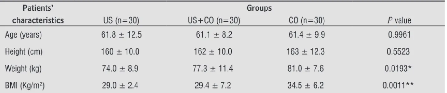
Age, height, weight and body mass index (BMI) of patients in different groups were measured and compared to one another (one-way ANOVA). The CO group (81 ± 7.6 Kg) presented a statistically significant difference when compared against the US group (74 ± 8.9 Kg) regarding the variable of weight with p = 0.0193. Regarding BMI, the CO group (34.5 ± 6.2 Kg/m²) was statistically different from the US group (29 ± 2.4 Kg/m²) and from the US+CO group (29.4 ± 7.2 Kg/m²), with p = 0.0011. The remainder variables did not present significant differences as shown in Table 1.
all groups were subject to assisted, active-assisted and resisted, free-active kinesiotherapy to supple-ment treatsupple-ment through stretching, strengthening and muscle proprioception. Exercises were the same in the three groups. Kinesiotherapy was included to ensure proper therapy to all patients, as consolidated in scientific literature for the pathology in question, since in our study hypothesis ultrasound associated with Copaíba oil was in experimentation with no as -surance of its efficacy.
The variables investigated in the study were evalu-ated in the first and tenth treatment session. Pain intensity was assessed using the Pain Visual Analog Scale (VAS) (29). Patients were instructed to mark their level of pain on the scale. Range of motion was measured using a goniometer. Goniometry is vali-dated and reliable for the measurement of knee joint angles (30). The muscle strength degree was evalu-ated through the Medical Research Council Scale. The examiner’s subjective perception was based on a scale ranging from 0 to 5, where 0, 1 and 2 degrees are tested in a position in favor of gravity. All the re-mainder degrees are tested in a position opposite to the force of gravity (31, 32).
The results achieved were statistically analyzed using Student’s t test and the Analysis of Variance (ANOVA), being represented by means and stan-dard deviation (SD) values. The t test was used for
size of the US+CO (d= –3.50) group was larger when compared to that of the US group (d= –2.90) and the CO group (c= –1.50).
The variable pain was assessed through VAS scores. Table 2 shows that VAS scores were signifi -cantly reduced after therapies (p<0.0001). The effect
Table 1 - Comparison of age, height, weght and BMI of patients in different groups
Patients’ Groups
characteristics US (n=30) US+CO (n=30) CO (n=30) P value
Age (years) 61.8 ± 12.5 61.1 ± 8.2 61.4 ± 9.9 0.9961
Height (cm) 160 ± 10.0 162 ± 10.0 163 ± 12.3 0.5523
Weight (kg) 74.0 ± 8.9 77.3 ± 11.4 81.0 ± 7.6 0.0193*
BMI (Kg/m²) 29.0 ± 2.4 29.4 ± 7.2 34.5 ± 6.2 0.0011**
447
Table 2 - Differences between pain intensity before and after therapy
VASScores
Groups Before After P value
US (n=30) 3.1 ± 1.0 0.8 ± 0.5 –2.90 <0.0001***
US + CO (n=30) 5.4 ± 1.9 0.4 ± 0.5 –3.50 <0.0001***
CO (n=30) 4.6 ± 2.2 1.2 ± 2.2 –1.50 <0.0001***
Note: Results are expresses through mean and standard deviation. D effect represents the effect size of each group (n*30). VAS = Visual Analog Scale US = therapeutic ultrasound. US+CO = therapeutic ultrasound + Copaíba oil. CO = Copaíba oil.
Table 4 - Muscle strength assessment. Comparison of muscle strength degrees of patients during knee flexion and extension movements, according with the Medical Research Council Scale
Muscle strength degrees for flexion
Groups Before After P value
US (n=30) 3.4 ± 0.5 4.1 ± 0.4 1.54 <0.0001***
US + CO (n=30) 3.7 ± 0.5 4.5 ± 0.5 1.60 <0.0001***
CO (n=30) 3.9 ± 0.3 4.1 ± 0.6 0.42 0.1079
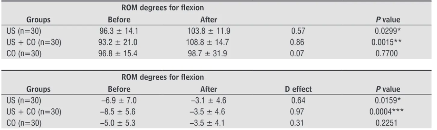
Table 3 - Range of motion assessment. Comparison of range of motion degrees of patients before and after therapy for knee flexion and extension movements
ROM degrees for flexion
Groups Before After P value
US (n=30) 96.3 ± 14.1 103.8 ± 11.9 0.57 0.0299*
US + CO (n=30) 93.2 ± 21.0 108.8 ± 14.7 0.86 0.0015**
CO (n=30) 96.8 ± 15.4 98.7 ± 31.9 0.07 0.7700
ROM degrees for flexion
Groups Before After D effect P value
US (n=30) –6.9 ± 7.0 –3.1 ± 4.6 0.64 0.0159*
US + CO (n=30) –8.5 ± 5.6 –3.5 ± 4.6 0.97 0.0004***
CO (n=30) –5.0 ± 5.3 –3.5 ± 4.1 0.31 0.2251
Note: Results are expresses through means and standard deviation. D effect represents the effect size of each group (n*30). ROM = range of motion. US = therapeutic ultrasound. US+CO = therapeutic ultrasound + Copaíba oil. CO = Copaíba oil.
Range of motion was evaluated by goniometry on joint movements of knee flexion and extension during the 1st and 10th therapy sessions. In the flexion movement, the US+CO (d=0.86) group pre -sented the largest size effect when compared to the US group (d=0.57) and the CO group (d=0.07). The US (p=0.0299) and the US+CO (p=0.0015) groups
presented significant differences before and after treatment (Table 3). Extension movement present -ed similar results. The US (p=0.0159) and US+CO (p= 0.0004) groups presented a statistical intra-group difference, where the US+CO intra-group (d= 0.97) presented the largest size effect.
The muscle strength degree was assessed through the Medical Research Council Scale, in knee flexion and extension movements. The US (d=1.54) and US+CO (d=1.60) groups provided better response to the therapy with size effects similar in flexion
448
Muscle strength degrees for flexion
Groups Before After P value
US (n=30) 3.6 ± 0.5 4.5 ± 0.5 1.80 <0.0001***
US + CO (n=30) 4.0 ± 0.4 4.5 ± 0.5 1.10 <0.0001***
CO (n=30) 3.9 ± 0.3 4.3 ± 0.7 0.74 0.0056**
Note: Results are expresses through mean and standard deviation. D effect represents the effect size of each group (n*30). US = therapeutic ultrasound. US+CO = therapeutic ultrasound + Copaíba oil. CO = Copaíba oil.
Discussion
Osteoarthritis therapy aims to reduce pain and improve patients’ functional state. Therapeutic ul-trasound has been used in osteoarthritis therapy but studies on its efficacy are controversial. This study analyzed the effect of ultrasound associated with Copaíba oil in patients with knee osteoarthritis grade II, comparing it before and after therapy, and found positive results regarding pain, range of motion and muscle strength.
Obesity, advanced age, overweight and female sex are considered risk factors to develop osteoarthritis (34). In this study, all patients were women, with age ranging from 45 to 75 years old, mean of 61.4 ± 10.2 years, and no significant differences between groups (p=0.9961). As regards body weight, patients in the CO group (81±7.6 Kg) reported having more weight than those in the remainder groups, with a significant difference (p=0.0193). Likewise, the BMI of patients in the CO group (34.5 ± 6.2) was significantly higher than that for the other groups (p=0.0011). Although a previous study shows that obese individuals pres-ent sharper degeneration than individuals of normal weight, all patients part of the study presented os-teoarthritis grade II (35).
Therapeutic ultrasound is one of the main non-pharmacological resources used to control pain. However, there are divergences about its efficacy (36). Ulus et al. (37) found no significant differences between the use of US and placebo in the VAS scores for patients with knee osteoarthritis. In our study, US reduced pain after therapy; however, effect presented larger size when compared with the use associated with Copaíba oil. Luk surapan et al. (38) found similar results. A gel traditionally used in association with US to treat musculoskeletal disorders proved to be more efficient to reduce pain than US alone. Copaíba oil pre -sented anti-inflammatory and analgesic properties in in vitro experiences; however, there is no evidence of such effects on patients with osteoarthritis.
The association of US and active substances to re-lieve pain and improve functionality is used in clinical practices, but there is little scientific evidence sup -porting this therapy (39). Anti-inflammatory sub -stances like Diclofenac and Ibuprofen are largely used. Rosim et al. (40) showed that previous use of US increased transdermal penetration of Diclofenac gel in healthy individuals. In patients with knee os-teoarthritis, the use of US associated with Diclofenac reduced pain and improved ROM in a more significant way than US alone (41). In our study, ultrasound com-bined with Copaíba oil presented larger effect than ultrasound or Copaíba oil alone to enhance range of motion both in flexion and extension.
Many factors are related to mobility deficit in knee osteoarthritis, including knee pain, strength of the quadriceps and age (42). Cubukcu et al. (43) assessed 114 patients with knee osteoarthritis and concluded that pain, rigidity and duration of the disease affect the patients’ functional incapacity. Observing our results, pain reduction and expan -sion of ROM were proportional among groups, ex -cept for the CO group that reported no significant difference between ROM degree before and after therapy. Besides being a factor that restrains the range of motion, pain may lead patients to develop kinesiophobia (44). Therefore, pain reduction may have favored increased ROM. The result for the CO group should be justified by the high BMI of patients in this group. Holla et al. (45) report that in addition to joint degeneration, pain, rigidity and male gender, BMI is also associated with range of motion degrees. Fat accumulation can limit ROM.
449
References
1. Coimbra IB, Rezende UM, Plapler PG. Osteoartrite
(artrose) — cenário atual e tendências no Brasil. São Paulo: Limay; 2012.
2. Seda H, Seda AC. Osteoartrite (Artrose). 2008 [cited 2014 Aug 10]. Available from: http://tinyurl.com/
jhz8few.
3. Rosis, RC, Massabki OS, Kairalla M. Osteoartrite: ava-liação clínica e epidemiológica de pacientes idosos em instituição de longa permanência. Rev. Bras. Clin.
Med. 2010;8(2):101-8.
4. Blagojevic M, Jinks C, Jeffery A, Jordan KP. Risk factors for onset of osteoarthritis of the knee in older adults: a systematic review and meta-analysis. Osteoarthritis
Cartilage. 2010;18(1):24-33.
5. Hochberg MC, Altman RD, April KT, Benkhalti M, Guy-att G, McGowan J, et al. American College of
Rheu-matology 2012 recommendations for the use of non
pharmacologic and pharmacologic therapies in os-teoarthritis of the hand, hip, and knee. Arthritis Care
Res (Hoboken). 2012;64(4):465-74.
6. ter Haar G. Therapeutic ultrasound. Eur J Ultrasound.
1999;9(1):3-9.
7. Ilter L, Dilek B, Batmaz I, Ulu MA, Sariyildiz MA, Nas
K, et al. Efficacy of Pulsed and Continuous Therapeu -tic Ultrasound in Myofascial Pain Syndrome: A Ran-domized Controlled Study. Am J Phys Med Rehabil.
2015;94(7):547-54.
8. Yildirim MA, Ones K, Celik EC. Comparision of Ultra-sound Therapy of Various Durations in the Treatment of Subacromial Impingement Syndrome. J Phys Ther
Sci. 2013;25(9):1151-4.
9. Binder A, Hodge G, Greenwood AM, Hazleman BL, Page Thomas DP. Is therapeutic ultrasound effective in treating soft tissue lesions? Br Med J (Clin Res Ed).
1985;290(6467):512-4.
10. Ebenbichler GR, Erdogmus CB, Resch KL, Funovics MA, Kainberger F, Barisani G, et al. Ultrasound therapy
for calcific tendinitis of the shoulder. N Engl J Med. 1999;340(20):1533-8.
11. Langer MD, Levine V, Taggart R, Lewis GK, Hernandez L, Ortiz R. Pilot clinical studies of long duration, low inten-sity therapeutic ultrasound for osteoarthritis. Proc IEEE
Annu Northeast Bioeng Conf. 2014;2014. pii: 14789673.
muscle section area (47). Our results show that ultra -sound alone and ultra-sound associated with Copaíba oil have significantly increased muscle strength of knee flexor and extensor muscles, whereas Copaíba oil alone presented no changes. Patients tend to avoid movements when they feel pain, and this causes muscle damages. Pisters et al. (49) had previously shown that fearing movements is common among pa-tients with osteoarthritis, leading to reduced muscle strength. Reduction of pain in the respective groups tend to reduce fear of movement as happened with the US and US+CO groups and, therefore, increase muscle strength.
Obesity is related with muscle weakness in pa-tients with osteoarthritis (50). The response of patients treated exclusively with Copaíba oil was weaker in all variables analyzed, including muscle strength. This result could be explained by the high BMI value of patients in this group, when compared against other groups. Conroy et al. (51) observed that patients with knee osteoarthritis, with or without pain, high BMI and high quantity of intramuscular fat show weaker torque specific to the quadriceps against individuals without osteoarthritis.
This study aimed to evaluate the effects of US associated with natural oil in patients with knee osteoarthritis and found positive results on pain, range of motion and muscle strength, in line with findings previously published. However, this study had some limitations: patients and researchers were not blind; effects observed were analyzed only before and immediately after therapy with no long-term monitoring. An important issue was the size of the sample used, i.e., 30 patients in each group. Samples with between 30 and 40 partici-pants are considered large enough to waive the use of normality tests, since these tend to be normally distributed (52).
Conclusion
450
23. Veiga Junior VF, Pinto AC.O gênero copaifera L. Quím
Nova. 2002;25(2):273-86.
24. Santos AO, Ueda-Nakamura T, Dias Filho BP, Veiga Jr VF, Pinto AC, Nakamura CV. Antimicrobial activity of Brazilian copaiba oil sobtained from different spe-cies of the Copaifera genus. Mem Inst Oswaldo Cruz.
2008;103(3):277-81.
25. Gomes NM, Rezende CM, Fontes SP, Matheus ME, Fer-nandes PD. Antinociceptive activity of Amazonian
Co-paiba oils. J Ethnopharmacol. 2007;109(3):486-92. 26. Gomes NM, Rezende CM, Fontes SP,Hovell AMC,
Landgraf RG, Matheus ME, et al. Antineoplasic activ-ity of Copaifera multijuga oil and fractions against ascitic and solidehrlich tumor. J. Ethnopharmacol.
2008;119(1):179-84.
27. Botelho NM, Silveira EL, Lopes LN, Santos FA, Teix -eira RK, Silva TT. Copaiba oil effect under diferente path ways in mice subject edtosepsis. Acta Cir Bras.
2014;29(8):528-31.
28. Galli M, De Santis V, Tafuro L. Reliability of the Ahlbäck
classification of knee osteoarthritis. Osteoarthritis Cartilage. 2003;11(8):580-4.
29. Mob O, Ra A, Ro A. Reliability and concurrent validit
yof visual analogue scaleand modified verbal rat -ing scale of pain assessment in adult patients with knee osteoarthritis in Nigeria. S Afr J Physiother.
2003;59(2):12-5.
30. Gogia PP, Braatz JH, Rose SJ, Norton BJ. Reliability and validity of goniometric measurements at the knee.
Phys Ther. 1987;67(2):192-5.
31. Medical Research Council. Aids to the examination
of theperipheral nervous system. Memorandum No.
45 (superseding War Memorandum No. 7). London: HerMajesty'sStationery Office; 1981.
32. Paternostro-Sluga T, Grim-Stieger M, Posch M, Schuhfried O, Vacariu G, Mittermaier C, et al. Reli-ability and validity of the Medical Research Council
(ROMC) scaleand a modified scale for testing muscles
trength in patients with radial palsy. J Rehabil Med.
2008;40(8):665-71.
33. Cohen J. Statistical power analysis for the behavioral
sciences (2. Auflage). Hillsdale, NJ: Erlbaum, 1988. 12. Ulus Y, Tander B, Akyol Y, Durmus D, Buyukakıncak
O, Gul U, et al. Therapeutic ultrasound versus sham ultrasound for the management of patients with knee osteoarthritis: a randomized double-blind controlled
clinical study. Int J Rheum Dis. 2012;15(2):197-206.
13. Rutjes AW, Nüesch E, Sterchi R, Jüni P. Therapeutic ultrasound for osteoarthritis of the knee or hip.
Co-chrane Database Syst Rev. 2010;(1):CD003132.
14. Welch V, Brosseau L, Peterson J, Shea B, Tugwell P, Wells G. Therapeutic ultrasound for osteoar-thritis of the knee. Cochrane Database Syst Rev.
2001;(3):CD003132.
15. Pires-de-Campos MSM, Polacow MLO, Granzotto TM,
Spadari-Bratfisch RC, Leonardi GR, Grassi-Kassisse DM. Influence of the ultrasound in cutaneous per -meation of the caffeine: in vitro study. Pharmacology
online. 2007;1:477-86.
16. Cagnie B, Vinck E, Vanderstraeten G. Phonoforesis versus topical application of Ketoprofen: compari-son between tissue and plasma levels. Phys Ther.
2003;83(8):707-12.
17. Kozanoglu E, Basaran S, Guzel R, Guler-Uysal F. Short
term efficacy of ibuprofen phonophoresis versus con -tinuous ultrasound therapy in knee osteoarthritis.
Swiss Med Wkly. 2003;133(23-24):333-8.
18. Toopchizadeh V, Javadi R, Sadat BE. Therapeutic
Ef-ficacy of Dexamethasone Phonophoresis on Symp -tomatic Knee Osteoarthritis in Elderly Women. Int J
Women's Health Reproduction Sci. 2014;2(3):168-77. 19. Byl NN. The use of ultrasound as an enhancer for
transcutaneous drug delivery: phonophoresis. Phys
Ther. 1995;75(6):539-53.
20. Viriato EP, Bianchetti ES, Santos KC, Vaz AF, Campos RMV, Pereira AP, et al. Study of high dilutions of
co-paíba oil oninflammatory process. Int J High Dilution Res. 2009;8(26):9-14.
21. Dias DS, Fontes LB, Crotti AE, Aarestrup BJ, Aarestrup FM, Silva Filho AA, et al. Copaiba oil suppresses
in-flammatory cytokines in splenocytesof C57Bl/6 mice induced with experimental autoimmune encephalo
-myelitis (EAE). Molecules. 2014;19(8):12814-26. 22. Pieri FA, Mussi MC, Moreira MAS. Óleo de copaíba
(Co-paifera sp.): histórico, extração, aplicações industriais
e propriedades medicinais. Rev. Bras Plantas Med.
451
45. Holla JF, Steultjens MP, van der Leeden M, Roorda LD, Bierma-Zeinstra SM, den Broeder AA, et al. Determi-nantsof range of joint motion in patients with early symptomatic osteoarthritis of the hip and/or knee: an
exploratory study in the CHECK cohort. Osteoarthritis Cartilage. 2011;19(4):411-9.
46. Petterson SC, Barrance P, Buchanan T, Binder-Macleod S, Snyder-Mackler L. Mechanisms Undlerlying Quadri-ceps Weakness in Knee Osteoarthritis. Med Sci Sports
Exerc. 2008;40(3):422-7.
47. Kumar D, Karampinos DC, MacLeod TD, Lin W, Nardo L, Li X, et al. Quadriceps intramuscular fat fraction ratherthna musclesizeis associated with knee
osteo-arthritis. Osteoarthritis Cartilage. 2014;22(2):226-34.
48. Slemenda C, Heilman DK, Brandt KD, Katz BP, Maz-zuca SA, Braunstein EM, et al. Reduced quadriceps strength relative to body weight: a risk factor for kne eosteoarthritis in women? Arthritis Rheum.
1998;41(11):1951-9.
49. Pisters MF, Veenhof C, van Dijk GM, Dekker J; CARPA Study Group. Avoidance of activity and limitations in activities in patients with osteoarthritis of the hip orknee: a 5 year follow-up study on the mediating role of reduced muscle strength. Osteoarthritis Cartilage.
2014;22(2):171-7.
50. Lementowski PW, Zelicof SB. Obesity and
osteoarthri-tis. Am J Orthop (BelleMead NJ).2008;37(3):148-51.
51. Conroy MB, Kwoh CK, Krishnan E, Nevitt MC, Boudreau R, Carbone LD et al. Musclestrength, mass, andqual-ity in older menand women with knee osteoarthritis.
Arthritis Care Res (Hoboken). 2012;64(1):15-21. 52. Ghasemi A, Zahediasl S. Normality Tests for Statistical
Analysis: A Guide for Non-Statisticians. Int J
Endocri-nol Metab. 2012;10(2):486-9.
Received in 09/03/2015
Recebido em 03/09/2015
Approved in 08/18/2016
Aprovado em 18/08/2016 34. Silverwood V, Blagojevic-Bucknall M, Jinks C,
Jor-dan JL, Protheroe J, JorJor-dan KP.Current evidence on risk factors for knee osteoarthritis in older adults: a systematic review and meta-analysis. Osteoarthritis
Cartilage. 2015;23(4):507-15.
35. Muehleman C, Margulis A, Bae WC, Masuda K. Re-lationship between knee and anklede generation in
a population of organdonors. BMC Med. 2010;8:48.
36. Robertson VJ, Baker KG. A review of therapeu-tic ultrasound: effectiveness studies. PhysTher.
2001;81(7):1339-50.
37. Ulus Y, Tander B, Akyol Y, Durmus D, Buyukakıncak O,
Gul U, et al. Therapeutic ultrasound versus shamultra-sound for the management of patients with kneeosteo-arthritis: a randomized double-blind controlled
clini-cal study. Int J Rheum Dis. 2012;15(2):197-206.
38. Luksurapan W, Boonhong J. Effects of phonophoresis of
piroxicamand ultrasound onsymptomatic knee osteo
-arthritis. Arch Phys Med Rehabil. 2013;94(2):250-5. 39. Hoppenrath T, Ciccone CD. Is the reevidence that
phonophoresisis more effective than ultrasound in treating pain associated with lateral epicondylitis?
Physi Ther. 2006;86(1):136-40.
40. Rosim GC, Barbieri CH, Lanças FM, Mazzer N. Diclof-enac phonophoresis in human volunteers. Ultrasound
Med Biol. 2005;31(3):337-43.
41. Akinbo S, Owoeye O, Adesegun S. Comparison of the
Therapeutic Efficacy of Diclofenac Sodiumand Methyl
Salicylate Phonophoresis in the Management of Knee
Osteoarthritis. Turk J Rheumatol. 2011;26(2):111-9. 42. McAlindon TE, Cooper C, Kirwan JR, Dieppe PA. De-terminants of disability in osteoarthritis of the knee.
Ann Rheum Dis. 1993;52(4):258-62.
43. Cubukcu D, Sarsan A, Alkan H. Relationships between Pain, Function and Radiographic Findings in Osteoar-thritis of the Knee: A Cross-Sectional Study. ArOsteoar-thritis.
2012;2012:1-5.
44. Hart HF, Collins NJ, Ackland DC, Crossley KM. Is
im-paired knee confidence related to worse kinesiopho -bia, symptoms, and physical function in people with knee osteoarthritis after anterior cruciate ligament