C
ASER
EPORT| R
ELATO DEC
ASOSchistosoma mansoni associated glomerulopathy with IgA
mesangial deposits: case report
Glomerulopatia esquistossomótica com depósitos mesangiais de IgA:
relato de caso
Authors Fabiana Oliveira Gonçalves 1,2
Tânia Maria de Souza Fontes 2,3,4
Ana Paula Pereira Santana Lemes Canuto 1,2,4,5,6
1 Escola Superior de Ciências da Saúde. 2 Secretaria e Estado de Saúde do Distrito Federal 3 Sociedade Brasileira de Nefrologia.
4 Hospital de Base do Distrito Federal.
5 Universidade Federal de São Paulo.
6 Hospital Universitário de Brasília.
Submitted on: 2/17/2016. Approved on: 6/20/2016.
Correspondence to:
Ana Paula Pereira Santana Lemes Canuto.
Setor de Nefrologia do Hospital de Base do Distrito Federal - Hospital de Ensino. SMHS - Área Especial - Q. 101, 9º andar, Brasília, DF, Brazil.
CEP: 70335-900
E-mail: drenfermeirafog@ gmail.com
DOI: 10.5935/0101-2800.20170015
Introdução: O acometimento renal é uma forma grave da esquistossomose e ocorre em 10% a 15% dos pacientes com a for-ma hepatoesplênica da doença. A síndrome nefrótica é a apresentação clínica mais co-mum. Trata-se de uma complicação causa-da por imunocomplexos (IC), sendo rara no contexto brasileiro apresentar-se com depó-sitos de imunoglobulina A (IgA). Quando instalada a lesão renal pelo Schistosoma mansoni, apresenta-se classicamente como glomerulonefrite membranoproliferativa (mesangiocapilar), com acentuação lobular.
Objetivo: Relatar caso de glomerulopatia esquistossomótica que se apresentou 7 anos após tratamento de esquistossomose hepa-toesplênica com padrão histológico de glo-merulonefrite proliferativa mesangial com depósitos de IgA em mesângio. Clinicamen-te, evoluiu com diminuição progressiva de proteinúria com bloqueador do receptor de angiotensina (BRA). Método: Foi relatado caso de paciente com 36 anos, parda, com quadro clássico de síndrome nefrótica (pro-teinúria > 3,5 g/24h, hipoalbuminemia e hi-percolesterolemia), no entanto, com histó-rico de esquistossomose hepatoesplênica há 7 anos e com hipertensão portal. Paciente foi submetida à biópsia renal, que apresen-tou depósitos de IgA em mesângio, sendo mais intensos que imunoglobulina G (IgG), acompanhados de C1q e C3, com 4/13 glo-mérulos esclerosados, padrão de lesão renal de glomerulopatia mesangial leve com de-pósitos de IgA. Paciente iniciou uso de BRA, com melhora progressiva da proteinúria.
Conclusão: Pacientes com glomerulopatia por schistosoma não apresentam melhora da progressão da doença com tratamento antiparasitário. Entretanto, o tratamento antiproteinúrico pode retardar a progressão da doença renal crônica terminal.
RESUMO
Palavras-chave: antagonistas de recepto-res de angiotensina; glomerulonefrite por IGA; Schistosoma mansoni.
Introduction: Renal involvement is a se-vere form of schistosomiasis and occurs in 10% to 15% of patients with the hepa-tosplenic form of the disease. Nephrotic syndrome is the most common clinical presentation. It is a complication caused by immune complexes (IC), it is rare to appear in the Brazilian context with a immunoglobulin A (IgA) deposits. When installed the renal injury by Schistosoma mansoni, classically presents as membra-noproliferative glomerulonephritis (me-sangiocapillary) with lobular accentua-tion. Objective: To report a case of schis-tosomiasis nephropathy that appeared 7 years after treatment of hepatosplenic schistosomiasis with histologic pattern of mesangial proliferative glomerulonephri-tis with IgA deposits in mesangium. Clini-cally developed with progressive decrease of proteinuria with angiotensin receptor blocker (ARB). Method: It was reported a case of a 36 years old patient, brown, with classical sintoms of nephrotic syndrome (proteinuria > 3.5 g/24h, hypoalbumin-emia and hypercholesterolhypoalbumin-emia), however with hepatosplenic schistosomiasis his-tory 7 years ago and portal hypertension. Patient underwent renal biopsy which showed IgA deposits in mesangial, be-ing more intense than immunoglobulin G (IgG), accompanied by C1q and C3, with 4/13 glomeruli sclerotic, standard light mesangial glomerulonephritis renal in-jury with IgA deposits. Patient began tak-ing ARB with progressive improvement in proteinuria. Conclusion: Patients with glomerulonephritis by schistosoma don't show improvement of disease progression with antiparasitic treatment. However the anti-proteinuric treatment can slow the progression of end stage kidney disease.
ABSTRACT
INTRODUCTION
Schistosomiasis is one of the most infectious-contagious parasitic diseases affecting humans.1,2 It is caused by worms of the genus Schistosoma, with freshwater snails of the genus Biomphalaria as intermediary hosts; and it may evolve from asymptomatic to extremely severe clinical forms.1,3 There are estimates of about 200 million people infected by the three major species of Schistosoma (mansoni, haematobium and japonicum).2
Renal impairment is a severe form of schistosomiasis and occurs in 10% to 15% of the patients with the hepatosplenic form, and the nephrotic syndrome (NS) is the most common clinical presentation. It is a complication caused by the deposition of immunocomplexes (IC);4-6 and mesangial deposits of immunoglobulin A (IgA)3 is rare in Brazil.
When the renal lesion is installed, the predominant histological finding is membranoproliferative glomerulonephritis (MPGN) (mesangiocapillary), with lobular accentuation. The second most frequent histological type is focal segmental glomerulosclerosis (FSGS).1,4,5,7
The prognosis of renal injury caused by schistosomiasis does not change with the proposed therapeutic regimens (schistosomicides alone or associated with immunosuppressants). The kidney damage is progressive and the disease evolves to chronic end-stage renal disease (ESRD).1,4,6
CASE PRESENTATION
A 36-year-old patient being followed at the gastroenterology clinic due to hepatosplenic schistosomiasis. She was submitted to a splenectomy in 2007. For about 6 months she had had proteinuria upon routine examinations. In 2015, she developed classic NS (proteinuria > 3.5 g/24h, hypoalbuminemia, dyslipidemia and edema) associated with systemic arterial hypertension (SAH), without hematuria, without renal dysfunction (Chart 1).
The patient had a normal physical examination, no comorbidities and no family history of ESRD.
Laboratory tests showed high levels of aminotransferases, normal complements, non-reactive anti-nuclear factor, negative viral serology (hepatitis B, hepatitis C and HIV), portal hypertension, non-alcoholic hepatitis (NASH) with advanced fibrosis and confirmation by Fibroscan, duodenal ulcer,
She was treated with losartan and atenolol, yielding a significant reduction of proteinuria - to subnephrotic levels (a renal biopsy was ordered), as shown in Figures 1 to 3.
We concluded that the presence of IgA deposits, being more intense than immunoglobulin G (IgG), together with C1q and C3, could correspond to a secondary IgA nephropathy, with 4/13 sclerosed glomeruli, mild hyaline arteriolesclerosis and moderate intimal fibroelastic thickening of the interlobular artery. Since there are C1q deposits, another possibility would be systemic lupus erythematosus (SLE), which was discarded because the patient did not meet other clinical and/or serological criteria. She also had mild focal tubular atrophy associated with mild interstitial fibrosis, and mild subintimal fibrosis in small gauge arteries.
With this lesion pattern shown in the biopsies (ML, IF and ME), it can be inferred that this is a case of schistosomal glomerulopathy with mesangial deposits of IgA.
DISCUSSION
Schistosoma glomerulopathy has six classes, ac-cording to Barsoum8 and Johnson9: Class I - me-sangial proliferative glomerulonephritis, micro-hematuria and subnephrotic proteinuria; Class II - diffuse exudative glomerulonephritis, acute NS and toxemia; Class III - membranoproliferative glomerulonephritis, hepatosplenomegaly, NS, SAH and kidney failure; Class IV - Segmental and fo-cal glomerulosclerosis, hepatosplenomegaly, NS, SAH, kidney failure; Class V - amyloidosis, hepa-tosplenomegaly, NS, SAH, kidney failure; Class VI - cryoglobulinemic glomerulonephritis (associated with the hepatitis C virus), hepatosplenomegaly, NS, purpura, vasculitis, arthritis, SAH, renal failu-re. According to the patient’s biopsy, she had class I histology and class III symptomatology.
In a Brazilian study, carried out by Rodrigues
CHART 1 PROGRESSIONOFTHETESTSBETWEENAUGUST, 2013 ANDNOVEMBEROF 2015
Tests/Dates Reference
values 08.16.13 03.20.14 27.03.15 24.08.15 28.09.15 14.10.15 23.11.15
Urea (mg/dl) 10.0 - 50.0 24.0 22.0 16.0 18,0 22,0 29,0 15,0
Creatinine (mg/
dl) 0.70 - 1.20 0.58 0.54 0.64 0,68 0,65 0,78 0,74
Glutamic pyruvic transaminase (GPT)(U/L)
0 - 31 100 75 57 84 129 74
Glutamic oxalacetic transaminase (GOT)(U/L)
0 - 32 68 56 49 111 141 106
Triglycerides
(mg/dl) 65 - 150 105 150 157 120 117
Total cholesterol
(mg/dl) < 200 308 472 535 302 254 319
Albumin 3.5 - 5.0
g/dL 2.9 2,4
Search for abnormal elements/sediments in the urine
Proteins Negative Positive(++++) Positive(+++) Positivo(+++) Positivo(+++) Positivo(++)
Hemoglobin Absent Absent Absent Absent Absent Absent
Red blood cells /field 0 0 0 3 rare
Leucocytes /field 2 8 4 10 3
Proteinuria and microalbuminuria
Urine volume/
24h mL 2000 2000 1960 1900
Microalbuminuria
(mg/24h) 0.0 - 30.0 3620.8 3048.2 2039.5
24h proteinuria
(mg/24h) 30 - 140 4372 2888 2760 1766
Source: Laboratory test results from the Base Hospital of the Federal District.
Figure 1. Light microscopy (LM).
Glomeruli (Figs. A to B): Two sclerotic. One with ischemic changes and changes in the basement membrane. There is M1 in two; there is E1 in one of them, with decreased capillary lumen and basal membrane duplication, in addition to S1. Tubules and interstitium: discrete foci with interstitial fibrosis associated with tubular atrophy, with mononuclear inflammatory infiltrate. Vessels: discreet hyaline deposit in an arteriole. Moderate fibroelastic-intimal thickening in an interlobular artery, there are other normal ones.
Anti-IgA serum (Fig. E): mild to moderate mesangial positive in the glomeruli. Positive in cylinders.
Anti-IgG serum: discrete positive in some mesangial axes.
Anti-IgM serum: moderate mesangial positive in the glomeruli.
Anti-Kappa Serum: accompanies Ig Immunoglobulins (IgA, IgG and IgM).
Anti-Lambda Serum: accompanies Ig Immunoglobulins (IgA, IgG and IgM).
Anti-C1q serum (Fig. F): moderate positive mesangial in the glomeruli.
Anti-C3 serum: discrete positive in some mesangial axes. Positive in the vascular wall and peritubular focal.
Antifibrinogenic serum: negative.
Figure 2. Immunofluorescence (IF).
Source: MEC Federal University of the Triângulo Mineiro (UFTM) - Department of Nephrology - Discipline of General Pathology - Uberaba, MG.
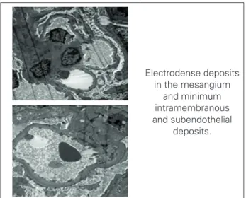
Electrodense deposits in the mesangium
and minimum intramembranous and subendothelial
deposits.
Figure 3. Electron microscopy.
Source: MEC Federal University of the Triângulo Mineiro (UFTM) - Department of Nephrology - Discipline of General Pathology - Uberaba, MG. General Pathology Discipline. Http://www.uftm.edu.br/ patge/ Federal University of the Triângulo Mineiro.
IgA deposits were seen in the renal tubules in all the cases, around the cylinders, and Kappa and Lambda in 75%. Electrodense deposits were found in the glomerular structures of three patients, all with type 1 MPGN. In summary, the sample showed: four patients with type 1 MPGN; one patient with mesangioproliferative segmental sclerosis and C3 deposits; one patient with mesangial deposits of IgA, C1q and C3, and one patient with segmental and mesangial sclerosis with of IgM, C3 and C1q deposits.2
Schistosomotic glomerulopathy occurs because of chronic infection by Schistosoma mansoni.1,6 According to the Department of Surveillance of Communicable Diseases of the Ministry of Health,1 Dos Santos et al.10 and Nussenzveig et al.,3 when renal injury is established, the histological picture is usually
The second type of glomerulopathy most found in these cases is FSGS; however, the relationship between FSGS and Schistosoma glomerulopathy has not yet been well established,3,8 and it is more associated with African descendants.
Our patient was positive for C1q, which according to Gusmão et al.,11 has been strongly associated with renal involvement due to systemic lupus erythematosus (SLE), although she did not have all the criteria for SLE, according to the Brazilian Society of Rheumatology.12
The histological diagnosis of IgA nephropathy (IgAN) is direct, and it is defined by the presence of IgA-dominant or codominant immune deposits within the glomerulus, as shown by immunohistochemistry and immunofluorescence.13,14 The product of the activation of the classical complement pathway: C1q, is not typically found in IgA nephropathy.
According to the Oxford classification,13 the intensity of IgA immunopositivity should be more than traces, and its distribution is characteristically mesangial, with or without deposits in capillary loops. IgG and IgM may be positive, but not at a greater intensity than IgA.
According to Barsoum et al.,15 IgA may be a late mediator of glomerular injury in these cases. The dominance of mesangial IgA deposits in the Egyptian context is common, suggesting significant glomerular pathogenicity by IgA, requiring a factor of comorbidity with concomitant exposure to schistosomal antigens.
The glomerular deposits of IgA that have been found in patients with portal hypertension in the hepatosplenic form of schistosomiasis can be considered as a factor of progression of renal disease, and are explained by the dysfunction in immunoglobulin clearance of by Kupffer cells.14
The importance of IgA in the pathogenesis of this nephropathy is unknown, and studies are needed to redefine the molecular structure of glomerular IgA deposits.14
Patients diagnosed with IgAN are treated with angiotensin converting enzyme inhibitors (ACEI) and/or ARB, and associations may be more effective when the patients have proteinuria ≥ 1 g/day, being hypertensive or not, with normal renal function and mild histological lesions;16,17 as it was the case in question, showing progressive proteinuria improvement.
CONCLUSION
Although global and national parameters present MPGN as the main histological lesion in Schistosoma mansoni infection with renal impairment,3,8,18 the case in question presents histology of a schistosomal GN with mesangial deposits of IgA.
The importance of IgA in the pathogenesis of this nephropathy is still unknown. Treatment with ARB resulted in improvements in proteinuria, contributing to retard the progression to ESRD.
Mesangial proliferative GN is the earliest and most frequent form, followed by MPGN, favoring the hypothesis of the mesangium as the initial target. The clinical presentation is variable, and glomerulopathy is asymptomatic in up to 35% of the patients. However, since the diagnosis is usually late, SN, SAH, and hypocomplementemia are common findings.7,19
Considering the national frequency of the disorder, it is necessary to understand the different histological presentations of schistosomal glomerulopathy.3,18
Patients with schistosomal glomerulopathy do not show improvements in terms of disease progression with antiparasitic treatment; 8,10,20 however, treatment with antiproteinuric agents can delay the progression of ESRD.8,21
REFERENCES
1. Brasil. Ministério da Saúde. Vigilância da Esquistossomose Mansoni: diretrizes técnicas. Secretaria de Vigilância em Saúde, Departamento de Vigilância das Doenças Transmissíveis. 4ª ed. Brasília: Ministério da Saúde; 2014.
2. Rodrigues VL, Otoni A, Voieta I, Antunes CM, Lambertucci JR. Glomerulonephritis in schistosomiasis mansoni: a time to reappraise. Rev Soc Bras Med Trop 2010;43:638-42 DOI: http://dx.doi.org/10.1590/S0037-86822010000600007 3. Nussenzveig I, De Brito T, Carneiro CR, Silva AM. Human
Schistosomomansoni-associated glomerulopathy in Brazil. Nephrol Dial Transplant 2002;17:4-7. DOI: http://dx.doi. org/10.1093/ndt/17.1.4
4. Nascimento GL. Formas graves da esquistossomose mansoni: carga epidemiológica e custos no Brasil em 2010. [Dissertação de mestrado]. Brasília: Universidade de Brasília, Faculdade de Medicina; 2013. 5. Abensur H, Nussenzveig I, Saldanha LB, Pestalozzi MS, Barros
MT, Marcondes M, et al. Nephrotic syndrome associated with hepatointestinal schistosomiasis. Rev Inst Med Trop Sao Paulo 1992;34:273-6. PMID: 1342083 DOI: http://dx.doi. org/10.1590/S0036-46651992000400002
6. Barros RT, Alves MAVFR, Dantas M, Kirsztajn GM, Sens YAS. Glomerulopatias: patogenia, clínica e tratamento. 3a ed. São Paulo: Sarvier; 2012.
7. Brito TD, Nussenzveig I, Carneiro CR, Silva AM. Schistosoma mansoni associated glomerulopathy. Rev Inst Med Trop Sao Paulo 1999;41:269-72. PMID: 10602539 DOI: http://dx.doi. org/10.1590/S0036-46651999000500001
8. Barsoum R. The changing face of schistosomal glomerulopathy. Kidney Int 2004;66:2472-84. PMID: 15569345 DOI: http:// dx.doi.org/10.1111/j.1523-1755.2004.66042.x
9. Johnson RJ, Feehally J, Floege J. Comprehensive clinical nephrology. 5th ed. Philadelphia: Elsevier Saunders; 2015. p. 657-64.
10. dos-Santos WL, Sweet GM, Bahiense-Oliveira M, Rocha PN. Schistosomal glomerulopathy and changes in the distribution of histological patterns of glomerular diseases in Bahia, Brazil. Mem Inst Oswaldo Cruz 2011;106:901-4. PMID: 22124564 DOI: http://dx.doi.org/10.1590/S0074-02762011000700017
11. Gusmão I, Moura CGG, Mangueira CLP, Rocha MS, Cruz CMS. Anticorpos anti-C1q e sua correlação com atividade do lúpus eritematoso sistêmico. Rev Soc Bras Clin Med 2014;12:149-53. 12. Jesus AA, Campos LMA, Liphaus BL, Carneiro-Sampaio
M, Mangueira CLP, Rosseto EA, et al. Anticorpos anti-C1q, anticromatina/nucleossomo e anti-dsDNA em pacientes com lúpus eritematoso sistêmico juvenil. Rev Bras Reumatol 2012;52:976-81. DOI: http://dx.doi.org/10.1590/S0482-50042012000600015 13. Working Group of the International IgA Nephropathy Network
and the Renal Pathology Society. Roberts IS, Cook HT, Troyanov S, Alpers CE, Amore A, Barratt J, et al. The Oxford classification of IgA nephropathy: pathology definitions, correlations, and reproducibility. Kidney Int 2009;76:546-56. PMID: 19571790 14. Alamartine E, Sauron C, Laurent B, Sury A, Seffert A, Mariat C.
The use of the Oxford classification of IgA nephropathy to predict renal survival. Clin J Am Soc Nephrol 2011;6:2384-8. DOI: http:// dx.doi.org/10.2215/CJN.01170211
15. Barsoum R, Nabil M, Saady G, Genin C, Saleh E, Francis M, et al. Immunoglobulin-A and the pathogenesis of schistosomal glomerulopathy. Kidney Int 1996;50:920-8. PMID: 8872967 DOI: http://dx.doi.org/10.1038/ki.1996.392
16. Sociedade Brasileira de Nefrologia. Tratamento da Nefropatia por IgA. J Bras Nefrol 2005;27:18-21.
17. Ramos EA, Andrade ZA. Chronic glomerulonephritis associated with hepatosplenic Schistosomiasis mansoni. Rev Inst Med Trop Sao Paulo 1987;29:162-7. DOI: http://dx.doi. org/10.1590/S0036-46651987000300008
18. Barsoum RS. Schistosomal glomerulopathies. Kidney Int 1993;44:1-12. PMID: 8355449 DOI: http://dx.doi.org/10.1038/ki.1993.205 19. Sociedade Brasileira de Nefrologia. Glomerulopatias associadas
a doenças parasitárias. J Bras Nefrol 2005;27:30-1.
20. Kidney Disease: Improving Global Outcomes (KDIGO) Glomerulonephritis Work Group. KDIGO Clinical Practice Guideline for Glomerulonephritis. Kidney Int Suppl 2012;2:139-74. 21. Gonzaga CC, Passarelli Jr. O, Amodeo C. Interações